Submitted Successfully!
To reward your contribution, here is a gift for you: A free trial for our video production service.
Thank you for your contribution! You can also upload a video entry or images related to this topic.
Version Summary Created by Modification Content Size Created at Operation
1 -- 2581 2023-08-17 11:29:39 |
2 layout + 10 word(s) 2591 2023-08-18 07:21:52 |

Video Upload Options

Do you have a full video?

Confirm

Are you sure to Delete?
Cite
If you have any further questions, please contact Encyclopedia Editorial Office.
Caturano, A.; D’angelo, M.; Mormone, A.; Russo, V.; Mollica, M.P.; Salvatore, T.; Galiero, R.; Rinaldi, L.; Vetrano, E.; Marfella, R.; et al. Oxidative Stress in Type 2 Diabetes. Encyclopedia. Available online: https://encyclopedia.pub/entry/48163 (accessed on 04 May 2024).
Caturano A, D’angelo M, Mormone A, Russo V, Mollica MP, Salvatore T, et al. Oxidative Stress in Type 2 Diabetes. Encyclopedia. Available at: https://encyclopedia.pub/entry/48163. Accessed May 04, 2024.
Caturano, Alfredo, Margherita D’angelo, Andrea Mormone, Vincenzo Russo, Maria Pina Mollica, Teresa Salvatore, Raffaele Galiero, Luca Rinaldi, Erica Vetrano, Raffaele Marfella, et al. "Oxidative Stress in Type 2 Diabetes" Encyclopedia, https://encyclopedia.pub/entry/48163 (accessed May 04, 2024).
Caturano, A., D’angelo, M., Mormone, A., Russo, V., Mollica, M.P., Salvatore, T., Galiero, R., Rinaldi, L., Vetrano, E., Marfella, R., Monda, M., Giordano, A., & Sasso, F.C. (2023, August 17). Oxidative Stress in Type 2 Diabetes. In Encyclopedia. https://encyclopedia.pub/entry/48163
Caturano, Alfredo, et al. "Oxidative Stress in Type 2 Diabetes." Encyclopedia. Web. 17 August, 2023.
Oxidative Stress in Type 2 Diabetes
Edit

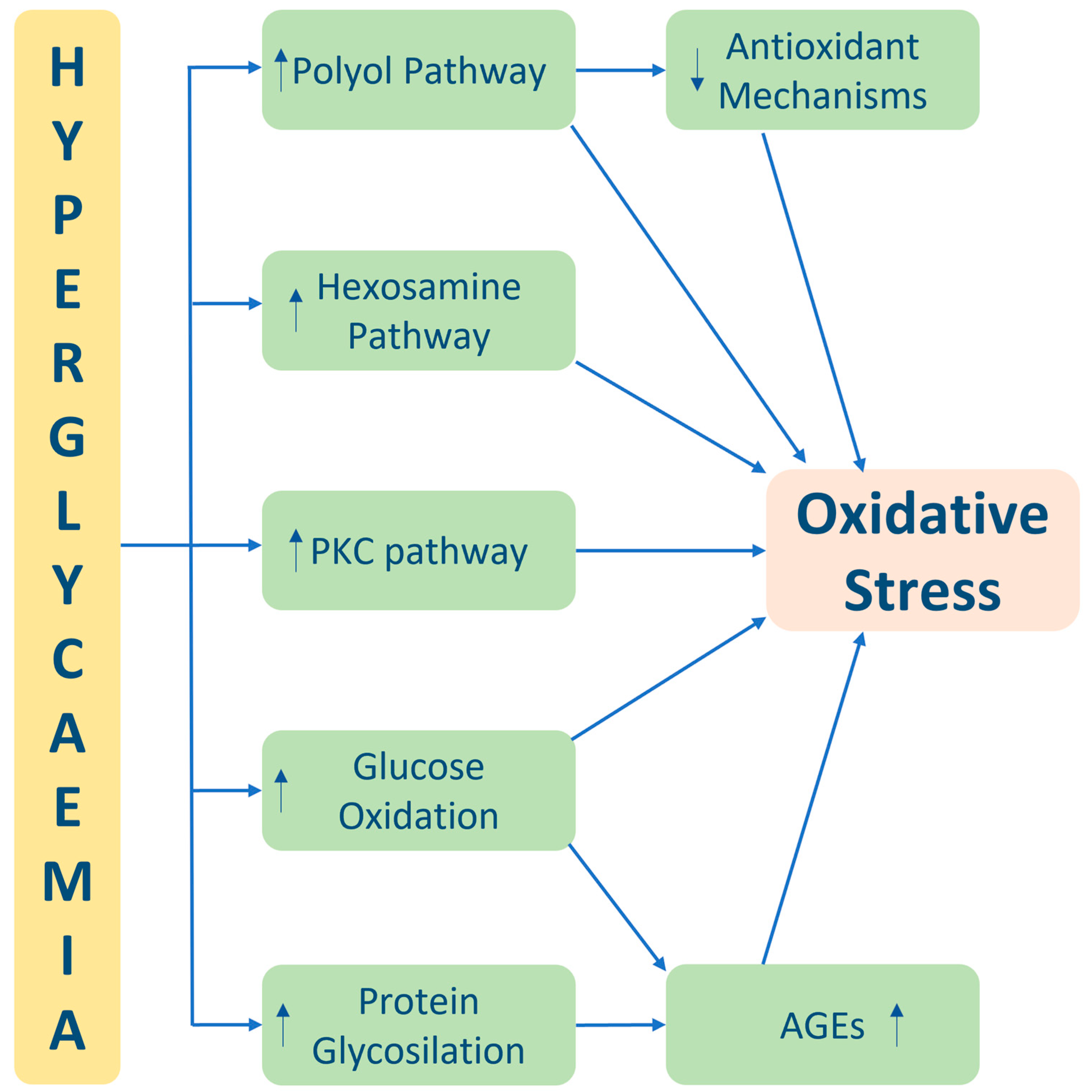
Oxidative stress is a critical factor in the pathogenesis and progression of diabetes and its associated complications. The imbalance between reactive oxygen species (ROS) production and the body’s antioxidant defence mechanisms leads to cellular damage and dysfunction. In diabetes, chronic hyperglycaemia and mitochondrial dysfunction contribute to increased ROS production, further exacerbating oxidative stress. This oxidative burden adversely affects various aspects of diabetes, including impaired beta-cell function and insulin resistance, leading to disrupted glucose regulation. Additionally, oxidative stress-induced damage to blood vessels and impaired endothelial function contribute to the development of diabetic vascular complications such as retinopathy, nephropathy, and cardiovascular diseases. Moreover, organs and tissues throughout the body, including the kidneys, nerves, and eyes, are vulnerable to oxidative stress, resulting in diabetic nephropathy, neuropathy, and retinopathy. Strategies to mitigate oxidative stress in diabetes include antioxidant therapy, lifestyle modifications, and effective management of hyperglycaemia

oxidative stress type 2 diabetes diet Mediterranean diet physical activity lifestyle modifications diabetes complications

1. Genesis of Oxidative Stress in Diabetes

Oxidative stress in diabetes arises from a complex interplay of various factors, including the accumulation of glycolysis intermediates, activation of the polyol pathway, formation of advanced glycation end products (AGEs), activation of Protein Kinase C (PKC), and activation of the hexosamine pathway (Figure 1) [1].
Figure 1. Main pathophysiological mechanism of hyperglycaemia induced oxidative stress. Abbreviations: PKC—Protein Kinase C and AGEs—advanced glycation end products.

1.1. Pentose Phosphate and Glycolytic Pathways and Oxidative Stress

The primary cause of oxidative stress is undoubtedly the elevation in blood glucose concentration. In fact, once glucose enters cells, it undergoes oxidation through either the pentose phosphate pathway, leading to the production of biosynthetic molecules and NADPH, or through the glycolytic pathway [2]. Glycolysis continues with the Krebs cycle, resulting in the generation of nicotinamide adenine dinucleotide (NADH) and reduced flavin adenine dinucleotide (FADH2), which are utilised in oxidative phosphorylation to produce ATP. This process generates reactive oxygen species (ROS) such as hydrogen peroxide (H2O2), superoxide anion (O2•-), and hydroxyl radicals (•OH). Under normal physiological conditions, the antioxidant defence system comprising enzymes such as superoxide dismutase (SOD), catalase (CAT), and glutathione peroxidase (GPx) effectively neutralises ROS [3]. However, when blood glucose concentration becomes excessively high, the production of radicals is upregulated, leading to the inhibition of antioxidant systems. Consequently, this downregulation results in DNA damage and the subsequent production of DNA repair enzymes such as Poly-ADP Ribose Polymerase-1 (PARP-1), which inactivates glyceraldehyde-3-phosphate dehydrogenase (GAPDH), leading to the accumulation of glyceraldehyde-3-phosphate (GAP), Glucose 6-phosphate (G-6-P), and Fructose 6-phosphate (F-6-P). The elevation of these three intermediates contributes to various reactions that converge on oxidative stress: G-6-P and GAP can undergo autoxidation, leading to the formation of AGE precursors; G-6-P and F-6-P can follow the Polyol pathway, while GAP induces the activation of PKC [4]. Notably, with the increased glucose concentration, the enzyme hexokinase becomes saturated and cannot catalyse the formation of G-6-P. Consequently, glucose is converted to sorbitol via aldose reductase, which is further converted to fructose by sorbitol dehydrogenase (SDH). This process consumes excess NADPH, which serves as a substrate for GPx to produce glutathione (GSH) [5]. Thus, the inhibition of antioxidant enzymes in this pathway also contributes to oxidative stress. Additionally, under hyperglycaemic conditions, SDH is upregulated, leading to increased fructose production, which is then converted into the triose-phosphates GAP and dihydroxyacetone-3-phosphate (DHA-3-P), ultimately resulting in the activation of PKC and oxidative stress [6].
Glucose in excess can undergo autoxidation to form glyoxal, while glucose-derived GAP and dihydroxyacetone-3-phosphate (DHAP) can undergo non-enzymatic dephosphorylation to form methylglyoxal. Both products, along with 3-deoxyglucosone (or Amadori product), serve as precursors for the formation of AGEs, which react with elements of the extracellular matrix (ECM), leading to AGE production. Specifically, the interaction of the carboxyl residue of a glucose molecule with the terminal ε-amino residue of a protein non-enzymatically results in the alteration of protein functionality. Once formed, AGEs interact with AGE receptors (RAGE), inducing oxidative stress and activating PKC, which upregulates NADPH oxidase and lipoxygenase, thereby generating ROS [7]. Moreover, the excess of GAP can be converted to dihydroxyacetone-3-phosphate (DHA-3-P), which is subsequently reduced to glycerol 3-phosphate. When combined with fatty acids, glycerol 3-phosphate forms diacylglycerol (DAG), capable of inducing PKC [8].

1.2. Inflammation and Oxidative Stress

There exists a strong correlation between inflammation and oxidative stress, as the immune system triggers the production of pro-inflammatory cytokines and chemokines, activating ROS-producing macrophages to eliminate pathogens. However, the chronic inflammatory state that arises in diabetes leads to continuous ROS production, resulting in cellular damage and the depletion of antioxidant systems [9]. In a reciprocal manner, ROS stimulate the expression of pro-inflammatory cytokines by activating transcription factors such as nuclear factor-kappa B (NF-κB) and activator protein-1 (AP-1). Additionally, the excess adipose tissue often observed in type II diabetes secretes pro-inflammatory cytokines, including tumour necrosis factor alpha (TNF-α) and interleukins 1 (IL-1) and 6 (IL-6), further amplifying oxidative stress [1]. Another mechanism implicated in diabetes complications is the hexosamine pathway. Hyperglycaemia leads to the accumulation of fructose, as mentioned earlier, which is converted to Glucosamine 6-phosphate and ultimately to UDP-N-Acetylhexosamine (UDP-GLCNac), potentiating O-Glucosamine-N-Acetyltransferase (OGT) activity [10]. OGT binds O-GlcNAc to serine and threonine residues of transcription factors, such as Sp1, thereby altering gene expression. Sp1, a transcription factor commonly implicated in diabetic complications, regulates the expression of various genes, including tissue-type plasminogen activator inhibitor-1 (PAI-1) and transforming growth factor-β1 (TGF-β1). PAI-1 is believed to play a role in diabetic neuropathy, although further studies are required to fully elucidate its function [11], as it likely impairs fibrinolysis in neural blood vessels, promoting nerve ischemia and oxidative stress. Conversely, the upregulation of TGF-β1 induces ROS production in vascular smooth muscle and endothelial cells by activating NADPH oxidase. TGF-β1 is involved in diabetic nephropathy, stimulating collagen formation and inhibiting mesangial cell mitosis [2][12]. Furthermore, the accumulation of ROS activates mitochondrial uncoupling protein-2 (UCP-2), reducing ATP production and initiating a cascade of events that ultimately impairs insulin secretion by pancreatic beta cells [13]. Moreover, oxidative stress induced by hyperglycaemia has been shown to inhibit the expression of the insulin gene and promote beta-cell apoptosis. These conditions collectively disrupt beta-cell function and insulin release, contributing to hyperglycaemia and exacerbating oxidative stress [14].

2. Oxidative Stress Role in the Development of Type 2 Diabetes Complications

In diabetic pathology, which is becoming progressively more prevalent worldwide [15], inflammation is playing an increasingly significant role. Navarro and Mora [16], building upon the hypothesis proposed by Pickup and Crook [17], suggest that diabetes is transitioning from a metabolic disorder to a genuine inflammatory pathology, where prolonged and improper activation of the immune system contributes to the development and progression of the disease. In addition to its involvement in the onset of diabetes [18][19][20], an inflammatory state plays a crucial role in the advancement of the disease and the development of both microvascular and macrovascular complications [8][21][22][23][24][25]. Chronic hyperglycaemia is the primary driver of this process, as it induces a persistent activation of the innate immune system and triggers oxidative stress, leading to the production of harmful free radicals [26][27] that adversely affect the body [28][29][30][31]. Free radicals, specifically ROS and reactive nitrogen species (RNS), are highly unstable molecules with unpaired electrons, making them potent oxidants. Although our body naturally produces these reactive species, excessive production, as observed in diabetes, is detrimental [32]. Many cells in our body, such as red blood cells and endothelial cells, are particularly vulnerable to free radicals due to their high levels of polyunsaturated fatty acids, molecular oxygen, and ferrous ions [33][34]. In diabetic pathology, chronic inflammation and oxidative stress create a vicious cycle: each stimulates the other, resulting in mutual amplification. ROS and RNS, produced as a consequence of inflammation, can promote the transcription of growth factors like Nf-Kb and AP-1, which in turn stimulate the production of inflammatory proteins [35].
Free radicals play a crucial role in the onset and progression of diabetic complications through three different pathways: the aldose reductase pathway, the PKC pathway, and the production of AGEs. The aldose reductase pathway normally converts glucose to sorbitol through the simultaneous oxidation of sorbitol to fructose in a nicotinamide adenine dinucleotide phosphate (NADPH)-dependent manner. In diabetes, chronic hyperglycaemia saturates this pathway early on, causing more than 30% of glucose to be metabolised through the polyol pathway [4][36]. The increased utilisation of this pathway depletes intracellular NADPH and increases extracellular NADH levels. The excess NADH serves as a substrate for the enzyme NADH oxidase, leading to the generation of excessive ROS [37]. This mechanism has been primarily implicated in the development of diabetic retinopathy due to sorbitol accumulation in the retina [38][39][40].
Excess glucose in the body can also react spontaneously with the amino groups of plasma proteins, forming Schiff bases that subsequently contribute to the formation of AGEs [41]. These AGEs react with other membrane molecules in endothelial cells and blood vessels, leading to structural and functional alterations in proteins and the activation of pro-inflammatory gene transcription [22].
Chronic hyperglycaemia induces oxidative stress through the PKC-dependent activation of the NADPH oxidase pathway. While this enzyme is normally present primarily in phagocytic cells, in diabetes, it is also expressed in fibroblasts, endothelial cells, and smooth muscle cells, where it becomes the main producer of ROS. PKC also activates endothelial nitric oxide synthase (eNOS), NADPH oxidase, phospholipase A2 (PLA2), endothelin-1 (ET-1), vascular endothelial growth factor-B (TGF-B), and NF-KB, all of which play a role in the development of complications [42]. PKC, when activated by diacylglycerol, also directly transcribes genes involved in protein synthesis, leading to direct cell damage, capillary occlusion, and reduced blood flow [43][44].
During periods of hyperglycaemia, the cytokine cascade resulting from the interaction between ROS and inflammation is characterised by an increase in monocyte chemoattractant protein-1 and a decrease in insulin-like growth factor-1 levels [45][46][47]. These proteins, as observed by Al Hannan et al. [48], can induce dedifferentiation of adipose tissue through macrophages, resulting in increased hyperinsulinemia and insulin resistance. It is also possible that, in this cellular crosstalk, a role can also be played by the opioid system, though more studies are needed [49]. Our body normally possesses various protection systems against ROS and oxidative stress, with the main ones being SOD, catalase, and glutathione peroxidase. However, in diabetes, these defence mechanisms are compromised due to chronic inflammation, contributing to the progression of damage and the disease itself.
Microvascular complications of diabetes include retinopathy, nephropathy, and neuropathy. Retinopathy, in particular, mainly develops due to damage to small retinal vessels and connective tissue, sometimes resulting in the formation of small haemorrhages. The retinal tissue is highly susceptible to damage mediated by oxidative stress due to its high concentration of polyunsaturated fats [50]. Diabetic nephropathy involves the interstitial and glomerular membranes, which are damaged by inflammatory cytokines and ROS, leading to the loss of proteins such as albumin and a decreased glomerular filtration rate [51][52]. Additionally, changes in kidney haemodynamics may occur due to thinning of the basement membrane, expansion of renal mesangial cells, and hyperplasia of the extracellular matrix. Increased ROS levels in the kidney cause vasoconstriction, endothelial dysfunction, and enhanced sodium reabsorption [38]. Diabetic neuropathy, the most common microvascular complication of diabetes, affects approximately 50% of patients after 20 years of the disease. Oxidative stress in the central nervous system can induce neuronal apoptosis [53] and impair the ability to repair and regenerate neurons effectively.

3. Physical Exercise and Oxidative Stress

3.1. Effects of Different Physical Exercise Protocols on Oxidative Stress Markers in Patients with Type 2 Diabetes Mellitus

Various physical exercise protocols have been recommended for patients with type 2 diabetes mellitus (T2DM), including continuous moderate-intensity exercise (CMIE) [54], resistance exercise (RE) [55], high-intensity interval exercise (HIIE) [56], and concurrent exercise (CE), which combines CMIE and RE [57].

3.2. Resistance Exercise

RE involves performing both monoarticular or polyarticular movements against resistance and returning to the start position [55]. While RE has been shown to improve metabolic health in T2DM patients [58], its impact on oxidative stress parameters in this population remains inconclusive [59]. Remarkably, the response to RE on oxidative stress markers seems to vary depending on the health status of the patients. In healthy individuals, RE protocols involving three sets at 65–70% of one repetition maximum (RM) performed three times a week for 6–8 weeks resulted in decreased plasma levels of malondialdehyde and increased blood activity of GPx [60][61]. Similarly, high-intensity protocols (3 × 3–6 repetitions at 85–90% of 1 RM) have been associated with an increase in blood SOD activity [60]. However, studies on healthy subjects [60][61] involved younger participants under the age of 30 years, compared to the trial enrolling diabetic patients [59], whose participants were over 50 years old. Due to this, it is worth noting that the effectiveness of T2DM might have been influenced by age-related factors or lower adaptive responses typical of this patient group.

3.3. Continuous Moderate-Intensity Exercise

CMIE, or “aerobic exercise”, involves cyclical modalities such as walking, jogging, and cycling, engaging large muscle groups [62]. Studies examining the effects of CMIE on oxidative stress biomarkers in T2DM patients generally show improvements in redox balance due to an increase in blood antioxidant biomarkers [60][63][64][65] and a decrease in blood protein oxidation biomarkers [65] and DNA in urine [66]. Moreover, these positive responses align with improvements in clinical parameters such as increased cardiovascular function [63][64][65], lipid profile control [60], body composition enhancement [64][66], and glycaemic control [64][66]. Of note, only one study reported no significant changes in antioxidant abundance/activity or markers of oxidative stress damage, specifically malondialdehyde levels, even though positive effects on clinical parameters like fasting glycaemia, HOMA index, and body fat percentage were confirmed [56]. Krause et al. noted a significant increase in blood CAT activity following a moderate-intensity free-walking protocol, along with a decrease in carbonylated protein levels with low/moderate-intensity protocols. Though reporting an approximately 2% body fat percentage reduction, this was not statistically significant, and there were no significant changes in fasting glycaemia and the HOMA index [65]. The response to CMIE on oxidative stress levels appears to depend on the intervention’s duration and the subjects’ age. It has been shown that participants over 60 years of age may require an intervention period of 16 weeks or more, while those below 60 years of age may experience changes with 12-week interventions, with an exercise duration equal to or greater than 30 min at moderate intensity [67].

3.4. High-Intensity Interval Exercise

HIIT involves the repeated performance of short bursts of intense exercise (ranging from 10 s to 4 min) at an intensity level exceeding the anaerobic threshold, followed by periods of recovery at low intensity or complete rest [68]. Limited evidence exists regarding the effects of HIIT on oxidative stress markers in patients with T2DM. Mitranun et al. [56] conducted a clinical trial enrolling 43 patients randomly assigned to three groups: HIIT, CMIE, and a control group with no exercise intervention. The HIIT group participated in 12 weeks of treadmill jogging with three weekly sessions, with a gradually increasing session duration from 30 to 60 min. Although no statistically significant changes in SOD concentrations were observed in any of the experimental groups, the HIIT group exhibited a significant decrease in malondialdehyde levels and an increase in GPx enzyme activity. These results correlated with a reduced percentage of HbA1c, fasting glycaemia, and HOMA index, as well as improvements in other clinical parameters related to cardiovascular function, lipid profile, and body composition [56].

3.5. Concurrent Exercise

The CE protocol typically combines CMIE with RE within the same training session [69]. CE has shown benefits in glycaemic control (both fasting glycaemia and HbA1c levels) and body composition in patients with T2DM, and it has been found to induce positive effects on oxidative stress parameters [70]. In this context, CE has been associated with enhancements in blood antioxidant biomarkers like GSH and SOD, along with a decrease in malondialdehyde levels [59]. These positive responses have been reported to be observed after 8–16 weeks of intervention [56], thus highlighting the potential of CE protocol in promoting a more favourable redox balance in T2DM.

References

  1. Oguntibeju, O.O. Type 2 diabetes mellitus, oxidative stress and inflammation: Examining the links. Int. J. Physiol. Pathophysiol. Pharmacol. 2019, 11, 45–63.
  2. Ighodaro, O.M. Molecular pathways associated with oxidative stress in diabetes mellitus. Biomed. Pharmacother. 2018, 108, 656–662.
  3. Makino, A.; Scott, B.T.; Dillmann, W.H. Mitochondrial fragmentation and superoxide anion production in coronary endothelial cells from a mouse model of type 1 diabetes. Diabetologia 2010, 53, 1783–1794.
  4. Funk, S.D.; Yurdagul, A., Jr.; Orr, A.W. Hyperglycemia and endothelial dysfunction in atherosclerosis: Lessons from type 1 diabetes. Int. J. Vasc. Med. 2012, 2012, 569654.
  5. Figueroa-Romero, C.; Sadidi, M.; Feldman, E.L. Mechanisms of disease: The oxidative stress theory of diabetic neuropathy. Rev. Endocr. Metab. Disord. 2008, 9, 301–314.
  6. Garg, S.S.; Gupta, J. Polyol pathway and redox balance in diabetes. Pharmacol. Res. 2022, 182, 106326.
  7. Twarda-Clapa, A.; Olczak, A.; Białkowska, A.M.; Koziołkiewicz, M. Advanced Glycation End-Products (AGEs): Formation, Chemistry, Classification, Receptors, and Diseases Related to AGEs. Cells 2022, 11, 1312.
  8. Giacco, F.; Brownlee, M. Oxidative stress and diabetic complications. Circ. Res. 2010, 107, 1058–1070.
  9. Luc, K.; Schramm-Luc, A.; Guzik, T.J.; Mikolajczyk, T.P. Oxidative stress and inflammatory markers in prediabetes and diabetes. J. Physiol. Pharmacol. 2019, 70, 809–824.
  10. Nie, H.; Yi, W. O-GlcNAcylation, a sweet link to the pathology of diseases. J. Zhejiang. Univ. Sci. B. 2019, 20, 437–448.
  11. Buse, M.G. Hexosamines, Insulin resistance, and the complications of diabetes: Current status. Am. J. Physiol. Endocrinol. Metab. 2006, 290, E1–E8.
  12. Darenskaya, M.A.; Kolesnikova, L.I.; Kolesnikov, S.I. Oxidative Stress: Pathogenetic Role in Diabetes Mellitus and Its Complications and Therapeutic Approaches to Correction. Bull. Exp. Biol. Med. 2021, 171, 179–189.
  13. Sreedhar, A.; Zhao, Y. Uncoupling protein 2 and metabolic diseases. Mitochondrion 2017, 34, 135–140.
  14. Sjöholm, Å.; Corbett, J.A.; Leung, P.S.; Portha, B. Editorial: Understanding the gene expression of β cell dysfunction in diabetes. , Front. Endocrinol. Lausanne 2022, 13, 1069991.
  15. Badawi, A.; Klip, A.; Haddad, P.; Cole, D.E.; Bailo, B.G.; El-Sohemy, A.; Karmali, M. Type 2 diabetes and inflammation: Prospects for biomarkers of risk and nutritional intervention. Diabetes Metab. Syndr. Obes. 2010, 3, 173–186.
  16. Navarro, J.F.; Mora, C. Role of inflammation in diabetic complications. Nephrol. Dial. Transplant. 2005, 20, 2601–2604.
  17. Crook, M. Is type 2 diabetes mellitus a disease of the innate immune system. Diabet. Med. 2004, 21, 203–207.
  18. Westermann, D.; Van Linthout, S.; Dhayat, S.; Dhayat, N.; Schmidt, A.; Noutsias, M.; Song, X.Y.; Spillmann, F.; Riad, A.; Schultheiss, H.P.; et al. Tumor necrosis factor-alpha antagonism protects from myocardial inflammation and fibrosis in experimental diabetic cardiomyopathy. Basic. Res. Cardiol. 2007, 102, 500–507.
  19. Zhang, P.; Zhang, X.; Brown, J.; Vistisen, D.; Sicree, R.; Shaw, J.; Nichols, G. Global healthcare expenditure on diabetes for 2010 and 2030. Diabetes Res Clin Pract. 2010, 87, 293–301.
  20. Derosa, G.; D’Angelo, A.; Bonaventura, A.; Bianchi, L.; Romano, D.; Maffioli, P. Effects of berberine on lipid profile in subjects with low cardiovascular risk. Expert. Opin. Biol. Ther. 2013, 13, 475–482.
  21. Butkowski, E.G.; Jelinek, H.F. Hyperglycemia, oxidative stress and inflammation markers. Redox Rep. 2017, 22, 257–264.
  22. Collier, B.; Dosssett, L.A.; May, A.K.; Diaz, J.J. Glucose control and the inflammatory response. Nutr. Clin. Pract. 2008, 23, 3–15.
  23. Palmiero, G.; Cesaro, A.; Vetrano, E.; Pafundi, P.C.; Galiero, R.; Caturano, A.; Moscarella, E.; Gragnano, F.; Salvatore, T.; Rinaldi, L.; et al. Impact of SGLT2 Inhibitors on Heart Failure: From Pathophysiology to Clinical Effects. Int. J. Mol. Sci. 2021, 22, 5863.
  24. Falco, L.; Tessitore, V.; Ciccarelli, G.; Malvezzi, M.; D’Andrea, A.; Imbalzano, E.; Golino, P.; Russo, V. Antioxidant Properties of Oral Antithrombotic Therapies in Atherosclerotic Disease and Atrial Fibrillation. Antioxidants 2023, 12, 1185.
  25. Russo, V.; Fabiani, D. Put out the fire: The pleiotropic anti-inflammatory action of non-vitamin K oral anticoagulants. Pharmacol. Res. 2022, 182, 106335.
  26. Salvatore, T.; Galiero, R.; Caturano, A.; Vetrano, E.; Rinaldi, L.; Coviello, F.; Di Martino, A.; Albanese, G.; Marfella, R.; Sardu, C.; et al. Effects of Metformin in Heart Failure: From Pathophysiological Rationale to Clinical Evidence. Biomolecules 2021, 11, 1834.
  27. Tousoulis, D.; Papageorgiou, N.; Androulakis, E.; Siasos, G.; Latsios, G.; Tentolouris, K.; Stefanadis, C. Diabetes mellitus-associated vascular impairment: Novel circulating biomarkers and therapeutic approaches. J. Am. Coll. Cardiol. 2013, 62, 667–676.
  28. Brownlee, M. Biochemistry and molecular cell biology of diabetic complications. Nature 2001, 414, 813–820.
  29. Esposito, K.; Nappo, F.; Marfella, R. Inflammatory cytokine concentrations are acutely increased by hyperglycemia in humans: Role of oxidative stress. Circulation 2002, 106, 2067–2072.
  30. Monnier, L.; Lapinski, H.; Colette, C. Contributions of fasting and postprandial plasma glucose increments to the overall diurnal hyperglycemia of type 2 diabetic patients: Variations within increasing levels of HbA(1c). Diabetes Care 2003, 26, 881–885.
  31. Huang, C.; Kim, Y.; Caramori, M.L.; Fish, A.J.; Rich, S.S.; Miller, M.E.; Russell, G.B.; Mauer, M. Cellular basis of diabetic nephropathy: II. The transforming growth factor-beta system and diabetic nephropathy lesions in type 1 diabetes. Diabetes 2002, 51, 3577–3581.
  32. Halliwell, B. The wanderings of a free radical. Free Radic. Biol. Med. 2009, 46, 531–542.
  33. Yadav, P.; Sarkar, S.; Bhatnagar, D. Lipid peroxidation and antioxidant enzymes in erythrocytes and tissues in aged diabetic rats. Indian. J. Exp. Biol. 1997, 35, 389–392.
  34. Hamden, K.; Carreau, S.; Jamoussi, K.; Miladi, S.; Lajmi, S.; Aloulou, D.; Ayadi, F.; Elfeki, A. 1Alpha, 25 dihydroxyvitamin D3: Therapeutic and preventive effects against oxidative stress, hepatic, pancreatic and renal injury in alloxan-induced diabetes in rats. J. Nutr. Sci. Vitaminol. 2009, 55, 215–222.
  35. Fialkow, L.; Wang, Y.; Downey, G.P. Reactive oxygen and nitrogen species as signaling molecules regulating neutrophil function. Free Radic. Biol. Med. 2007, 42, 153–164.
  36. González, R.G.; Barnett, P.; Aguayo, J.; Cheng, H.M.; Chylack, L.T., Jr. Direct measurement of polyol pathway activity in the ocular lens. Diabetes. 1984, 33, 196–199.
  37. Morré, D.M.; Lenaz, G.; Morré, D.J. Surface oxidase and oxidative stress propagation in aging. J. Exp. Biol. 2000, 203, 1513–1521.
  38. Ishill, N.; Patel, K.P.; Lane, P.H. Nitric oxide synthesis and oxidative stress in the renal cortex of rats with diabetes mellitus. J. Am. Soc. Nephrol. 2001, 12, 1630–1639.
  39. Etoh, T.; Inoguchi, T.; Kakimoto, M.; Sonoda, N.; Kobayashi, K.; Kuroda, J.; Sumimoto, H.; Nawata, H. Increased expression of NAD(P)H oxidase subunits, NOX4 and p22phox, in the kidney of streptozotocin-induced diabetic rats and its reversibility by interventive insulin treatment. Diabetologia 2003, 46, 1428–1437.
  40. Wu, L.J.; Wu, G.; Akhavan Sharif, M.R.; Baker, A.; Jia, Y.; Fahey, F.H.; Luo, H.R.; Feener, E.P.; Clapham, D.E. The voltage-gated proton channel, Hv1, enhances brain damage from ischemic stroke. Nat. Neurosci. 2012, 15, 565–573.
  41. Thallas-Bonke, V.; Thorpe, S.R.; Coughlan, M.T.; Fukami, K.; Yap, F.Y.; Sourris, K.C.; Penfold, S.A.; Bach, L.A.; Cooper, M.E.; Forbes, J.M. Inhibition of NADPH oxidase prevents advanced glycation end product-mediated damage in diabetic nephropathy through a protein kinase C-alpha-dependent pathway. Diabetes 2008, 57, 460–469.
  42. Noh, H.; King, G.L. The role of protein kinase C activation in diabetic nephropathy. Kidney Int. 2007, 72, S49–S53.
  43. Way, K.J.; Katai, N.; King, G.L. Protein kinase C and the development of diabetic vascular complications. Diabet. Med. 2001, 18, 945–959.
  44. Harani, H.; Otmane, A.; Makrelouf, M.; Ouadahi, N.; Abdi, A.; Berrah, A.; Zenati, A.; Alamir, B.; Koceir, E.A. The relationship between inflammation, oxidative stress and metabolic risk factors in type 2 diabetic patients. Ann. Biol. Clin. 2012, 70, 669–677.
  45. Salvatore, T.; Galiero, R.; Caturano, A.; Vetrano, E.; Loffredo, G.; Rinaldi, L.; Catalini, C.; Gjeloshi, K.; Albanese, G.; Di Martino, A.; et al. Coronary Microvascular Dysfunction in Diabetes Mellitus: Pathogenetic Mechanisms and Potential Therapeutic Options. Biomedicines 2022, 10, 2274.
  46. Esser, N.; Legrand-Poels, S.; Piette, J.; Scheen, A.J.; Paquot, N. Inflammation as a link between obesity, metabolic syndrome, and type 2 diabetes. Diabetes Res. Clin. Pract. 2014, 105, 141–150.
  47. Elmarakby, A.A.; Sullivan, J.C. Relationship between oxidative stress and inflammatory cytokines in diabetic nephropathy. Cardiovasc. Ther. 2012, 30, 49–59.
  48. Al Hannan, F.; Culligan, K.G. Human resistin and the RELM of inflammation in diabesity. Diab Metab. Syndr. 2015, 7, 54.
  49. Cozzolino, D.; Sessa, G.; Salvatore, T.; Sasso, F.C.; Giugliano, D.; Lefebvre, P.J.; Torella, R. The involvement of the opioid system in human obesity: A study in normal weight relatives of obese people. J. Clin. Endocrinol. Metab. 1996, 81, 713–718.
  50. Du, Y.; Miller, C.M.; Kern, T.S. Hyperglycemia increases mitochondrial superoxide in the retina and retinal cells. Free Rad. Biol. Med. 2003, 35, 1491–1499.
  51. Kafle, D.; Singh, N.; Singh, S.K.; Islam, N. Relationship between hyperglycemia, inflammation, and oxidative stress in type 2 diabetic nephropathy subjects. Int. J. Pharm. Biol. Arch. 2012, 3, 1203–1206.
  52. Sasso, F.C.; Carbonara, O.; Persico, M.; Iafusco, D.; Salvatore, T.; D’Ambrosio, R.; Torella, R.; Cozzolino, D. Irbesartan reduces the albumin excretion rate in microalbuminuric type 2 diabetic patients independently of hypertension: A randomised double-blind placebo-controlled crossover study. Diabetes Care 2002, 25, 1909–1913.
  53. Russell, J.W.; Sullivan, K.A.; Windebank, A.J.; Herrmann, D.N.; Feldman, E.L. Neurons undergo apoptosis in animal and cell culture models of diabetes. Neurobiol. Dis. 1999, 6, 347–363.
  54. Gómez-Cabrera, M.; Domenech, E.; Viña, J. Moderate exercise is an antioxidant: Upregulation of antioxidant genes by training. Free Radic. Biol. Med. 2008, 44, 126–131.
  55. Gacitua, T.; Karachon, L.; Romero, E.; Parra, P.; Poblete, C.; Russell, J.; Rodrigo, R. Effects of resistance training on oxidative stress-related biomarkers in metabolic diseases: A review. Sport. Sci. Health 2017, 14, 1–7.
  56. Mitranun, W.; Deerochanawong, C.; Tanaka, H.; Suksom, D. Continuous vs interval training on glycemic control and macro- and microvascular reactivity in type 2 diabetic patients. Scand. J. Med. Sci. Sports 2014, 24, 69–76.
  57. Vinetti, G.; Mozzini, C.; Desenzani, P.; Boni, E.; Bulla, L.; Lorenzetti, I.; Romano, C.; Pasini, A.; Cominacini, L.; Assanelli, D. Supervised exercise training reduces oxidative stress and cardiometabolic risk in adults with type 2 diabetes: A randomised controlled trial. Sci. Rep. 2015, 5, 9238.
  58. Gordon, B.A.; Benson, A.C.; Bird, S.R.; Fraser, S.F. Resistance training improves metabolic health in type 2 diabetes: A systematic review. Diabetes Res. Clin. Pract. 2009, 83, 157–175.
  59. de Oliveira, V.N.; Bessa, A.; Jorge, M.L.; Oliveira, R.J.; de Mello, M.T.; De Agostini, G.G.; Jorge, P.T.; Espindola, F.S. The effect of different training programs on antioxidant status, oxidative stress, and metabolic control in type 2 diabetes. Appl. Physiol. Nutr. Metab. 2012, 37, 334–344.
  60. Azizbeigi, K.; Azarbayjani, M.A.; Atashak, S.; Stannard, S.R. Effect of moderate and high resistance training intensity on indices of inflammatory and oxidative stress. Res. Sports Med. 2015, 23, 73–87.
  61. Cakir-Atabek, H.; Demir, S.; PinarbaŞili, R.D.; Gündüz, N. Effects of different resistance training intensity on indices of oxidative stress. J. Strength. Cond. Res. 2010, 24, 2491–2497.
  62. Quílez Llopiz, P.; Reig García-Galbis, M. Glycemic control through physical exercise in type 2 diabetes systematic review. Nutr. Hosp. 2015, 31, 1465–1472.
  63. Kurban, S.; Mehmetoglu, I.; Yerlikaya, H.F.; Gonen, S.; Erdem, S. Effect of chronic regular exercise on serum ischemia-modified albumina levels and oxidative stress in type 2 diabetes mellitus. Endocr. Res. 2011, 36, 116–123.
  64. Moghaddam, D.A.; Heber, A.; Capin, D.; Kreutz, T.; Opitz, D.; Lenzen, E.; Bloch, W.; Brixius, K.; Brinkmann, C. Training increases peroxiredoxin 2 contents in the erythrocytes of overweight/obese men suffering from type 2 diabetes. Wien. Med. Wochenschr. 2011, 161, 511–518.
  65. Krause, M.; Rodrigues-Krause, J.; O’Hagan, C.; Medlow, P.; Davison, G.; Susta, D.; Boreham, C.; Newsholme, P.; O’Donnell, M.; Murphy, C.; et al. The effects of aerobic exercise training at two different intensities in obesity and type 2 diabetes: Implications for oxidative stress, low-grade inflammation and nitric oxide production. Eur. J. Appl. Physiol. 2014, 114, 251–260.
  66. Nojima, H.; Watanabe, H.; Yamane, K.; Kitahara, Y.; Sekikawa, K.; Yamamoto, H.; Yokoyama, A.; Inamizu, T.; Asahara, T.; Kohno, N.; et al. Effect of aerobic exercise training on oxidative stress in patients with type 2 diabetes mellitus. Metabolism 2008, 57, 170–176.
  67. Poblete-Aro, C.; Russell-Guzmán, J.; Parra, P.; Díaz, E.; Reyes, D.; Saavedra, M.; Lanas, F. Efecto del ejercicio físico sobre marcadores de estrés oxidativo en pacientes con diabetes mellitus tipo 2 . Rev. Med. Chil. 2018, 146, 362–372.
  68. Buchheit, M.; Laursen, P.B. High-intensity interval training, solutions to the programming puzzle: Part I: Cardiopulmonary emphasis. Sports Med. 2013, 43, 313–338.
  69. Coffey, V.G.; Hawley, J.A. Concurrent exercise training: Do opposites distract? J. Physiol. 2016, 595, 2883–2896.
  70. Oliveira, C.; Simões, M.; Carvalho, J.; Ribeiro, J. Combined exercise for people with type 2 diabetes mellitus: A systematic review. Diabetes Res. Clin. Pract. 2012, 98, 187–198.
More
Information
Contributors MDPI registered users' name will be linked to their SciProfiles pages. To register with us, please refer to https://encyclopedia.pub/register : , , , , , , , , , , , ,
View Times: 217
Revisions: 2 times (View History)
Update Date: 18 Aug 2023
1000/1000