Submitted Successfully!
To reward your contribution, here is a gift for you: A free trial for our video production service.
Thank you for your contribution! You can also upload a video entry or images related to this topic.
Version Summary Created by Modification Content Size Created at Operation
1 -- 3471 2023-08-17 10:23:18 |
2 Reference format revised. Meta information modification 3471 2023-08-17 11:16:56 |

Video Upload Options

Do you have a full video?

Confirm

Are you sure to Delete?
Cite
If you have any further questions, please contact Encyclopedia Editorial Office.
Radulska, A.; Pelikant-Małecka, I.; Jendernalik, K.; Dobrucki, I.T.; Kalinowski, L. Proteomic and Metabolomic Changes in Psoriasis. Encyclopedia. Available online: https://encyclopedia.pub/entry/48158 (accessed on 27 July 2024).
Radulska A, Pelikant-Małecka I, Jendernalik K, Dobrucki IT, Kalinowski L. Proteomic and Metabolomic Changes in Psoriasis. Encyclopedia. Available at: https://encyclopedia.pub/entry/48158. Accessed July 27, 2024.
Radulska, Adrianna, Iwona Pelikant-Małecka, Kamila Jendernalik, Iwona T. Dobrucki, Leszek Kalinowski. "Proteomic and Metabolomic Changes in Psoriasis" Encyclopedia, https://encyclopedia.pub/entry/48158 (accessed July 27, 2024).
Radulska, A., Pelikant-Małecka, I., Jendernalik, K., Dobrucki, I.T., & Kalinowski, L. (2023, August 17). Proteomic and Metabolomic Changes in Psoriasis. In Encyclopedia. https://encyclopedia.pub/entry/48158
Radulska, Adrianna, et al. "Proteomic and Metabolomic Changes in Psoriasis." Encyclopedia. Web. 17 August, 2023.
Proteomic and Metabolomic Changes in Psoriasis
Edit

Skin diseases such as psoriasis (Ps) and psoriatic arthritis (PsA) are immune-mediated inflammatory diseases. Overlap of autoinflammatory and autoimmune conditions hinders diagnoses and identifying personalized patient treatments due to different psoriasis subtypes and the lack of verified biomarkers. Proteomics and metabolomics have been intensively investigated in a broad range of skin diseases with the main purpose of identifying proteins and small molecules involved in the pathogenesis and development of the disease. 

psoriasis proteomics metabolomics

1. Introduction

Psoriasis is a common and recurrent immune-mediated disease, mainly manifested by skin lesions of well-demarcated and erythematous plaques usually covered with silver scale. The presence of this pathological feature results from epidermal hyperproliferation, abnormal keratinocyte differentiation, neovascularization, and extensive inflammatory infiltration. The etiology of psoriasis is multifactorial and still insufficiently known, but the dominant role is indicated by the dysregulation of the immune system with genetic susceptibilities. Furthermore, a variety of environmental factors, such as stress, infections, smoking cigarettes, and alcohol consumption, are also widely related to the development of psoriasis [1].
Psoriasis is a heterogeneous disease with several different clinical types depending on the morphology and anatomical location of lesions. The most widespread and well-recognized type of psoriasis with its skin manifestation is vulgaris (PsV) or plaque psoriasis (PsP). Several other rare clinical psoriasis subtypes have been described, such as guttate, erythrodermic, or pustular psoriasis, but their frequency of occurrence is less than 10% of cases [2]. The extracutaneous manifestation of psoriasis is psoriatic arthritis (PsA), which leads to chronic, systematic inflammation and debilitation of joints, ligaments, and tendons. Psoriatic arthritis affects 30–35% of patients with dermal subtypes of psoriasis as a result of an exacerbation of the disease [3]. Psoriasis is strongly associated with the development of comorbidities, such as cardiovascular, metabolic, and mental health diseases, often referred to as systemic diseases [4]. The variety of clinical symptoms and the severity of the disease requires complex diagnostics and treatment regimens, which is a great challenge for clinicians. Standard clinical procedure is based on the physical examination of the skin lesions. The hallmark of psoriasis is skin plaques that are usually located on the scalp, trunk, and limbs, especially in the flexural area, but in some cases, the nails are also involved. The hallmark of plaque psoriasis is red, demarcated plaques that are usually covered by silvery scales. The detachment of the adherent scales can lead to the appearance of punctate bleeding spots, known as the Auspritz’s sign. Plaques are usually located symmetrically on the scalp, trunk, and limbs, especially on the elbows and knees. Severe forms of psoriasis can also lead to nail involvement. The characteristic feature of patients with psoriasis is the presence of the Koebner’s phenomenon, which is the appearance of new lesions on previously unchanged skin areas as a result of trauma. Even minor exposure to triggers, such as skin injuries, infections, or tattoos can trigger the development of Koebner’s phenomenon [5][6]. In doubtful and atypical cases, a skin biopsy can be performed, but it is not a routine procedure. Recommended laboratory tests, such as C-reactive protein or red blood cell sedimentation rate (ESR), are mainly used to monitor inflammation and indirectly predict the effectiveness of treatment. Nonetheless, available diagnostic tests are insufficient; therefore, the search for specific biomarkers at the early stage of the disease development is needed. 

2. Rodents as a Model for Psoriasis

2.1. Spontaneous Mouse Models

The first psoriasis-like dermatitis model has been identified as spontaneous mouse mutation in the BALB/c strain, characterized by, e.g., hyperkeratosis, alopecia, and single hair-follicles [7]. Mice with spontaneous genetic mutations, such as Asebia (AB), flaky tail, or flaky skin (Fsn), possess psoriasis-like symptoms that are not fully comparable to humans, including parakeratosis, acanthosis, changes in vascularity, and infiltration of mast cells. However, lack of T-cells has been observed in infiltrates in psoriasis-like lesion spontaneous mouse models [8][9][10][11][12][13]. The interleukin 17 family plays a crucial role in the pathogenesis of human psoriasis. Neutralization of one of the members—IL-17C—has the effect of reducing skin inflammation in the flaky tail mice model [14].

2.2. Induced Modulation of the Skin Environment

Changes in the skin environment can be reached by repeated topical application of imiquimod (IQ) or dermal injection of IL-23. The first model is the most common and convenient to use in the psoriasis-like inflammation mice model. IQ is a commercially available Toll-like receptor (TLR) 7/8 agonist which can be used for testing the therapeutic potential of a new drug or searching for psoriasis disease mechanisms. Pathogenic psoriasis disease underlines the significant role of IL-23 in the development and progression of this disease [15]. The in vivo model of acute inflammation in IQ mice manifested elevated levels of IL-23 and showed mediation throw axis IL-23/IL-17, which is similar to human psoriasis [16][17][18][19][20].

2.3. Psoriatic Human Skin Xenograft Model

Xenotransplantation of a human psoriatic skin mouse model with immunodeficiency has been widely used for new anti-psoriasis drug research [21][22]. This mouse model possesses preserved immunological and phenotypic properties of human psoriasis [23]. Active T-cells in donor grafts allow for the analyzed activity of candidates for new anti-psoriasis drugs, which target T-cell pathways [23][24]. It should be mentioned that, in this model, T-cells activity in donor grafts decreases over time [25][26]. Moreover, data show that active NK cells, which have normally been active in scid/scid mice, have been responsible for graft rejection [27][28].

2.4. Transgenic Models

Transgenic mouse models (TM) are typically characterized by specific genetic changes that result in overexpression or knockout (KO) of a defined protein. In standard TM models, these changes can be observed in all cell types throughout the mouse’s body. However, with advancements in engineering techniques, it is now possible to restrict these genetic modifications to specific tissues in the rodent’s body. Furthermore, these genetic modifications can be controlled by specific gene promoters or gene expression modulators, such as tamoxifen or tetracycline (doxycycline) [29][30][31][32][33]. Those chronic inflammation models with psoriasis-like changes in mice skin condition can include acanthosis, hyperparakeratosis, altered keratinocyte differentiation, epidermal hyperplasia, hypervascularity, and mixed inflammatory infiltrate. Despite the fact that TM does not fully correspond to human psoriasis on histological and immunological levels, during past years, genetic mouse models have been used to understand the role of cytokines, factors, and inflammatory mediators in the development of psoriasis, which can be potential biomarkers of psoriasis [33].

2.5. T-Cell Transfer Rodents Model

Shifting immunological balance can result in the development of psoriasis-like symptoms in mice models. Models based on T-cell transfer are more difficult to use and are affected by the genetic properties of the recipient. Typically, T-cells from HLA-B27 transgenic rats [34], CD+/CD45RBhi T-cells (naive T cells, e.g., from B10.D2 mice) [35], or Th17 cells from desmoglein 3-specific–tg mice (Dsg3H1-Th17) can be used [36]. As recipients, immunocompromised nontransgenic rats [37], scid/scid mice [34][38], or mice that produce no mature T cells or B cells (recombination activating gene 2, Rag2−/− KO mice) can be used [39]. Shifting the immunological balance using T-cells gives the opportunity to analyze changes in immune cell response and pathways involved in these processes. Moreover, this transfer model gives the opportunity to confirm that both Th-17 cells and related cytokines, such as IL-17 and IL-23 (biomarkers of psoriasis), are involved in the pathogenesis of psoriasis [39].

2.6. Genome Editing-Models Based on CRISPR/Cas9 Technology

A new biotechnological tool, the Clustered Regularly Interspaced Short Palindromic Repeats associated protein 9 (CRISPR/Cas9), changed the perception of genetic engineering of psoriasis in mice models. This Nobel prize winning technology allows gene editing in eukaryotic cells [40][41]. Transgenic mice with desmogelin 1 knockout exhibited peeling and denuded skin, typical for skin inflammation in the psoriasis-like type [42][43]. Another set of experiments with Tip1flox/flox mice showed that Tnip1 gene, which codes TNFAIP3-interacting protein 1, has an impact on the regulation of IL-17 [44].

3. Proteomic and Metabolomic in Biomarker Discovery

Recently, proteomics and metabolomics have been intensively investigated in a broad range of skin diseases. The main purpose is the identification of proteins and small molecules involved in the pathogenesis and development of disease. There are two main strategies: non-targeted and targeted analysis. Strategy selection depends on several factors, such as instrumentation, data processing software, type of research, sample type availability, laboratory capabilities, and specialized staff. The limiting factor in the non-targeted proteomic and metabolic analysis is the availability of high-resolution mass spectrometers based on Time-Of-Flight (TOF) with electrospray ionization (ESI) or matrix-assisted laser desorption ionization (MALDI), or as hybrid instruments coupled with quadrupole (qQTOF) or ion traps (Orbitrap) [45][46]. Manufacturers are competing to provide more sensitive and efficient devices. High sensitivity and resolution mass spectrometers give the power for non-targeted ‘shotgun’ analysis used mainly by researchers. The proteomic screening in psoriasis was performed using iTRAQ [47][48][49] or TMT labeling [50] and non-labeled strategies [51][52][53][54], obtaining relative quantitation results of differentially expressed proteins (DEPs) as the difference between the control and psoriasis group. Identified biomarkers in psoriasis and psoriasis arthritis mainly belong to a few function categories. Most explored biomarkers are concerned with systemic inflammation, acute immune response (e.g., IL-6, IL-23, anti-factor VIII, and immunoglobulin GCT-A3) [51], cytoskeletal (profilin-1, kallikrein-8, component C3) [55], and Ca2+-binding proteins (S100A7, S100A8, S100A9) [53][55][56] in plasma. Palvina et al. applied the intact low molecular weight peptidome in psoriasis plasma to evaluate the concentrations of endogenous peptides, which allowed them to evaluate proteolytic activity. The results of peptidome analysis agreed with the protein identified in proteomic analysis. Increased concentrations of cytoskeletal proteins and their peptides in psoriatic plasma emphasize the combability of these two strategies in biomarker discovery [56]. It is worth pointing out that sample preparation in peptidomic methods is less challenging, and the method is easier to convert for quantitative analysis. However, the method’s equivalence still needs to be validated, and the clinical value of the identified proteins and peptides must be determined in longitudinal studies of psoriasis. Peptidomic analysis was also applied for the identification of disease-related peptides. Comparison of Ps, PsA, and control subjects (HC) showed significant differences in some peptides between Ps + PsA and HC groups. They identified the difference in the number and intensity of peptides between Ps and PsA, which could be used for the diagnosis of disease progression and differentiation [57].
Most metabolomic studies have been performed as untargeted discovery screenings to identify small molecules involved in psoriasis pathologies [58][59][60][61][62][63]. Obtained results underline the importance of polyunsaturated fatty acids and amino acid levels, which were involved in several metabolic pathways, such as immune activity, urea cycle, cell turnover, cardiovascular disease, nitric oxide synthase, collagen synthesis, or protein synthesis. Targeted metabolomics based on specific metabolite analysis is popular in some classes of metabolites, such as lipidomic [64][65][66] or amino acids [67][68][69]. Only a few reports present comprehensive data from untargeted discovery studies with results verification in targeted analysis. Non-targeted metabolomics was performed to identify significantly altered amino acids and carnitines in psoriasis patients. Based on that, carnitine and amino acid-targeted metabolomic profiling were investigated in plasma samples of mice induced by imiquimod (IMQ) to explore the role of metabolism in psoriasis. Studies identified 23 upregulated amino acids, including essential amino acids (EAAs) and branched-chain amino acids (BCAAs), whereas glutamine, cysteine, and asparagine were significantly downregulated. In the carnitine-targeted metabolomic analysis, 40 significantly altered carnitines were identified. Hexanoylcarnitine (C6) and 3-OH-octadecenoylcarnitine (C18:1-OH) were significantly upregulated, and 14 carnitines, included palmitoylcarnitine (C16), were downregulated in psoriasis [69]. The lipidomic analysis demonstrated changes in phospholipid profiles associated with changes in fatty acids composition. Ambrożewicz et al. showed the decreased concentration of phospholipid LA (18:2, 18:3), free AA (20:4) and DHA (22:6) in Ps and PsA patients compared with healthy subjects. In PsA, fatty acid levels were significantly reduced compared to Ps [65]
Proteomic/metabolomic studies are very expensive because of the biological material collection (transport, storage), and procedure for sample preparation and analysis. Each step needs special consumable material, reagents, and analytical devices. Complex data analysis using special proteomics software and libraries programs needs time and specialists to reject the false positive results. The biggest advantage of non-targeted analysis is the verification of possible diagnostic markers and therapeutic targets, as well as molecular mechanisms and signaling pathways in skin disease development. From a thousand proteins/ molecules identified as potential biomarkers, only a few of them have the chance to be approved in clinical practice. Most publications present proteomic/metabolomic data using relative quantitation methods to determine protein/molecule expression as up or downregulated without the level of physiological norm limits. This could be crucial for final biomarker clinical validation.
Figure 1. (A) Basic techniques used in proteomic/metabolomic analysis and their utility in psoriasis. (B) Catalogue of future candidate proteomic and metabolomic biomarkers differentiating psoriasis and psoriatic arthritis [70]. Black font represents biomarkers identified in both diseases and green/yellow fonts correspond, respectively, to Ps and PsA.
Data comparisons between different study groups must be completed very carefully. Different instruments, study groups, biological material, sample preparation, analytical and acquisition methodology or data processing, programs, and statistical analysis are usually used. This all has an impact on the final results of non-targeted proteomic/metabolomic analysis, and it is challenging to harmonize workflow to obtain reproducible results. The next step should be method modification for targeted analysis, where the use of internal standards will allow for quantitative analysis. In this case, multi-reaction monitoring (MRM) workflow, internal standard correction, and calibration curves enable method standardization and validation for standard clinical analysis. The targeted analysis is much less costly and time-consuming, providing fast results for patients. The targeted metabolomics approach is much easier to implement because of the availability of metabolite standards and internal standards. This approach is often used in psoriasis and psoriasis arthritis analysis of lipids [64][65][66] and amino acids [67][68][69]. The targeted proteomics approach is more complicated to implement because of the low availability of internal standards and their high costs. 
The latest publication summarizing current proteomics and metabolomics knowledge of biomarkers with future potential utility for predicting psoriasis severity and psoriasis arthritis was prepared by the international experts BIOMAP consortium. Presented results were based on 181 reports including only studies with more than 50 participants. They analyzed studies published mostly in the last decade, in which almost 49% were dominated by studies of proteomic biomarkers. In summary, of the studies of biomarkers in psoriasis and psoriasis arthritis, around 60% of them concentrated on the immune system, especially in cytokines and chemokines and acute case response, and less in immune cells and their signaling, antigen presentation, and innate immune response. Similar interest was taken (around 15–20%) in metabolism (mainly fat and iron metabolism), tissue homeostasis (angiogenesis, tissue remodeling, and skin barrier function), and intracellular signaling (hormonal signaling).

3.1. Post-Translational Modifications (PTMs) of Proteins

Post-translational modifications (PTMs) change the structure, stability, and protein–protein interactions by the covalent adding of a functional group to the protein substrates. Psoriasis is an inflammatory skin disease characterized by keratinocyte hyperproliferation and infiltration of immune cells that are subjected to various PTMs. The most common types of PTMs in psoriasis are acetylation/deacetylation, glycosylation, citrullination, and PARylation. Most reports present the importance of post-translational modifications in single protein/enzyme studies on a genetically induced model of psoriasis in mice [71][72]. Fewer reports present PTMs in the human sample [73][74], where they explain how deacetylation/acetylation and phosphorylation processes are involved in SIRT1, STAT3 activity regulation in psoriatic keratinocytes. They underline the proinflammatory cytokine IFN-γ responsibility for SIRT1 reduction and STAT3 acetylation in the pathogenesis of psoriasis. PARylation is a PTM that relies on ADP-ribose moiety in addition to the amino acid in protein catalyzed by PARP, which plays a crucial role in inflammation. It was found that PARP1 regulates the expression of NF-κB in inflammation [75].

3.2. Biological Material Selection for Proteomics/Metabolomics Studies

The selection of biological material for proteomic and metabolomic analysis in skin diseases depends on the research purpose, such as biomarker discovery, disease progression, metabolite turnover, or drug response therapies. Each type has different sample complexity, collection, and preparation procedure to overcome to obtain appropriate results. Plasma/serum is the most popular biological sample for proteomic/metabolomic studies in psoriasis (Ps) and psoriasis arthritis (PsA), especially when investigating disease progression and conversion of Ps to PsA [51]. Blood samples collected using the standard clinical procedure are the most useful biological material because of their complexity and because they provide data about organism’s whole metabolism. In some cases, such as psoriasis/psoriasis arthritis, analysis of blood components could be useful, as changes in white blood cells give information about inflammatory reactions and increased keratinocyte proliferation. This was found to be crucial in lipidomic studies of samples from psoriatic patients, where proteome and lipidome, skin, and its individual cell types, as well as blood and blood cells, were analyzed [76]. The advantage of a blood sample is reproducible and easy to compare between the samples from different stages of the disease progression. The disadvantage that could have an impact on the results is a hemolyzed sample from problematic blood collection or a lipemic sample from patients with hypercholesterolemia. Another important biological material in skin disease is skin analysis, which provides information on local changes caused by psoriasis [47][50][77]. Skin sample collection exists as a standard clinical procedure, such as a biopsy, blister fluid, or scraping. Depending on the method used, the tissue size, dip, and skin layers will be different, which has a significant impact on the results. Identical patient skin samples are difficult to collect, causing a problem with gathering a representative group of patients.

4. Clinical Trials in Proteomic and Metabolomic Biomarkers Discovery

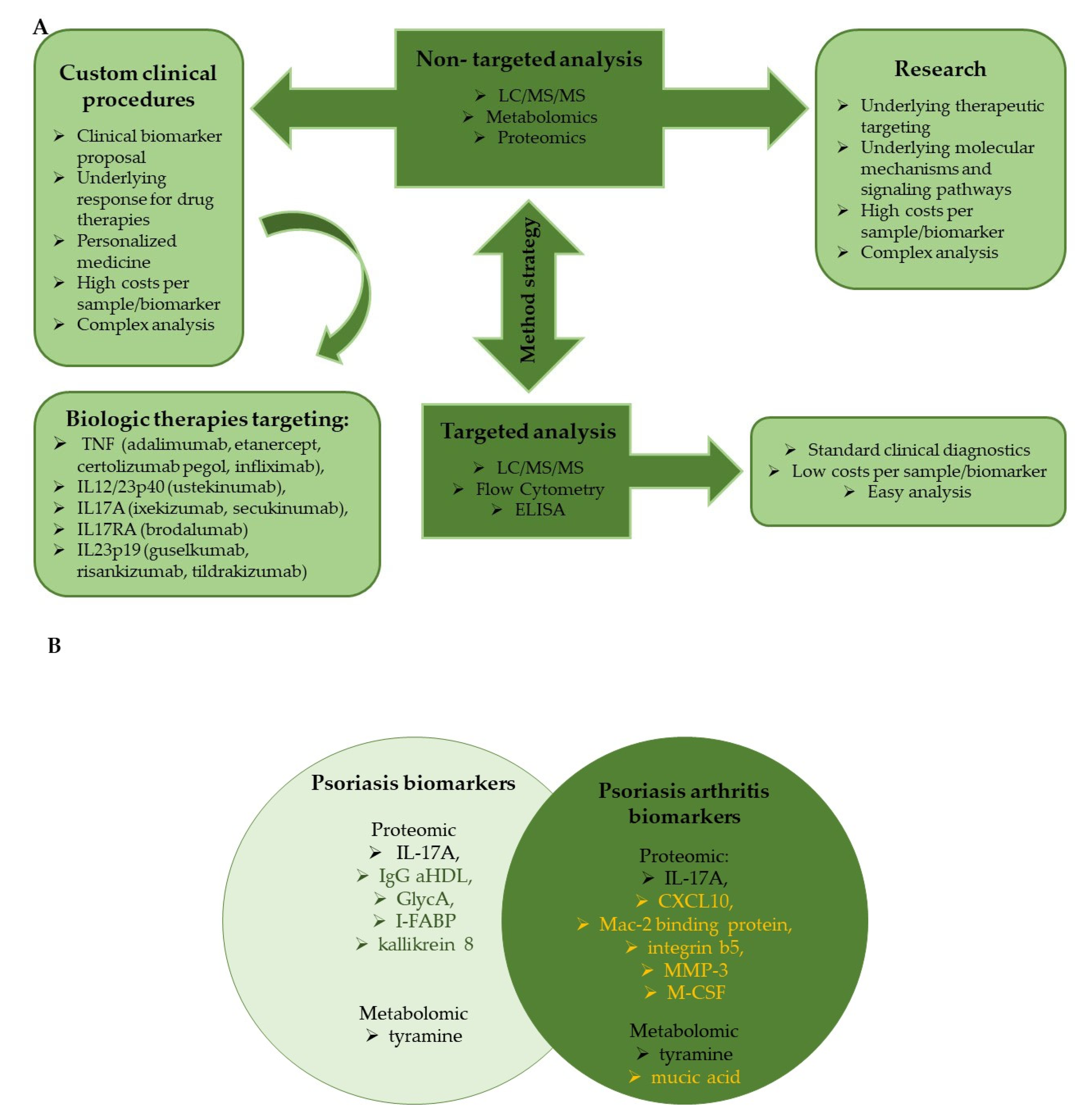
Many of the selected clinical trials focused on biomarkers that could have the ability to predict responses to treatment with biologic disease modifying antirheumatic drugs (DMARDs), such as secukinumab, apremilast, and adalimumab. The primary purpose of the trials was to investigate inflammatory pathways and report changes in the levels of inflammatory markers at baseline and after treatment. To achieve the assumed goal, various analytical methods were used, such as RT-PCR and qPCR to assess the gene inflammatory biomarkers, flow cytometry to reveal the aberrant inflammatory profiles of cells, and histological examination to evaluate the reversing of lesion skin inflammation. The association between putative biomarkers and the progression and severity of psoriasis and psoriatic arthritis was investigated in several trials. Three independent clinical trials have identified matrix metalloproteinase (MMP-3) as a potential biomarker. This finding is consistent with the results presented by Ramessur et al., who identified MMP-3 as a proteomic candidate for predicting the development of psoriatic arthritis (PsA) from psoriasis lesions based on an analysis of 181 scientific articles [70]. Serum level of cytokines implicated in the Th17 pathway was measured in three clinical trials, indicating that IL 17A was a candidate for predicting psoriasis severity. In the study ‘Identification of New Prognostic Markers in Psoriatic Arthritis’, the concentration of IL 17A and other cytokines was correlated with markers of bone remodeling, identifying the molecular pathways involved in psoriatic arthropathy. The progression of psoriasis leads to the development of many comorbidities other than psoriatic arthritis, such as metabolic syndrome and cardiovascular diseases [78]. The study ‘Psoriasis Inflammation and Systemic Co Morbidities’ was intended to explore the pathophysiology of psoriasis and its comorbidities, but it also provided guidance on how long-term treatment of inflammation can reduce or prevent cardiovascular events.

It should be highlighted that the completed clinical trials were mostly characterized by a limited number of participants, and the largest of them had only 100 subjects, while the ongoing recruitment for the trials estimates the number of participants to be 200–300 and even 20,000 subjects. The presented studies show that attempts to research candidate biomarkers take into account a variety of biological materials, not only serum or plasma, but also urine, synovial fluid, or skin biopsies. The broad extent of measurements, methods and multifarious characteristics of study groups provide the increased clinical value and usefulness in clinical practice to the biomarkers.

5. Conclusions

In vivo psoriasis mouse models do not fully reflect the human pathophysiology of psoriasis. However, they can be a powerful tool for preclinical application and pick out biomarkers for future research in human psoriasis. Non-targeted mass spectrometry analysis is mainly used by scientists to discover and explain some molecular mechanisms of skin disease, thus proposing the possible therapeutic target and monitoring of treatment. However, this is usually performed on small patient groups and needs to be validated during longitudinal clinical trials. The availability of multiple biological therapies did not give the expected results in all patients because of heterogeneity in efficacy and tolerability. That is why untargeted proteomics should be available for patients that did not respond to treatments. Identifying robust biomarkers as representative of various clinical psoriasis phenotypes would allow the stratification of patients into subgroups and treatments to be tailored to them as personalized medicine. The most significant conclusion is that the majority of presented psoriasis-like dermatitis in vivo models, academic research, and clinical trials focus on intensive research on proteomic and metabolic markers in psoriasis diagnosis, their severity, and in drug response treatment. The use of multiomic technologies is currently essential and is one of the most promising directions in the identification of biomarkers associated with psoriasis and psoriatic arthritis.

References

  1. Yan, D.; Gudjonsson, J.E.; Le, S.; Maverakis, E.; Plazyo, O.; Ritchlin, C.; Scher, J.U.; Singh, R.; Ward, N.L.; Bell, S.; et al. New Frontiers in Psoriatic Disease Research, Part I: Genetics, Environmental Triggers, Immunology, Pathophysiology, and Precision Medicine. J. Investig. Dermatol. 2021, 141, 2112–2122.e3.
  2. Rendon, A.; Schäkel, K. Psoriasis Pathogenesis and Treatment. Int. J. Mol. Sci. 2019, 20, 1475.
  3. Zachariae, H. Prevalence of Joint Disease in Patients with Psoriasis: Implications for Therapy. Am. J. Clin. Dermatol. 2003, 4, 441–447.
  4. Bu, J.; Ding, R.; Zhou, L.; Chen, X.; Shen, E. Epidemiology of Psoriasis and Comorbid Diseases: A Narrative Review. Front. Immunol. 2022, 13, 880201.
  5. Griffiths, C.E.M.; Armstrong, A.W.; Gudjonsson, J.E.; Barker, J.N.W.N. Psoriasis. Lancet 2021, 397, 1301–1315.
  6. Sanchez, D.P.; Sonthalia, S. Koebner Phenomenon; StatPearls Publishing: Treasure Island, FL, USA, 2023.
  7. Gates, A.H.; Karasek, M. Hereditary Absence of Sebaceous Glands in the Mouse. Science 1965, 148, 1471–1473.
  8. Sundberg, J.P.; Dunstan, R.W.; Roop, D.R.; Beamer, W.G. Full-Thickness Skin Grafts from Flaky Skin Mice to Nude Mice: Maintenance of the Psoriasiform Phenotype. J. Investig. Dermatol. 1994, 102, 781–788.
  9. Sundberg, J.P.; Beamer, W.G.; Shultz, L.D.; Dunstan, R.W. Inherited Mouse Mutations as Models of Human Adnexal, Cornification, and Papulosquamous Dermatoses. J. Investig. Dermatol. 1990, 95, 62S–63S.
  10. Sundberga, J.P.; Francea, M.; Boggessa, D.; Sundberga, B.A.; Jenson’, A.B.; Beamera, W.G.; Shultza, L.D.; Words, K. Development and Progression of Psoriasiform Dermatitis and Systemic Lesions in the Flaky Skin (Fsn) Mouse Mutant. Pathobiology 1997, 65, 271–286.
  11. Brown, W.R.; Hardy, M.H. A Hypothesis on the Cause of Chronic Epidermal Hyperproliferation in Asebia Mice. Clin. Exp. Dermatol. 1988, 13, 74–77.
  12. Fallon, P.G.; Sasaki, T.; Sandilands, A.; Campbell, L.E.; Saunders, S.P.; Mangan, N.E.; Callanan, J.J.; Kawasaki, H.; Shiohama, A.; Kubo, A.; et al. A Homozygous Frameshift Mutation in the Mouse Flg Gene Facilitates Enhanced Percutaneous Allergen Priming. Nat. Genet. 2009, 41, 602–608.
  13. Wilkinson, D.I.; Karasek, M.A. Skin Lipids of a Normal and Mutant (Asebic) Mouse Strain. J. Investig. Dermatol. 1966, 47, 449–455.
  14. Vandeghinste, N.; Klattig, J.; Jagerschmidt, C.; Lavazais, S.; Marsais, F.; Haas, J.D.; Auberval, M.; Lauffer, F.; Moran, T.; Ongenaert, M.; et al. Neutralization of IL-17C Reduces Skin Inflammation in Mouse Models of Psoriasis and Atopic Dermatitis. J. Investig. Dermatol. 2018, 138, 1555–1563.
  15. Chan, T.C.; Hawkes, J.E.; Krueger, J.G. Interleukin 23 in the Skin: Role in Psoriasis Pathogenesis and Selective Interleukin 23 Blockade as Treatment. Ther. Adv. Chronic Dis. 2018, 9, 111–119.
  16. van der Fits, L.; Mourits, S.; Voerman, J.S.A.; Kant, M.; Boon, L.; Laman, J.D.; Cornelissen, F.; Mus, A.-M.; Florencia, E.; Prens, E.P.; et al. Imiquimod-Induced Psoriasis-Like Skin Inflammation in Mice Is Mediated via the IL-23/IL-17 Axis. J. Immunol. 2009, 182, 5836–5845.
  17. Nakahara, T.; Kido-Nakahara, M.; Ulzii, D.; Miake, S.; Fujishima, K.; Sakai, S.; Chiba, T.; Tsuji, G.; Furue, M. Topical Application of Endothelin Receptor a Antagonist Attenuates Imiquimod-Induced Psoriasiform Skin Inflammation. Sci. Rep. 2020, 10, 9510.
  18. Mohammed, S.S.; Kadhim, H.M.; Al-Sudani, I.M.; Musatafa, W.W. Anti-Inflammatory Effects of Topically Applied Azilsartan in a Mouse Model of Imiquimod-Induced Psoriasis. Int. J. Drug Deliv. Technol. 2022, 12, 1249–1255.
  19. Yang, Y.; Zhao, Y.; Lai, R.; Xian, L.; Lei, Q.; Xu, J.; Guo, M.; Xian, D.; Zhong, J. An Emerging Role of Proanthocyanidins on Psoriasis: Evidence from a Psoriasis-Like Mouse Model. Oxid. Med. Cell. Longev. 2022, 2022, 5800586.
  20. Schafer, P.H.; Chen, P.; Fang, L.; Wang, A.; Chopra, R. The Pharmacodynamic Impact of Apremilast, an Oral Phosphodiesterase 4 Inhibitor, on Circulating Levels of Inflammatory Biomarkers in Patients with Psoriatic Arthritis: Substudy Results from a Phase III, Randomized, Placebo-Controlled Trial (PALACE 1). J. Immunol. Res. 2015, 2015, 906349.
  21. Svensson, L.; Røpke, M.A.; Norsgaard, H. Psoriasis Drug Discovery: Methods for Evaluation of Potential Drug Candidates. Expert Opin. Drug Discov. 2012, 7, 49–61.
  22. Haftek, M.; Ortonne, J.P.; Staquet, M.J.; Viac, J.; Thivolet, J. Normal and Psoriatic Human Skin Grafts on “nude” Mice: Morphological and Immunochemical Studies. J. Investig. Dermatol. 1981, 76, 48–52.
  23. Raychaudhuri, S.; Raychaudhuri, S. Scid Mouse Model of Psoriasis: A Unique Tool for Drug Development of Autoreactive T-Cell and TH-17 Cell-Mediated Autoimmune Diseases. Indian J. Dermatol. 2010, 55, 157.
  24. Di Domizio, J.; Conrad, C.; Gilliet, M. Xenotransplantation Model of Psoriasis. In Inflammation: Methods in Molecular Biology; Clausen, B., Laman, J., Eds.; Humana Press: New York, NY, USA, 2017; Volume 1559, pp. 83–90.
  25. Tiirikainen, M.L.; Woetmann, A.; Norsgaard, H.; Santamaria-Babí, L.F.; Lovato, P. Ex Vivo Culture of Lesional Psoriasis Skin for Pharmacological Testing. J. Dermatol. Sci. 2020, 97, 109–116.
  26. Norsgaard, H.; Svensson, L.; Hagedorn, P.H.; Moller, K.; Olsen, G.M.; Labuda, T. Translating Clinical Activity and Gene Expression Signatures of Etanercept and Ciclosporin to the Psoriasis Xenograft SCID Mouse Model. Br. J. Dermatol. 2012, 166, 649–652.
  27. Gourlay, W.A.; Chambers, W.H.; Monaco, A.P.; Maki, T. Importance of natural killer cells in the rejection of hamster skin xenografts. Transplantation 1998, 65, 727–734.
  28. Ashkar, A.A.; Di Santo, J.P.; Croy, B.A. Interferon γ Contributes to Initiation of Uterine Vascular Modification, Decidual Integrity, and Uterine Natural Killer Cell Maturation during Normal Murine Pregnancy. J. Exp. Med. 2000, 192, 259–270.
  29. Chen, L.; Deshpande, M.; Grisotto, M.; Smaldini, P.; Garcia, R.; He, Z.; Gulko, P.S.; Lira, S.A.; Furtado, G.C. Skin Expression of IL-23 Drives the Development of Psoriasis and Psoriatic Arthritis in Mice. Sci. Rep. 2020, 10, 8259.
  30. Van Nuffel, E.; Staal, J.; Baudelet, G.; Haegman, M.; Driege, Y.; Hochepied, T.; Afonina, I.S.; Beyaert, R. MALT 1 Targeting Suppresses CARD 14-induced Psoriatic Dermatitis in Mice. EMBO Rep. 2020, 21, e49237.
  31. Schonthaler, H.B.; Huggenberger, R.; Wculek, S.K.; Detmar, M.; Wagner, E.F.; Karin, M. Systemic Anti-VEGF Treatment Strongly Reduces Skin Inflammation in a Mouse Model of Psoriasis. Proc. Natl. Acad. Sci. USA 2009, 106, 21264–21269.
  32. Retser, E.; Schied, T.; Skryabin, B.V.; Vogl, T.; Kanczler, J.M.; Hamann, N.; Niehoff, A.; Hermann, S.; Eisenblätter, M.; Wachsmuth, L.; et al. Doxycycline-Induced Expression of Transgenic Human Tumor Necrosis Factor α in Adult Mice Results in Psoriasis-like Arthritis. Arthritis Rheum. 2013, 65, 2290–2300.
  33. Voskas, D.; Jones, N.; Van Slyke, P.; Sturk, C.; Chang, W.; Haninec, A.; Olya Babichev, Y.; Tran, J.; Master, Z.; Chen, S.; et al. A Cyclosporine-Sensitive Psoriasis-Like Disease Produced in Tie2 Transgenic Mice. Am. J. Pathol. 2005, 166, 843–855.
  34. SchÖn, P.; Detmar, M.; Parker’, C.M. Murine Psoriasis-like Disorder Induced by Naive CD4+ T Cells. Nat. Med. 1997, 3, 183–188.
  35. Davenport, C.M.; Mcadams, H.A.; Kou, J.; Mascioli, K.; Eichman, C.; Healy, L.; Peterson, J.; Murphy, S.; Coppola, D.; Truneh, A. Inhibition of Pro-Inf Lammatory Cytokine Generation by CTLA4-Ig in the Skin and Colon of Mice Adoptively Transplanted with CD45RB Hi CD4 + T Cells Correlates with Suppression of Psoriasis and Colitis. Int. Immunopharmacol. 2002, 2, 653–672.
  36. Takahashi, H.; Kouno, M.; Nagao, K.; Wada, N.; Hata, T.; Nishimoto, S.; Iwakura, Y.; Yoshimura, A.; Yamada, T.; Kuwana, M.; et al. Desmoglein 3-Specific CD4+ T Cells Induce Pemphigus Vulgaris and Interface Dermatitis in Mice. J. Clin. Investig. 2011, 121, 3677–3688.
  37. Breban, M.; Fernández-Sueiro, J.L.; Richardson, J.A.; Hadavand, R.R.; Maika, S.D.; Hammer, R.E.; Taurog, J.D. T Cells, but Not Thymic Exposure to HLA-B27, Are Required for the Inflammatory Disease of HLA-B27 Transgenic Rats. J. Immunol. 1996, 156, 794–803.
  38. Hong, K.; Chu, A.; Lú, B.R.; Berg, E.L.; Ehrhardt, R.O. IL-12, Independently of IFN-gamma, Plays a Crucial Role in the Pathogenesis of a Murine Psoriasis-Like Skin Disorder. J. Immunol. 1999, 162, 7480–7491.
  39. Nishimoto, S.; Kotani, H.; Tsuruta, S.; Shimizu, N.; Ito, M.; Shichita, T.; Morita, R.; Takahashi, H.; Amagai, M.; Yoshimura, A. Th17 Cells Carrying TCR Recognizing Epidermal Autoantigen Induce Psoriasis-like Skin Inflammation. J. Immunol. 2013, 191, 3065–3072.
  40. Dort, E.N.; Tanguay, P.; Hamelin, R.C. CRISPR/Cas9 Gene Editing: An Unexplored Frontier for Forest Pathology. Front. Plant Sci. 2020, 11, 1126.
  41. Strzyz, P. CRISPR–Cas9 Wins Nobel. Nat. Rev. Mol. Cell Biol. 2020, 21, 714.
  42. Roth-Carter, Q.R.; Godsel, L.; Koetsier, J.L.; Broussard, J.A.; Burks, H.E.; Fitz, G.; Huffine, A.L.; Amagai, S.; Lloyd, S.; Kweon, J.; et al. 225 Desmoglein 1 Deficiency in Knockout Mice Impairs Epidermal Barrier Formation and Results in a Psoriasis-like Gene Signature in E18.5 Embryos. J. Investig. Dermatol. 2020, 140, S26.
  43. Godsel, L.M.; Roth-Carter, Q.R.; Koetsier, J.L.; Tsoi, L.C.; Broussard, J.A.; Fitz, G.N.; Lloyd, S.M.; Kweon, J.; Huffine, A.L.; Burks, H.E.; et al. Th17-Skewed Inflammation Due to Genetic Deficiency of a Cadherin Stress Sensor. bioRxiv 2020.
  44. Ippagunta, S.K.; Gangwar, R.; Finkelstein, D.; Vogel, P.; Pelletier, S.; Gingras, S.; Redeckea, V.; Häckera, H. Keratinocytes Contribute Intrinsically to Psoriasis upon Loss of TNIP1 Function. Proc. Natl. Acad. Sci. USA 2016, 113, E6162–E6171.
  45. Kowalczyk, T.; Ciborowski, M.; Kisluk, J.; Kretowski, A.; Barbas, C. Mass Spectrometry Based Proteomics and Metabolomics in Personalized Oncology. Biochim. Biophys. Acta (BBA)—Mol. Basis Dis. 2020, 1866, 165690.
  46. Rozanova, S.; Barkovits, K.; Nikolov, M.; Schmidt, C.; Urlaub, H.; Marcus, K. Quantitative Mass Spectrometry-Based Proteomics: An Overview. In Methods in Molecular Biology; Humana: New York, NY, USA, 2021; Volume 2228, pp. 85–116.
  47. Zhou, Y.; Wang, P.; Yan, B.X.; Chen, X.Y.; Landeck, L.; Wang, Z.Y.; Li, X.X.; Zhang, J.; Zheng, M.; Man, X.Y. Quantitative Proteomic Profile of Psoriatic Epidermis Identifies OAS2 as a Novel Biomarker for Disease Activity. Front. Immunol. 2020, 11, 1432.
  48. Schonthaler, H.B.; Guinea-Viniegra, J.; Wculek, S.K.; Ruppen, I.; Ximénez-Embún, P.; Guío-Carrión, A.; Navarro, R.; Hogg, N.; Ashman, K.; Wagner, E.F. S100A8-S100A9 Protein Complex Mediates Psoriasis by Regulating the Expression of Complement Factor C3. Immunity 2013, 39, 1171–1181.
  49. Yan, K.X.; Meng, Q.; He, H.; Zhu, H.W.; Wang, Z.C.; Han, L.; Huang, Q.; Zhang, Z.H.; Yawalkar, N.; Zhou, H.; et al. ITRAQ-Based Quantitative Proteomics Reveals Biomarkers/Pathways in Psoriasis That Can Predict the Efficacy of Methotrexate. J. Eur. Acad. Dermatol. Venereol. 2022, 36, 1784–1795.
  50. Li, Y.; Lin, P.; Wang, S.; Li, S.; Wang, R.; Yang, L.; Wang, H. Quantitative Analysis of Differentially Expressed Proteins in Psoriasis Vulgaris Using Tandem Mass Tags and Parallel Reaction Monitoring. Clin. Proteom. 2020, 17, 30.
  51. Gęgotek, A.; Domingues, P.; Wroński, A.; Wójcik, P.; Skrzydlewska, E. Proteomic Plasma Profile of Psoriatic Patients. J. Pharm. Biomed. Anal. 2018, 155, 185–193.
  52. Gęgotek, A.; Domingues, P.; Wroński, A.; Ambrożewicz, E.; Skrzydlewska, E. The Proteomic Profile of Keratinocytes and Lymphocytes in Psoriatic Patients. Proteom. Clin. Appl. 2019, 13, 1800119.
  53. Gęgotek, A.; Domingues, P.; Wroński, A.; Skrzydlewska, E. Changes in Proteome of Fibroblasts Isolated from Psoriatic Skin Lesions. Int. J. Mol. Sci. 2020, 21, 5363.
  54. Xu, M.; Deng, J.; Xu, K.; Zhu, T.; Han, L.; Yan, Y.; Yao, D.; Deng, H.; Wang, D.; Sun, Y.; et al. In-Depth Serum Proteomics Reveals Biomarkers of Psoriasis Severity and Response to Traditional Chinese Medicine. Theranostics 2019, 9, 2475–2488.
  55. Reindl, J.; Pesek, J.; Krüger, T.; Wendler, S.; Nemitz, S.; Muckova, P.; Büchler, R.; Opitz, S.; Krieg, N.; Norgauer, J.; et al. Proteomic Biomarkers for Psoriasis and Psoriasis Arthritis. J. Proteom. 2016, 140, 55–61.
  56. Plavina, T.; Hincapie, M.; Wakshull, E.; Subramanyam, M.; Hancock, S.W. Increased Plasma Concentrations of Cytoskeletal and Aa2+-Binding Proteins and Their Peptides in Psoriasis Patients. Clin. Chem. 2008, 54, 1805–1814.
  57. Matsuura, T.; Sato, M.; Nagai, K.; Sato, T.; Arito, M.; Omoteyama, K.; Suematsu, N.; Okamoto, K.; Kato, T.; Soma, Y.; et al. Serum Peptides as Putative Modulators of Inflammation in Psoriasis. J. Dermatol. Sci. 2017, 87, 36–49.
  58. Li, S.S.; Liu, Y.; Li, H.; Wang, L.-P.; Xue, L.-F.; Yin, G.-S.; Wu, X.-S. Identification of Psoriasis Vulgaris Biomarkers in Human Plasma by Non-Targeted Metabolomics Based on UPLC-Q-TOF/MS. Eur. Rev. Med. Pharmacol. Sci. 2019, 23, 3940–3950.
  59. Kishikawa, T.; Arase, N.; Tsuji, S.; Maeda, Y.; Nii, T.; Hirata, J.; Suzuki, K.; Yamamoto, K.; Masuda, T.; Ogawa, K.; et al. Large-Scale Plasma-Metabolome Analysis Identifies Potential Biomarkers of Psoriasis and Its Clinical Subtypes. J. Dermatol. Sci. 2021, 102, 78–84.
  60. Sun, L.; Guo, X.; Qin, Y.; Li, P.; Yu, C.; Gao, X.; Xie, X.; Xu, X. Serum Intestinal Metabolites Are Raised in Patients with Psoriasis and Metabolic Syndrome. Clin. Cosmet. Investig. Dermatol. 2022, 15, 879–886.
  61. Armstrong, A.W.; Wu, J.; Johnson, M.A.; Grapov, D.; Azizi, B.; Dhillon, J.; Fiehn, O. Metabolomics in Psoriatic Disease: Pilot Study Reveals Metabolite Differences in Psoriasis and Psoriatic Arthritis. F1000Research 2014, 3, 248.
  62. Kang, H.; Li, X.; Zhou, Q.; Quan, C.; Xue, F.; Zheng, J.; Yu, Y. Exploration of Candidate Biomarkers for Human Psoriasis Based on Gas Chromatography-Mass Spectrometry Serum Metabolomics. Br. J. Dermatol. 2017, 176, 713–722.
  63. Looby, N.; Roszkowska, A.; Reyes-Garcés, N.; Yu, M.; Bączek, T.; Kulasingam, V.; Pawliszyn, J.; Chandran, V. Serum Metabolic Fingerprinting of Psoriasis and Psoriatic Arthritis Patients Using Solid-Phase Microextraction—Liquid Chromatography—High-Resolution Mass Spectrometry. Metabolomics 2021, 17, 59.
  64. Mysliwiec, H.; Harasim-Symbor, E.; Baran, A.; Szterling-Jaworowska, M.; Milewska, A.J.; Chabowski, A.; Flisiak, I. Abnormal Serum Fatty Acid Profile in Psoriatic Arthritis. Arch. Med. Sci. 2019, 15, 1407–1414.
  65. Ambrożewicz, E.; Wójcik, P.; Wroński, A.; Łuczaj, W.; Jastrzab, A.; Žarković, N.; Skrzydlewska, E. Pathophysiological Alterations of Redox Signaling and Endocannabinoid System in Granulocytes and Plasma of Psoriatic Patients. Cells 2018, 7, 159.
  66. Tsoukalas, D.; Fragoulakis, V.; Sarandi, E.; Docea, A.O.; Papakonstaninou, E.; Tsilimidos, G.; Anamaterou, C.; Fragkiadaki, P.; Aschner, M.; Tsatsakis, A.; et al. Targeted Metabolomic Analysis of Serum Fatty Acids for the Prediction of Autoimmune Diseases. Front. Mol. Biosci. 2019, 6, 120.
  67. Bilgiç, Ö.; Altınyazar, H.C.; Baran, H.; Ünlü, A. Serum Homocysteine, Asymmetric Dimethyl Arginine (ADMA) and Other Arginine–NO Pathway Metabolite Levels in Patients with Psoriasis. Arch. Dermatol. Res. 2015, 307, 439–444.
  68. Sikora, M.; Kiss, N.; Stec, A.; Giebultowicz, J.; Samborowska, E.; Jazwiec, R.; Dadlez, M.; Olszewska, M.; Rudnicka, L. Trimethylamine N-Oxide, a Gut Microbiota-Derived Metabolite, Is Associated with Cardiovascular Risk in Psoriasis: A Cross-Sectional Pilot Study. Dermatol. Ther. 2021, 11, 1277–1289.
  69. Chen, C.; Hou, G.; Zeng, C.; Ren, Y.; Chen, X.; Peng, C. Metabolomic Profiling Reveals Amino Acid and Carnitine Alterations as Metabolic Signatures in Psoriasis. Theranostics 2020, 11, 754–767.
  70. Ramessur, R.; Corbett, M.; Marshall, D.; Acencio, M.L.; Barbosa, I.A.; Dand, N.; Di Meglio, P.; Haddad, S.; Jensen, A.H.M.; Koopmann, W.; et al. Biomarkers of Disease Progression in People with Psoriasis: A Scoping Review. Br. J. Dermatol. 2022, 187, 481–493.
  71. Shelef, M.A.; Sokolove, J.; Lahey, L.J.; Wagner, C.A.; Sackmann, E.K.; Warner, T.F.; Wang, Y.; Beebe, D.J.; Robinson, W.H.; Huttenlocher, A. Peptidylarginine Deiminase 4 Contributes to Tumor Necrosis Factor α-Induced Inflammatory Arthritis. Arthritis Rheumatol. 2014, 66, 1482–1491.
  72. Kiss, B.; Szántó, M.; Hegedűs, C.; Antal, D.; Szödényi, A.; Márton, J.; Méhes, G.; Virág, L.; Szegedi, A.; Bai, P. Poly(ADP-Ribose) Polymerase-1 Depletion Enhances the Severity of Inflammation in an Imiquimod-Induced Model of Psoriasis. Exp. Dermatol. 2020, 29, 79–85.
  73. Sestito, R.; Madonna, S.; Scarponi, C.; Cianfarani, F.; Failla, C.M.; Cavani, A.; Girolomoni, G.; Albanesi, C. STAT3-dependent Effects of IL-22 in Human Keratinocytes Are Counterregulated by Sirtuin 1 through a Direct Inhibition of STAT3 Acetylation. FASEB J. 2011, 25, 916–927.
  74. Fan, X.; Yan, K.; Meng, Q.; Sun, R.; Yang, X.; Yuan, D.; Li, F.; Deng, H. Abnormal Expression of SIRTs in Psoriasis: Decreased Expression of SIRT 1-5 and Increased Expression of SIRT 6 and 7. Int. J. Mol. Med. 2019, 44, 157–171.
  75. Bohio, A.A.; Sattout, A.; Wang, R.; Wang, K.; Sah, R.K.; Guo, X.; Zeng, X.; Ke, Y.; Boldogh, I.; Ba, X. C-Abl–Mediated Tyrosine Phosphorylation of PARP1 Is Crucial for Expression of Proinflammatory Genes. J. Immunol. 2019, 203, 1521–1531.
  76. Łuczaj, W.; Gęgotek, A.; Skrzydlewska, E. Analytical Approaches to Assess Metabolic Changes in Psoriasis. J. Pharm. Biomed. Anal. 2021, 205, 114359.
  77. Lundberg, K.C.; Fritz, Y.; Johnston, A.; Foster, A.M.; Baliwag, J.; Gudjonsson, J.E.; Schlatzer, D.; Gokulrangan, G.; McCormick, T.S.; Chance, M.R.; et al. Proteomics of Skin Proteins in Psoriasis: From Discovery and Verification in a Mouse Model to Confirmation in Humans. Mol. Cell. Proteom. 2015, 14, 109–119.
  78. Boehncke, W.H. Systemic Inflammation and Cardiovascular Comorbidity in Psoriasis Patients: Causes and Consequences. Front. Immunol. 2018, 9, 579.
More
Information
Subjects: Dermatology
Contributors MDPI registered users' name will be linked to their SciProfiles pages. To register with us, please refer to https://encyclopedia.pub/register : , , , ,
View Times: 281
Revisions: 2 times (View History)
Update Date: 17 Aug 2023
1000/1000
Video Production Service