Submitted Successfully!
To reward your contribution, here is a gift for you: A free trial for our video production service.
Thank you for your contribution! You can also upload a video entry or images related to this topic.
Version Summary Created by Modification Content Size Created at Operation
1 -- 3636 2023-06-26 10:19:32 |
2 references update Meta information modification 3636 2023-06-29 08:08:01 | |
3 layout + 14 word(s) 3650 2023-07-10 03:12:43 |

Video Upload Options

Do you have a full video?

Confirm

Are you sure to Delete?
Cite
If you have any further questions, please contact Encyclopedia Editorial Office.
Lafon-Hughes, L. SARS-CoV-2 Interactions with the Host Cell Nucleus. Encyclopedia. Available online: https://encyclopedia.pub/entry/46029 (accessed on 27 July 2024).
Lafon-Hughes L. SARS-CoV-2 Interactions with the Host Cell Nucleus. Encyclopedia. Available at: https://encyclopedia.pub/entry/46029. Accessed July 27, 2024.
Lafon-Hughes, Laura. "SARS-CoV-2 Interactions with the Host Cell Nucleus" Encyclopedia, https://encyclopedia.pub/entry/46029 (accessed July 27, 2024).
Lafon-Hughes, L. (2023, June 26). SARS-CoV-2 Interactions with the Host Cell Nucleus. In Encyclopedia. https://encyclopedia.pub/entry/46029
Lafon-Hughes, Laura. "SARS-CoV-2 Interactions with the Host Cell Nucleus." Encyclopedia. Web. 26 June, 2023.
SARS-CoV-2 Interactions with the Host Cell Nucleus
Edit

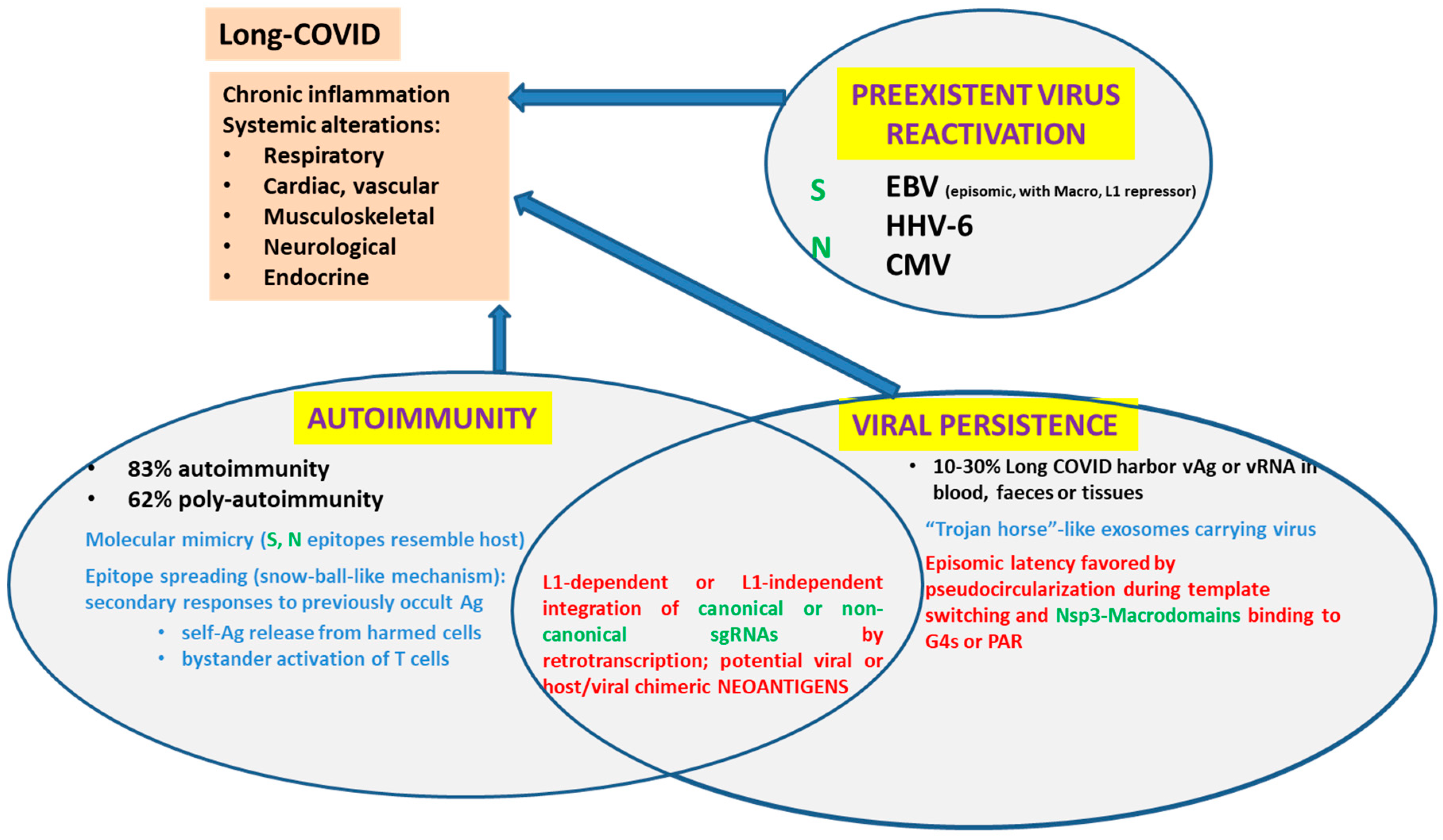
SARS-CoV-2 components disturb the transport of certain proteins through the nuclear pores. Some SARS-CoV-2 structural proteins such as Spike (S) and Nucleocapsid (N), most non-structural proteins (remarkably, Nsp1 and Nsp3), as well as some accessory proteins (ORF3d, ORF6, ORF9a) can reach the nucleoplasm either due to their nuclear localization signals (NLS) or taking a shuttle with other proteins. A percentage of SARS-CoV-2 RNA can also reach the nucleoplasm. Remarkably, controversy has recently been raised by proving that-at least under certain conditions-, SARS-CoV-2 sequences can be retrotranscribed and inserted as DNA in the host genome, giving rise to chimeric genes. In turn, the expression of viral-host chimeric proteins could potentially create neo-antigens, activate autoimmunity and promote a chronic pro-inflammatory state.

SARS-CoV-2 nucleocytoplasmic shuttling NLS

1. Introduction

Severe Acute Respiratory Syndrome Coronavirus 2 (SARS-CoV-2) is a coronavirus that caused the COVID-19 pandemic. Table 1, Table 2 and Table 3 show the involvement of SARS-CoV-2 non-structural, structural and accessory proteins (column I) in the diminution of the inflammatory/antiviral response (columns II and III), interactions with host NTRs or Nups (column IV), detection in the nucleus (column V) and highlights. Columns II and III refer to the impairment of IFN signaling. Upon viral infection, pattern recognition receptors such as Toll-like receptors (TLR) or RIG-I receptors are activated in immune cells (macrophages, monocytes, neutrophils, dendritic, epithelial) by foreign viral molecules, leading to NF-KB and IRF-dependent transcription of inflammatory genes and IFN-I/III. Then, IFNs act as autocrine or paracrine signals, leading to STAT1/2-dependent transcription of IFN-stimulated genes (ISGs). Column II is focused on the impairment of signals from receptor activation to transcription factor phosphorylation. “True” means that the viral protein inhibits the signaling cascade at a point upstream of transcription factor (TF) phosphorylation. When phosphorylation itself is inhibited, it is indicated as No: TF-P (e.g., noNF-KB-P). Once phosphorylated, the transcription factor is ready to be imported to the nucleus. Column III refers to known cases of modulation of nuclear import of these pro-inflammatory transcription factors by individual SARS-CoV proteins. Additional mechanisms of immune evasion exist and are explained in other reviews [1]. Column (IV) lists some known interactions of SARS-CoV-2 proteins with NTRs or Nups. Column (IV) states whether each NSp has ever been detected in the nucleus, either after protein transfection and overexpression [2] or in other circumstances. TLR: Toll-like receptor; IFN: interferon; ISG: interferon-stimulated genes; TF: transcription factor. NTR: nuclear transport receptor), including Importins, Exportins or bifunctional receptors); Nups: nuclear pore proteins; PAR: poly (ADP-ribose). G4s: G-quadruplexes.
Table 2. SARS-CoV-2 structural proteins IFN signaling, nucleocytoplasmic traffic and localization.
Table 3. SARS-CoV-2 accessory proteins IFN signaling, nucleocytoplasmic traffic and localization.

2. SARS-CoV-2 Modulates IFN Signalling

Several signaling pathways activated by human coronaviruses, which modulate the antiviral immune response and contribute to the pathogenesis, had already been studied in 2019 [19] and seemed to have their counterpart in SARS-CoV-2. Upon infection, immune cells recognize foreign viral antigens and molecules (PAMPs) via pattern recognition receptors (PRRs), such as Toll-like receptors (TLRs) and RIG-I-like receptors (RLRs), thus stimulating the NF-KB and IRF3-dependent transcription of cytokines and IFNs, and subsequently inducing host immune responses. In turn, secreted IFNs bind to their cell surface receptors, activating STATs signaling to promote antiviral responses through IFN-stimulated genes (ISGs). Several SARS-CoV-2 proteins contribute to host immune escape. Nsps block both IFN synthesis and IFN-dependent transcription cascades at several points, from the PRRs to phosphorylation and/or nuclear translocation of the transcription factors NF-KB, IRF3 or STATs [3][6][20]. Nevertheless, a functional luciferase assay testing 23 SARS-CoV-2 proteins indicated that certain ones would increase IFN-β synthesis (Spike protein and Nsp2) or IFN-β-dependent transcription [20]. Interestingly, the same transcription factors (e.g., NF-KB) are involved in the regulatory sequences of an endogenous retrovirus LTR69 locus, termed Dup69, which has been activated by SARS-CoV-2 [21]. Regarding modulation of the host immune response, according to Zhao et al. [22], the SARS-CoV-2 N protein (which they did not detect inside the nucleus) has a biphasic effect on IFN-I production: while low-dose N reduces the phosphorylation and nuclear translocation of IRF3, STAT1, and STAT2, high-dose N protein has the opposite effects. NF-KB hyperactivation is a well-known mechanism to promote cytokine storms [9]. This could contribute to an explanation of how COVID-19 patients undergo insufficient IFN-I production (immunodepression) in early infection, which can be followed later by a cytokine storm (overactive immune response) [22]. Interestingly, the biphasic effect of N protein may be at least in part explained by liquid–liquid demixing phenomena, which occurs only at high N concentrations.
In turn, certain SARS-CoV-2 Nsps (NSP8 and NSP5) could be linked to oncogenic pathways [8].

3. SARS-CoV-2 Regulates Nucleocytoplasmic Shuttling

The main elements involved in nucleocytoplasmic transport are the Nucleoporins (Nups) that constitute the Nuclear Pore Complexes (NPCs), the Nuclear Transport Receptors (NTRs) which include importins, exportins and Bidirectional NTRs and RanGTP/GDP gradients. Viral hijacking of nuclear transport is known to be used by different viruses [23].
It was already known that SARS-CoV and MERS interacted with certain importins, thus blocking the nuclear translocation of NF-KB or STAT. In turn, SARS-CoV-2 interacts with Nup 37, Nup54, Nup58, Nup62, Nup88, Nup93, Nup160, Nup188, Nup210, Nup214, Nup98-RAE1, NUTF2, IPO5, IPO8, RanBP6, importin-β1, CRM1, XPOT, THOC3 and RanBP2/Nup358 [9]. SARS-CoV-2 infection reduced RanBP2/Nup358 protein. Thus, SARS-CoV-2 increased NF-KB, leading to cytokine storms [9]. Ivermectin, which reduces viral load 5000 fold, not only binds strongly to SARS-CoV-2 Spike protein glycan sites, thus diminishing their interaction with blood and epithelial cells (thus inhibiting hemagglutination induced by Spike) [24] but also destabilizes Imp1 [25].
As reviewed by Shen et al. [9], SARS-CoV-2 infection blocks the nuclear export of host antiviral mRNAs and nuclear translocation of STAT1 through the interaction with several Nups and nuclear transport receptors on the cytoplasmic side of the nuclear pore (such as Nup88, Nup214, Nup98-RAE1, importin-β1 or RanBP2/Nup358) as well as on the nuclear pore lumen (as Nup54, Nup58, Nup93). SARS-CoV-2 infection also alters the shuttling of CRM1 exportin. For example, ORF6 inhibits IRF3 activation and STAT1 nuclear translocation. ORF6 interacts with Nup98-RAE1 as well as RanBP2/Nup358, Nup160, Nup 188, Nup210, Nup 37, Nup93, importins and exportin such as CRM1 and XPO3. As ORF6 interacts with nuclear pore complex proteins such as NUP98 and RAE1, the authors hypothesize that this may be a way to avoid nuclear translocations [20]. Another good example is Nsp1. Nsp1 has been observed near the nuclear pore complexes (NPCs) and directly binds the mRNA export factor NXF1, decreasing the availability of host cell mRNAs for the translation machinery, thus favoring viral mRNAs translation. For this reason, the overall transcriptome profile is altered by Nsp1 in infected cells [7]. A third example is the 98-aminoacids SARS-CoV-2 accessory protein 9b, small enough to enter the nucleus by passive transport. Furthermore, 9b interacts with CRM1 and gets exported out of the nucleus using an active NES. NES activity influences the half-life of 9b, and blocking the nuclear export of 9b induces apoptosis [18]. Interestingly, Ivermectin, which binds to and destabilizes nuclear importin Imp/1 heterodimer, prevents the suppression of antiviral response reducing viral load by ~5000 folds [25].
To sum up, certain SARS-CoV-2 proteins can hamper nucleocytoplasmic shuttling affecting nuclear export (e.g., affecting host mRNAs) or nuclear import (e.g., of host transcription factors involved in the antiviral response) [25]. Avoiding nuclear entry of a host cell transcription factor can be easily done from the cytoplasmic side of the nuclear pore. Affecting nuclear export requires a further step. Either the export is blocked through cytoplasmic-side sequestration/degradation of transport proteins such as CRM1 (which are otherwise constantly exiting the nucleus and then being recycled, entering the nucleus again) or—maybe more parsimoniously—the export is blocked through obstruction from the nuclear side of the pore (which requires nuclear entry of viral proteins). Do some viral proteins enter the nucleus?

4. SARS-CoV-2 Proteins: Nuclear Localization

Most viruses with ss(+) RNA genomes undergo replication in the cytoplasm, but some of their structural proteins localize to the nucleus, possibly inhibiting the host antiviral response [26]. N protein of some RNA viruses localizes to the nucleus/nucleolus of some infected cells. The coronavirus N protein is abundantly produced within infected cells and is one of the first clearly recognized as a Multifunctional Protein. CoV N proteins can localize to the host cell cytoplasm alone or to both the cytoplasm and the nucleus/nucleolus. Protein N has multiple roles, including virus replication, transcription, translation and ribonucleocapsid formation [27][28][29]. In host cells, N proteins have been shown to induce cell-cycle deregulation, inhibit the production of interferon, up-regulate the production of COX2 and up-regulate the activity of AP1. N interacts with numerous host cell proteins, including hCypA, proteasome subunit p42, the B23 phosphoprotein, Smad3, nRNP-A1, the chemokine CXCL16, translation elongation factor-1 alpha, cellular pyruvate kinase, 14-3-3 and nucleolin [19][30][31]. Nuclear translocation of the Nucleoprotein (N) has been demonstrated in several coronaviruses but has not been reported in SARS-CoV-2.
Several viral proteins contain NLS and/or NES and localize to the nucleus. Unsurprisingly, SARS-CoV-2 Nsp1, involved in host mRNA export blocking, can reach the nucleus and interacts with DNA polymerase alpha (Pol a), an essential complex involved in DNA replication that couples cycle cell progression to DRR [8]. Nsp1 protein of SARS-CoV-2 prevents nuclear export of host mRNAs dependent on the receptor heterodimer NXF1-NXT1 through the interference with binding of NXF1 to mRNA export adaptors and NXF1 docking at the nuclear pore complex. NXF1 overexpression reverts Nsp1-mediated mRNA export block and reduction in mRNA levels. Although most Nsp1 is cytoplasmic, it does also colocalize with a nucleoporin (Nup358) and is even detected by ICF and confocal microscopy inside the nucleus near the nuclear envelope [5].
The SARS-CoV-2 surface glycoprotein Spike, coded by ORF2, presents five special features. First, it is optimized for human Angiotensin-converting enzyme 2 receptor (hACE2) binding [32]. Second, the Spike bears 4 HIV-like inserts with a high-density positive charge, very similar to HIV-1 surface proteins gp120 and Gag (as stated by Pradhan and Zhang in papers that were retracted for other reasons [33][34]), absent in other coronaviruses (absent even in SARS). For this reason, SARS-CoV-2 Spike, similar to HIV, binds CLEC4M (or CD299) and DC-SIGNR (or CD209), facilitating the infection of the immune system [35]. T-cells suffer exhaustion, and counts of total T cells are negatively correlated with patient survival [36]. Third, one of the insertions creates a furin-cleavable polybasic cleavage site (“PRRAR”). As both ACE2 receptors and furin protease are ubiquitous, this facilitates viral spreading across human tissues (and probably also across species) [37][38]. Fourth, the same insertion creates a superantigen (SAg) motif, namely CASYQTQT_NSPRRARSVASQSI, which was mapped due to its sequence similarity to the classical Sag called Staphylococcus enterotoxin B (SEB). Superantigens (SAgs) are a class of antigens produced by some pathogenic microbes as a defense mechanism against the host immune system. SAg cause the non-specific polyclonal activation of T-cells, massive cytokine release and hyperactivation of the immune system, which may lead to autoimmunity, multiple organ failure, and even death. The binding of this SAg to the T-cell receptor (TCR) may trigger the cytotoxic adaptive immune responses observed in multi-system inflammatory syndrome in children (MIS-C) as well as cytokine storms in adults with SARS-CoV-2 infection [12]. Finally, the same insertion creates a nuclear localization signal (NLS, specifically “PRRARSV”) overlapped with the polybasic cleavage site. Although Spike (S) is a glycoprotein, unlike in other coronaviruses, in the special case of SARS-CoV-2, Spike can reach the nucleus. In fact, if a highly differentiated pseudostratified airway epithelium is exposed to infection with SARS-CoV-2 (MOI: 0.1), fixed 4 days later and subject to immunocytofluorescence, 10% Spike protein is detected inside the host cell nucleus. In turn, 15% S protein is on the nuclear surface, and the remnant is cytoplasmic or membrane-bound. Consistently, 72 h after transfection of A549 cells with SARS-CoV-2 plasmid, SARS-CoV-2 protein is detected in cell homogenates in both cytoplasmic (CDC42+) and nuclear (Lamin A/C+) fractions [10][11].
The detection of nuclear Spike protein is a game changer, providing a new paradigm by which a direct effect on transcription by nuclear Spike protein would not be impossible, forcing us to re-interpret the experiments showing phenotypic changes in cells expressing ectopic Spike. Relevant experiments have been carried out. For example, the ectopic expression of Spike in cardiomyocytes derived from human induced pluripotent stem cells (hiPSCs) alters their metabolic profile and dampens their functions. Thus, the authors hypothesize that Spike can alter the transcriptional regulation in cardiac gene programs [13]. In the same line, SARS-CoV-2 infection of human lung cancer cell line transfected with ACE2 receptor (A549-ACE2 cells, MOI: 0.1) induced various pro-oncogenic signaling cascades including TGF-β signaling and epithelial to mesenchymal transition (EMT). As virus-induced metastasis has been found in many cancers, the authors focused on EMT mechanisms. In the MCF-7 breast cancer cell line, which has high ACE-2 expression [39], the ectopic expression of SARS-CoV-2 Spike, unlike other structural proteins (N, M, E), induces Snail-dependent (EMT), with E-cadherin downregulation, N-cadherin upregulation, increased migration and invasion [14]. Moreover, the ectopic expression of either SARS-CoV-2 Spike or SARS-CoV-2 Nucleoprotein is enough to induce lytic reactivation of Kaposi’s sarcoma-associated herpesvirus (KSHV), one of the major human oncogenic viruses in iSLK.219 cells [15].
In the extremely pathogenic SARS-CoV-2, SARS-CoV and highly related CoV found in bats but not other CoVs, Nsp3 protein (which harbors PLPRO activity) contains a SARS Unique Domain (SUD) characterized by Macrodomains. Viral Macrodomains are considered unique mediators of viral replication and pathogenesis [40]. Canonical Macrodomains are “readers” of a post-translational protein modification called poly-ADP-ribosylation and also “erasers” of mono-ADP-ribosylation [41]. The crystal structure of the SARS-CoV macro domain was determined at 1.8-Å resolution in complex with ADP-ribose. Similar to other viral macro domains, from hepatitis E virus and Semliki Forest virus, it has poor ADP-ribose 1-phosphohydrolase activity but does efficiently bind poly (ADP-ribose) in vitro [42]. SARS-CoV Nsp3 SUD contains a canonical Macrodomain plus two atypical Macrodomains that bind G-quadruplexes (G4s) [43]; at least one of the G4-binding Macrodomains is essential for the activity of the SARS-CoV replication/transcription complex [44]. G4s are a particular structure of the nucleic acids that can arise in G-rich regions. Some viruses present G4s in their genome, while SARS-CoV could recognize G4s in host nucleic acids, for example, in 3′-nontranslated regions of mRNAs coding for host-cell proteins involved in apoptosis or signal transduction. G4s exist in the human genome, especially in telomeres and oncogene promoters [45]. In other viral infections, the recognition of G4s by viral proteins is involved in latency [46].
According to a systemic approach to reveal the subcellular locations of SARS-CoV-2 FLAGged proteins transfected in HEp-2 cells, some proteins were detected just in the cytoplasm, but the following ones were also present in the nucleus: NSP1, NSP3N, NSP5, NSP6, NSP7, NSP9, NSP10, NSP12, NSP13, NSP14, NSP15, NSP16, E and ORF9a [4]. Nsp13 is enriched in the splicing compartment. ORF3d has also been detected in the nucleus [25]. Many of these proteins are small and probably enter the nucleus by passive diffusion. NES may promote the nuclear export of these proteins or allow them to interact with exportins to hijack nuclear transportation. Anyway, pharmacological inhibition of nuclear export leads to nuclear accumulation of viral proteins and significantly decreases viral infection. 

5. SARS-CoV-2 RNA: Nuclear Localization

After SARS-CoV-2 infection, not only SARS-CoV-2 Spike protein but also Spike mRNA is detected in the host cell nucleus. Moreover, Spike mRNA protein and mRNA exhibit certain colocalization. Nuclear translocation of Spike mRNA and protein is undoubtedly a novel feature of SARS-CoV-2 biology [11].
Single-molecule fluorescence in situ hybridization (smFISH) has allowed the detection of SARS-CoV-2 +gRNA, +sgRNA, –gRNA and dsRNA. Maximum z-projections of confocal images were required to quantify the RNA dispersion index of cytoplasmic signals (excluding DAPI-positive ROIs) on [47][48]. Some of the images suggest that it would be worth doing an analysis on successive single confocal images to reveal whether the few signals that colocalize with DAPI on maximum intensity z-projections should be interpreted as intranuclear or supranuclear (cytoplasmic, too) signals [47].

6. Evidence of SARS-CoV-2 sgRNA Retrotranscription and Insertion in the Host Genome

Chimeric viral-host reads in RNA-seq represent around 0.004–0.14% of the total SARS-CoV-2 reads in human samples across published datasets [49]. However, as RNA-seq library preparation is inherently error prone due to random template switching during reverse transcription of RNA to cDNA (with up to 1% of RNA-seq chimeric reads putatively artefactual) [50], the identification of genuine chimeric viral–cellular RNA transcripts is compromised by the generation of artifactual chimeras [49][50]. As retrotranscription and viral genomic integration, if happening, would be a low-frequency event, an experiment was designed by Zhang et al. using human Long Interspersed Nuclear Elements (LINEs) to increase the likelihood of detection of such events. LINEs are an endogenous cellular source of retrotranscriptase (RT) and are commonly over-expressed upon viral infection. LINEs are able to retrotranspose themselves and other non-autonomous elements, thus facilitating the integration in vertebrate genomes of DNA copies of non-retroviral RNA viruses. Therefore, HEK293T cells were transfected with LINE1 expression plasmids prior to infection with SARS-CoV-2. Then, genomic DNA was isolated from those cells 2 days after infection. DNA sequencing revealed target site duplications flanking the viral sequences and consensus LINE1 endonuclease recognition sequences at the integration sites, consistent with a LINE1 retrotransposon-mediated, target-primed reverse transcription and retroposition mechanism. Thus, SARS-CoV-2 sequences can be reverse-transcribed and integrated into the DNA of infected human cells in culture. Although infectious viruses cannot be produced from the integrated sub-genomic SARS-CoV-2 sequences, transcription of the integrated DNA copies could be responsible for positive PCR tests long after the initial infection. As a reference, only 1 in 1000 to 1 in 100,000 mouse cells infected with LCMV in culture or in vivo carried viral DNA copies integrated into the genome. Thus, if true, only a small fraction of cells in any patient tissues would be expected to be positive for viral sequences. No matter how challenging it may be, the authors propose “it will be important, in follow-up studies, to demonstrate the presence of SARS-CoV-2 sequences integrated into the host genome in patient tissues” [49].
Smits et al. pose some reasonable doubts regarding Zhang et al.’s work related to SARS-CoV-2 insertion length, abundance and structure. In fact, Smits et al. [51] have tried to replicate Zhang et al. study, using the same cell line (HEK293T) relying on endogenous LINEs with no exit. Although the authors recognize that widespread cell death post-infection reduces the probability of SARS-CoV-2 integration persistence and recovery, they use a relatively high viral load for HEK293T (MOI:1). (As a reference, Zhang used 0.5 for HEK293T cells and Mehedi used MOI:0.1 in airway epithelium). Smits et al. argue they have a positive control which works as expected. The positive control is an HBV-positive cancer tissue, and they recovered a single HBV insertion [51]. HBV has been studied for a long while. It is known that cumulative HBV integrations in the human genome disrupt regulatory genes, drive aberrant gene expression and induce genomic rearrangements. For these reasons, HBV favors oncogenic transformation. According to Podlaha et al., although it is still unknown the proportion of hepatocytes that carry viral integrations, the HBV virus integrates with a lower-bound frequency of 0.84 per diploid genome in hepatitis B positive hepatocellular cancer patients, and it is calculated that integrated viral DNA generates ~80% of the HBsAg transcripts in these patients. Such viral or viral-host chimeric antigens may be driving chronic inflammation and/or autoimmunity [52]. If just a single HBV insertion was identified in the positive control, it does not seem strange not having been able to find small SARS-CoV-2 insertions corresponding to canonical or non-canonical sgRNAs retrotranscription. Thus, further studies are required to reach an agreement and solve this extremely relevant issue, represented with question marks in Figure 1 bottom nuclear region.
Figure 1. Main current hypotheses to explain Long-COVID and their relation to the direct or indirect interaction of SARS-CoV-2 proteins or RNAs with the host cell nucleus.

References

  1. Masters, P.S. The Molecular Biology of Coronaviruses. Adv. Virus Res. 2006, 66, 193–292.
  2. Brierley, I.; Digard, P.; Inglis, S.C. Characterization of an efficient coronavirus ribosomal frameshifting signal: Requirement for an RNA pseudoknot. Cell 1989, 57, 537–547.
  3. Low, Z.Y.; Zabidi, N.Z.; Yip, A.J.W.; Puniyamurti, A.; Chow, V.T.K.; Lal, S.K. SARS-CoV-2 Non-Structural Proteins and Their Roles in Host Immune Evasion. Viruses 2022, 14, 1991.
  4. Zhang, J.; Cruz-cosme, R.; Zhuang, M.-W.; Liu, D.; Liu, Y.; Teng, S.; Wang, P.-H.; Tang, Q.A. A systemic and molecular study of subcellular localization of SARS-CoV-2 proteins. Signal Transduct. Target. Ther. 2020, 5, 269.
  5. Zhang, K.; Miorin, L.; Makio, T.; Dehghan, I.; Gao, S.; Xie, Y.; Zhong, H.; Esparza, M.; Kehrer, T.; Kumar, A.; et al. Nsp1 protein of SARS-CoV-2 disrupts the mRNA export machinery to inhibit host gene expression. Sci. Adv. 2021, 7, eabe7386.
  6. Pizzato, M.; Baraldi, C.; Boscato Sopetto, G.; Finozzi, D.; Gentile, C.; Gentile, M.D.; Marconi, R.; Paladino, D.; Raoss, A.; Riedmiller, I.; et al. SARS-CoV-2 and the Host Cell: A Tale of Interactions. Front. Virol. 2022, 1, 815388.
  7. Kamitani, W.; Huang, C.; Narayanan, K.; Lokugamage, K.G.; Makino, S. A two-pronged strategy to suppress host protein synthesis by SARS coronavirus Nsp1 protein. Nat. Struct. Mol. Biol. 2009, 16, 1134–1140.
  8. Rapti, V.; Tsaganos, T.; Vathiotis, I.; Syrigos, N.; Li, P.; Poulakou, G. New Insights into SARS-CoV-2 and Cancer Cross-Talk: Does a Novel Oncogenesis Driver Emerge? Vaccines 2022, 10, 1607.
  9. Shen, Q.; Wang, Y.E.; Palazzo, A.F. Crosstalk between nucleocytoplasmic trafficking and the innate immune response to viral infection. J. Biol. Chem. 2021, 297, 100856.
  10. Osan, J.K.; DeMontigny, B.A.; Mehedi, M. Immunohistochemistry for protein detection in PFA-fixed paraffin-embedded SARS-CoV-2-infected COPD airway epithelium. STAR Protoc. 2021, 2, 100663.
  11. Sattar, S.; Kabat, J.; Jerome, K.; Feldmann, F.; Bailey, K.; Mehedi, M. Nuclear translocation of spike mRNA and protein is a novel feature of SARS-CoV-2. Front. Microbiol. 2023, 14, 1073789.
  12. Vojdani, A.; Vojdani, E.; Saidara, E.; Maes, M. Persistent SARS-CoV-2 Infection, EBV, HHV-6 and Other Factors May Contribute to Inflammation and Autoimmunity in Long COVID. Viruses 2023, 15, 400.
  13. Zhang, P.; Liu, Y.; Li, C.; Stine, L.D.; Wang, P.-H.; Turnbull, M.W.; Wu, H.; Liu, Q. Ectopic expression of SARS-CoV-2 S and ORF-9B proteins alters metabolic profiles and impairs contractile function in cardiomyocytes. Front. Cell Dev. Biol. 2023, 11, 1110271.
  14. Lai, Y.-J.; Chao, C.-H.; Liao, C.-C.; Lee, T.-A.; Hsu, J.-M.; Chou, W.-C.; Wang, J.; Huang, H.-C.; Chang, S.-J.; Lin, Y.-L.; et al. Epithelial-mesenchymal transition induced by SARS-CoV-2 required transcriptional upregulation of Snail. Am. J. Cancer Res. 2021, 11, 2278–2290.
  15. Chen, J.; Dai, L.; Barrett, L.; James, J.; Plaisance-Bonstaff, K.; Post, S.R.; Qin, Z. SARS-CoV-2 proteins and anti-COVID-19 drugs induce lytic reactivation of an oncogenic virus. Commun. Biol. 2021, 4, 682.
  16. Kashyap, T.; Murray, J.; Walker, C.J.; Chang, H.; Tamir, S.; Hou, B.; Shacham, S.; Kauffman, M.G.; Tripp, R.A.; Landesman, Y. Selinexor, a novel selective inhibitor of nuclear export, reduces SARS-CoV-2 infection and protects the respiratory system in vivo. Antivir. Res. 2021, 192, 105115.
  17. Cascarina, S.M.; Ross, E.D. Phase separation by the SARS-CoV-2 nucleocapsid protein: Consensus and open questions. J. Biol. Chem. 2022, 298, 101677.
  18. Sharma, K.; Åkerström, S.; Sharma, A.K.; Chow, V.T.K.; Teow, S.; Abrenica, B.; Booth, S.A.; Booth, T.F.; Mirazi-mi, A.; Lal, S.K. SARS-CoV 9b protein diffuses into nucleus, undergoes active Crm1 mediated nucleocytoplasmic export and triggers apoptosis when retained in the nucleus. PLoS ONE 2011, 6, e19436.
  19. Fung, T.S.; Liu, D.X. Human Coronavirus: Host-Pathogen Interaction. Annu. Rev. Microbiol. 2019, 73, 529–557.
  20. Lei, X.; Dong, X.; Ma, R.; Wang, W.; Xiao, X.; Tian, Z.; Wang, C.; Wang, Y.; Li, L.; Ren, L.; et al. Activation and evasion of type I interferon responses by SARS-CoV-2. Nat. Commun. 2020, 11, 3810.
  21. Arora, A.; Kolberg, J.E.; Badarinarayan, S.S.; Munot, D.; Müller, M.; Sauter, D.; Bansal, V. SARS-CoV-2 infection activates endogenous retroviruses of the LTR69 subfamily. Mol. Biol. 2023, preprint.
  22. Zhao, Y.; Sui, L.; Wu, P.; Wang, W.; Tan, G.; Wang, Z.; Yu, Y.; Hou, Z.; Wang, G.; Liu, Q. SARS-CoV-2 nucleocapsid protein dually regulates innate immune responses. Microbiology 2021, preprint.
  23. Paci, G.; Caria, J.; Lemke, E.A. Cargo transport through the nuclear pore complex at a glance. J. Cell Sci. 2021, 134, jcs247874.
  24. Boschi, C.; Scheim, D.E.; Bancod, A.; Militello, M.; Bideau, M.L.; Colson, P.; Fantini, J.; Scola, B.L. SARS-CoV-2 Spike Protein Induces Hemagglutination: Implications for COVID-19 Morbidities and Therapeutics and for Vaccine Adverse Effects. Int. J. Mol. Sci. 2022, 23, 15480.
  25. Yadav, R.; Chaudhary, J.K.; Jain, N.; Chaudhary, P.K.; Khanra, S.; Dhamija, P.; Sharma, A.; Kumar, A.; Handu, S. Role of Structural and Non-Structural Proteins and Therapeutic Targets of SARS-CoV-2 for COVID-19. Cells 2021, 10, 821.
  26. Timani, K.A.; Liao, Q.; Ye, L.; Zeng, Y.; Liu, J.; Zheng, Y.; Ye, L.; Yang, X.; Lingbao, K.; Gao, J.; et al. Nuclear/nucleolar localization properties of C-terminal nucleocapsid protein of SARS coronavirus. Virus Res. 2005, 114, 23–34.
  27. Wu, H.-Y.; Brian, D.A. 5′-Proximal Hot Spot for an Inducible Positive-to-Negative-Strand Template Switch byCoronavirus RNA-Dependent RNA Polymerase. J. Virol. 2007, 81, 3206–3215.
  28. Wu, C.-H.; Chen, P.-J.; Yeh, S.-H. Nucleocapsid Phosphorylation and RNA Helicase DDX1 Recruitment Enables Coronavirus Transition from Discontinuous to Continuous Transcription. Cell Host Microbe 2014, 16, 462–472.
  29. Sola, I.; Almazán, F.; Zúñiga, S.; Enjuanes, L. Continuous and Discontinuous RNA Synthesis in Coronaviruses. Annu. Rev. Virol. 2015, 2, 265–288.
  30. McBride, R.; van Zyl, M.; Fielding, B. The Coronavirus Nucleocapsid Is a Multifunctional Protein. Viruses 2014, 6, 2991–3018.
  31. Wulan, W.N.; Heydet, D.; Walker, E.J.; Gahan, M.E.; Ghildyal, R. Nucleocytoplasmic transport of nucleocapsid proteins of enveloped RNA viruses. Front. Microbiol. Sec. Virol. 2015, 6, 553.
  32. Andersen, K.G.; Rambaut, A.; Lipkin, W.I.; Holmes, E.C.; Garry, R.F. The proximal origin of SARS-CoV-2. Nat. Med. 2020, 26, 450–452.
  33. Pradhan, P.; Pandey, A.K.; Mishra, A.; Gupta, P.; Tripathi, P.K.; Menon, M.B.; Gomes, J.; Vivekanandan, P.; Kundu, B. Uncanny similarity of unique inserts in the 2019-nCoV spike protein to HIV-1 gp120 and Gag. Evol. Biol. 2020; withdrawn preprint.
  34. Zhang, C.; Zheng, W.; Huang, X.; Bell, E.W.; Zhou, X.; Zhang, Y. Protein Structure and Sequence Reanalysis of 2019-nCoV Genome Refutes Snakes as Its Intermediate Host and the Unique Similarity between Its Spike Protein Insertions and HIV-1. J. Proteome Res. 2020, 19, 1351–1360.
  35. Feng, Z.; Diao, B.; Wang, R.; Wang, G.; Wang, C.; Tan, Y.; Liu, L.; Wang, C.; Liu, Y.; Liu, Y.; et al. The Novel Severe Acute Respiratory Syndrome Coronavirus 2 (SARS-CoV-2) Directly Decimates Human Spleens and Lymph Nodes. Infect. Dis. (Except. HIV/AIDS), 2020; preprint.
  36. Diao, B.; Wang, C.; Tan, Y.; Chen, X.; Liu, Y.; Ning, L.; Chen, L.; Li, M.; Liu, Y.; Wang, G.; et al. Reduction and Functional Exhaustion of T Cells in Patients with Coronavirus Disease 2019 (COVID-19). Infect. Dis. (Except. HIV/AIDS), 2020; preprint.
  37. Millet, J.K.; Whittaker, G.R. Host cell proteases: Critical determinants of coronavirus tropism and pathogenesis. Virus Res. 2015, 202, 120–134.
  38. Braun, E.; Sauter, D. Furin-mediated protein processing in infectious diseases and cancer. Clin. Transl. Immunol. 2019, 8, e1073.
  39. Zhang, Q.; Lu, S.; Li, T.; Yu, L.; Zhang, Y.; Zeng, H.; Qian, X.; Bi, J.; Lin, Y. ACE2 inhibits breast cancer angiogenesis via suppressing the VEGFa/VEGFR2/ERK pathway. J. Exp. Clin. Cancer Res. 2019, 38, 173.
  40. AFehr, R.; Jankevicius, G.; Ahel, I.; Perlman, S. Viral Macrodomains: Unique Mediators of Viral Replication and Pathogenesis. Trends Microbiol. 2018, 26, 598–610.
  41. Hottiger, M.O. SnapShot: ADP-Ribosylation Signaling. Mol. Cell 2015, 58, 1134.
  42. Egloff, M.-P.; Malet, H.; Putics, Á.; Heinonen, M.; Dutartre, H.; Frangeul, A.; Gruez, A.; Campanacci, V.; Cam-billau, C.; Ziebuhr, J.; et al. Structural and Functional Basis for ADP-Ribose and Poly(ADP-Ribose) Binding by Viral Macro Domains. J. Virol. 2006, 80, 8493–8502.
  43. Tan, J.; Vonrhein, C.; Smart, O.S.; Bricogne, G.; Bollati, M.; Kusov, Y.; Hansen, G.; Mesters, J.R.; Schmidt, C.L.; Hilgenfeld, R. The SARS-Unique Domain (SUD) of SARS Coronavirus Contains Two Macrodomains That Bind G-Quadruplexes. PLoS Pathog. 2009, 5, e1000428.
  44. Kusov, Y.; Tan, J.; Alvarez, E.; Enjuanes, L.; Hilgenfeld, R. A G-quadruplex-binding macrodomain within the ‘SARS-unique domain’ is essential for the activity of the SARS-coronavirus replication–transcription complex. Virology 2015, 484, 313–322.
  45. Ruggiero, E.; Richter, S.N. G-quadruplexes and G-quadruplex ligands: Targets and tools in antiviral therapy. Nucleic Acids Res. 2018, 46, 3270–3283.
  46. Lieberman, P.M. Epigenetics and Genetics of Viral Latency. Cell Host Microbe 2016, 19, 619–628.
  47. Lee, J.Y.; Wing, P.A.; Gala, D.S.; Noerenberg, M.; Järvelin, A.I.; Titlow, J.; Zhuang, X.; Palmalux, N.; Iselin, L.; Thompson, M.K.; et al. Absolute quantitation of individual SARS-CoV-2 RNA molecules provides a new paradigm for infection dynamics and variant differences. eLife 2022, 11, e74153.
  48. Stueland, M.; Wang, T.; Park, H.Y.; Mili, S. RDI Calculator: An Analysis Tool to Assess RNA Distributions in Cells. Sci. Rep. 2019, 9, 8267.
  49. Zhang, L.; Richards, A.; Barrasa, M.I.; Hughes, S.H.; Young, R.A.; Jaenisch, R. Reverse-transcribed SARS-CoV-2 RNA can integrate into the genome of cultured human cells and can be expressed in patient-derived tissues. Proc. Natl. Acad. Sci. USA 2021, 118, e2105968118.
  50. Yan, B.; Chakravorty, S.; Mirabelli, C.; Wang, L.; Trujillo-Ochoa, J.L.; Chauss, D.; Kumar, D.; Lionakis, M.S.; Olson, M.R.; Wobus, C.E.; et al. Host-Virus Chimeric Events in SARS-CoV-2-Infected Cells Are Infrequent and Artifactual. J. Virol. 2021, 95, e00294-21.
  51. Smits, N.; Rasmussen, J.; Bodea, G.O.; Amarilla, A.A.; Gerdes, P.; Sanchez-Luque, F.J.; Ajjikuttira, P.; Modhiran, N.; Liang, B.; Faivre, J.; et al. No evidence of human genome integration of SARS-CoV-2 found by long-read DNA sequencing. Cell Rep. 2021, 36, 109530.
  52. Podlaha, O.; Wu, G.; Downie, B.; Ramamurthy, R.; Gaggar, A.; Subramanian, M.; Ye, Z.; Jiang, Z. Genomic modeling of hepatitis B virus integration frequency in the human genome. PLoS ONE 2019, 14, e0220376.
More
Information
Contributor MDPI registered users' name will be linked to their SciProfiles pages. To register with us, please refer to https://encyclopedia.pub/register :
View Times: 365
Revisions: 3 times (View History)
Update Date: 10 Jul 2023
1000/1000
Video Production Service