Submitted Successfully!
To reward your contribution, here is a gift for you: A free trial for our video production service.
Thank you for your contribution! You can also upload a video entry or images related to this topic.
Version Summary Created by Modification Content Size Created at Operation
1 -- 3842 2023-06-20 14:35:44 |
2 layout + 12 word(s) 3854 2023-06-21 03:01:42 |

Video Upload Options

Do you have a full video?

Confirm

Are you sure to Delete?
Cite
If you have any further questions, please contact Encyclopedia Editorial Office.
Fontana, D.; Elli, E.M.; Pagni, F.; Piazza, R. Myelodysplastic Syndromes/Myeloproliferative Overlap Neoplasms. Encyclopedia. Available online: https://encyclopedia.pub/entry/45865 (accessed on 27 July 2024).
Fontana D, Elli EM, Pagni F, Piazza R. Myelodysplastic Syndromes/Myeloproliferative Overlap Neoplasms. Encyclopedia. Available at: https://encyclopedia.pub/entry/45865. Accessed July 27, 2024.
Fontana, Diletta, Elena M. Elli, Fabio Pagni, Rocco Piazza. "Myelodysplastic Syndromes/Myeloproliferative Overlap Neoplasms" Encyclopedia, https://encyclopedia.pub/entry/45865 (accessed July 27, 2024).
Fontana, D., Elli, E.M., Pagni, F., & Piazza, R. (2023, June 20). Myelodysplastic Syndromes/Myeloproliferative Overlap Neoplasms. In Encyclopedia. https://encyclopedia.pub/entry/45865
Fontana, Diletta, et al. "Myelodysplastic Syndromes/Myeloproliferative Overlap Neoplasms." Encyclopedia. Web. 20 June, 2023.
Myelodysplastic Syndromes/Myeloproliferative Overlap Neoplasms
Edit

The myelodysplastic syndromes/myeloproliferative overlap neoplasms (MDS/MPN) comprise a heterogeneous group of myeloid neoplastic diseases with clinical and pathologic overlapping features of both myelodysplastic and myeloproliferative neoplasms. The 2016 World Health Organization (WHO) classification included five entities: chronic myelomonocytic leukemia (CMML), atypical CML BCR-ABL1 (aCML), juvenile myelomonocytic leukemia (JMML), MDS/MPN with ring sideroblasts and thrombocytosis (MDS/MPN-RS-T), and MDS/MPN unclassifiable (MDS/MPN-U). In 2022, there emerged two competing classifications for myeloid neoplasms: the International Consensus Classification (ICC) and the fifth edition of the WHO classification. Both classifications now expand on these categories; in particular, MDS/MPN with ring sideroblasts and thrombocytosis (MDS/MPN-RS-T) has been split into two entities in the ICC 2022 based on the presence/absence of the SF3B1 mutation. Moreover, both the WHO 2022 classification and the ICC move JMML to be grouped with pediatric and/or germline mutation-associated disorders.

WHO 2022 ICC MDS/MPN CMML aCML neutrophilia

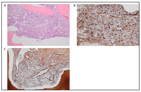
1. Myelodysplastic Syndromes/Myeloproliferative Neoplasms with Neutrophilia (Also Known as Atypical CML)

The fifth edition of the WHO Classification has updated the name of atypical chronic myeloid leukemia, BCR::ABL1-negative (aCML), and replaced it with myelodysplastic syndrome/myeloproliferative neoplasms with neutrophilia (MDS/MPN with neutrophilia) [1], while the ICC has kept the original name [2]. This change highlights the MDS/MPN nature of the disease, while the absence of the terms “atypical CML” and “BCR::ABL1-negative” in the WHO classification avoids potential confusion with both classical CML and other Philadelphia-negative disorders [1].
Myelodysplastic syndromes/myeloproliferative neoplasms (MDS/MPN) with neutrophilia is a rare clonal hematopoietic stem cell disorder of the elderly, with a median age at presentation between 60 and 76 years [3]. Its estimated incidence is 1 out of 100 cases of t(9;22)(q34;q11), BCR::ABL1-positive CML, meaning approximately 1 case per 1,000,000 persons per year [4][5][6][7]. Although a female predominance or no sex predominance has been reported in early studies [5][8], in more recent years, a slight male predominance has been evidenced in reports that analyze larger cohorts of patients [9][10][11]. MDS/MPN with neutrophilia is a disorder that presents clinical features similar to BCR::ABL1-positive CML, including splenomegaly and neutrophilic leukocytosis. Moreover, this leukemia predominantly affects the neutrophilic lineage associated with neutrophilic leukocytosis and circulating immature granulocytic precursors constituting ≥10% of all leukocytes [12]. MDS/MPN with neutrophilia also shows prominent granulocytic dysplasia (e.g., hypogranular and hypolobated neutrophils, abnormal chromatin clumping, and pseudo Pelger–Huet neutrophils) [13]. For these reasons, a main differential diagnosis of MDS/MPN with neutrophilia is BCR::ABL1-positive CML. To detect the presence of BCR::ABL1 translocation, the most therapeutically relevant diagnostic test is karyotype analysis, completed with molecular testing, such as reverse transcription polymerase chain reaction (RT-PCR) and fluorescence in situ hybridization (FISH) techniques, which are both able to reveal the presence of cryptic Ph translocations [14]. The lack of the BCR::ABL1 fusion gene prevents treatment with tyrosine kinase inhibitors, such as imatinib or second- or third-generation Abelson inhibitors. Therefore, its prognosis remains very poor, with a median overall survival of 24 months from diagnosis [15].
Since the clinical features of MDS/MPN with neutrophilia overlap with other myeloproliferative and myelodysplastic malignancies, the diagnosis is still challenging and relies primarily on morphologic criteria. Indeed, besides the absence of the Philadelphia chromosome and of the BCR::ABL1 translocation, the presence of dysgranulopoiesis in BM or PB is the major criterion that can be used to distinguish it from BCR::ABL1-positive CML.
In MDS/MPN with neutrophilia, basophilia is not as prominent as in CML, as basophils represent <2% of all leukocytes [13][16][17]; the diagnostic hallmark for aCML, as defined by the ICC and WHO classification, is the presence of leukocytosis ≥ 13 × 109/L, with ≥10% of immature granulocytes and <20% blasts in the PB and the BM [13][16][18][19]. Of note, ICC requires PB cytopenias, with similar thresholds as MDS, for a diagnosis of aCML [2].
In MDS/MPN with neutrophilia, it is also common to observe an absolute monocyte count > 1 × 10⁹/L; however, the percentage of monocytes at onset must be lower than 10% of the total leukocytes. This characteristic is crucial to discriminate MDS/MPN with neutrophilia from CMML, as in CMML, the presence of both absolute and relative monocytosis is required for diagnosis [16][20]. The proportion of immature myeloid cells (promyelocytes, myelocytes, and metamyelocytes) in PB, as well as the presence of dysplasia, are key criteria for the differential diagnosis against a rare BCR::ABL1-negative myeloproliferative neoplasm named chronic neutrophilic leukemia (CNL), as in CNL, the dysplasia is absent, the percentage of immature elements is always <10%, and persistent peripheral blood neutrophilia (WBC > 25 × 109/L, with >80% segmented neutrophils plus banded neutrophils) is present [7][16]. The higher frequency of CSF3R mutations in CNL [21] has been proposed as an additional criterion to orient the diagnosis; however, CSF3R mutations can be seen also in MDS/MPN with neutrophilia with highly variable frequency in different studies [22][23][24][25]. Therefore the real impact of CSF3R variants for the differential diagnosis of the two disorders is currently a matter of debate [26].
MDS/MPN with neutrophilia exhibits a hypercellular BM with myeloid hyperplasia and prominent granulocytic dysplasia (Figure 1A,B). In addition, trilineage dysplasia may be present [20][22][27]. Conversely, it is not characterized by eosinophilia; hence, eosinophils are expected to be less than 10% in the differential count [2][28]. This feature usually makes the separation between MDS/MPN with neutrophilia and chronic eosinophilic leukemia, not otherwise specified (CEL, NOS), quite straightforward [29]. In addition, as myeloid/lymphoid neoplasms with eosinophilia and tyrosine kinase gene fusions (MLN-TK) are characterized by genetic abnormalities such as PDGFRA, PDGFRB, FGFR1, JAK2, FLT3 rearrangements, or tyrosine kinase fusions (e.g., ETV6::ABL1, ETV6::FGFR2, ETV6::LYN, ETV6::NTRK3, RANBP2::ALK, BCR::RET, FGFR1OP::RET) [1], their differential diagnosis is largely based on molecular features.
Figure 1. MDS/MPN with neutrophilia. (A) At H&E (20×), a mildly hypercellular bone marrow shows minimal megakaryocytes dysplasia. No significant prevalence of immature cells in the other lines. (B) H&E at 40×. (C) Silver staining demonstrates a mild increase of the reticular fibers (MF1, WHO score 2018).
Regarding the myeloproliferative disorders, it is known that JAK2, CALR, and MPL genes, which are usually associated with MPN disorders such as PMF, PV, and essential thrombocythemia (ET), have to be absent in MDS/MPN with neutrophilia [30]. On the one hand, the absence of mutations occurring in these genes can support the differentiation between MDS/MPN with neutrophilia and PMF, but this is usually the most arduous (Figure 2C); on the other hand, the identification of the rare myeloproliferative cases negative for all the three marker genes, named triple-negative myeloproliferative disorders, becomes very difficult. In such cases, genetic analysis can facilitate the diagnosis. Indeed, as reported by the 2016 WHO revision [16], in up to one-third of cases, MDS/MPN with neutrophilia is characterized by the presence of recurrent mutations in ETNK1 (3.7–13.3%) [31][32][33] and SETBP1 (7.4–48%) [34][35][36] genes, often in association with ASXL1 (20–81%), whose presence has been linked to a more aggressive disease [2][3][10][28][37][38]. Besides these genes, other frequent somatic mutations involve NRAS and KRAS (11–27%), SRSF2 (14–65%), EZH2 (19–30%), RUNX1 (11–15%), TET2 (27–33%), and CBL (8–11%) [3][10][15][19][22][28][31][34][36][38][39][40][41][42][43][44][45][46][47]. According to NGS analysis, the fifth edition of the WHO criteria outlines supportive somatic mutations, such as ASXL1 and ETNK1 for diagnosis [1]. In contrast, the ICC has also added molecular supportive criteria, focusing on the presence of somatic mutations involving ASXL1 and SETBP1 [2].

2. Myelodysplastic Syndromes/Myeloproliferative Overlap Neoplasms with SF3B1 Mutation and Thrombocytosis and MDS/MPN with Ring Sideroblasts and Thrombocytosis

The “myelodysplastic syndrome/myeloproliferative neoplasm with ring sideroblasts and thrombocytosis (MDS/MPN-RS-T)” in the 2016 WHO classification [16] is renamed MDS/MPN with SF3B1 mutation and thrombocytosis (MDS/MPN-T) in the WHO 2022 classification [1], and split into two entities in the ICC [2], represented by MDS/MPN with thrombocytosis and SF3B1 mutation (MDS/MPN-T SF3B1), and MDS/MPN with ring sideroblasts and thrombocytosis, not otherwise specified, in the absence of SF3B1 mutation (MDS/MPN RS-T, NOS).
Key elements of this clonal disorder were in the WHO 2016 classification for the presence of dysplasia (Figure 2A–D), either involving the sole erythroid lineage and causing anemia, or involving multiple lineages, with >15% ring sideroblasts and coexisting with thrombocytosis. From a molecular point of view, the most relevant element is the frequent co-occurrence of somatic SF3B1 mutations, found in approximately 80% of the MDS/MPN-T cases, together with the JAK2 V617F mutation, found in 50–60%, or less commonly, with CALR or MPL mutations, globally found in less than 10% of the MDS/MPN-T cases. Clonal cytogenetic abnormalities are less common, as they are found in 20% of cases.
Figure 2. MDS/MPN with SF3B1 mutation and thrombocytosis. (AC) At H&E ((A): 20×; (B): 40×), moderate multilineage dysplasia with significant dyserythropoiesis and megalbostastosis, as demonstrated by the glycophorin staining (C). (D) At MGG, other dysplastic findings in the erythroid series, which correspond to Perls’ positive ring sideroblasts at bone marrow aspirate.
As mutations occurring in the spliceosome gene SF3B1 are known to be enriched in patients affected by MDS/MPN-RS-T, the identification of ring sideroblasts is not considered to be required to confirm the diagnosis in the most recent WHO classification [1], as well as in the ICC [2], provided that SF3B1 somatic mutation is found with a VAF greater than 10%.
According to the WHO 2022 classification [1], the term MDS/MPN with ring sideroblasts and thrombocytosis has been kept as an acceptable term to be used for cases with wild-type SF3B1 and ≥15% ring sideroblasts. Conversely, this entity is separated from SF3B1 mutated MDS/MPN forms in the ICC and designed as “MDS/MPN with sideroblasts and thrombocytosis, NOS”.
In agreement with the criteria defined for other MDS/MPN, and for MDS/MPN-T-SF3B1 and MDS/MPN-RS-T NOS, both thrombocytosis and anemia must be present at onset [2].
Differential diagnosis for MDS/MPN-T comprises myeloproliferative disorders, MDS, and other MDS/MPN neoplasms. Differential diagnosis against ET, sharing with MDS/MPN-T the presence of thrombocytosis, can be based on morphological as well as molecular criteria. From a morphological point of view, in the presence of mutated SF3B1, the evidence of anemia, optionally associated with erythroid dysplasia in the bone marrow, definitely supports the diagnosis of MDS/MPN-T. In the absence of SF3B1, anemia must be associated with erythroid-lineage dysplasia and ring sideroblasts to support the diagnosis of MDS/MPN-RS-T, NOS. At the molecular level, the evidence of SF3B1 mutations, either in the presence or absence of activating JAK2 mutations, determines the diagnosis of MDS/MPN-T SF3B1, regardless of the identification of ring sideroblasts, if SF3B1 is detectable with VAF > 10%. In contrast, the presence of JAK2, CALR, or MPL mutations in the absence of SF3B1 should orient the diagnosis to a myeloproliferative disorder.
Finally, cases of MDS-SF3B1 that later develop thrombocytosis are now considered to represent the thrombocytotic progression of MDS-SF3B1.

3. Myelodysplastic/Myeloproliferative Neoplasm, Not Otherwise Specified

MDS/MPN, unclassifiable is now termed MDS/MPN, not otherwise specified MDS/MPN, NOS) in both the WHO classification and the ICC [1][2]. This fact is in line with the international effort to remove the qualifier “unclassifiable” from the classification. MDS/MPN, NOS is still mainly a diagnosis of exclusion; however, it is now better described by the inclusion of diagnostic requirements. These criteria include a need for the presence of cytopenia in association with myeloproliferative features in PB and a lack of specific gene rearrangements/fusions of M/LN-Eo with tyrosine kinase gene fusions. Therefore, although the diagnosis can be made in the absence of clonality or mutations, even in the case of histopathologic evidence of MDS/MPN and the exclusion of other MDS/MPN entities, the establishment of clonality is expected [2].

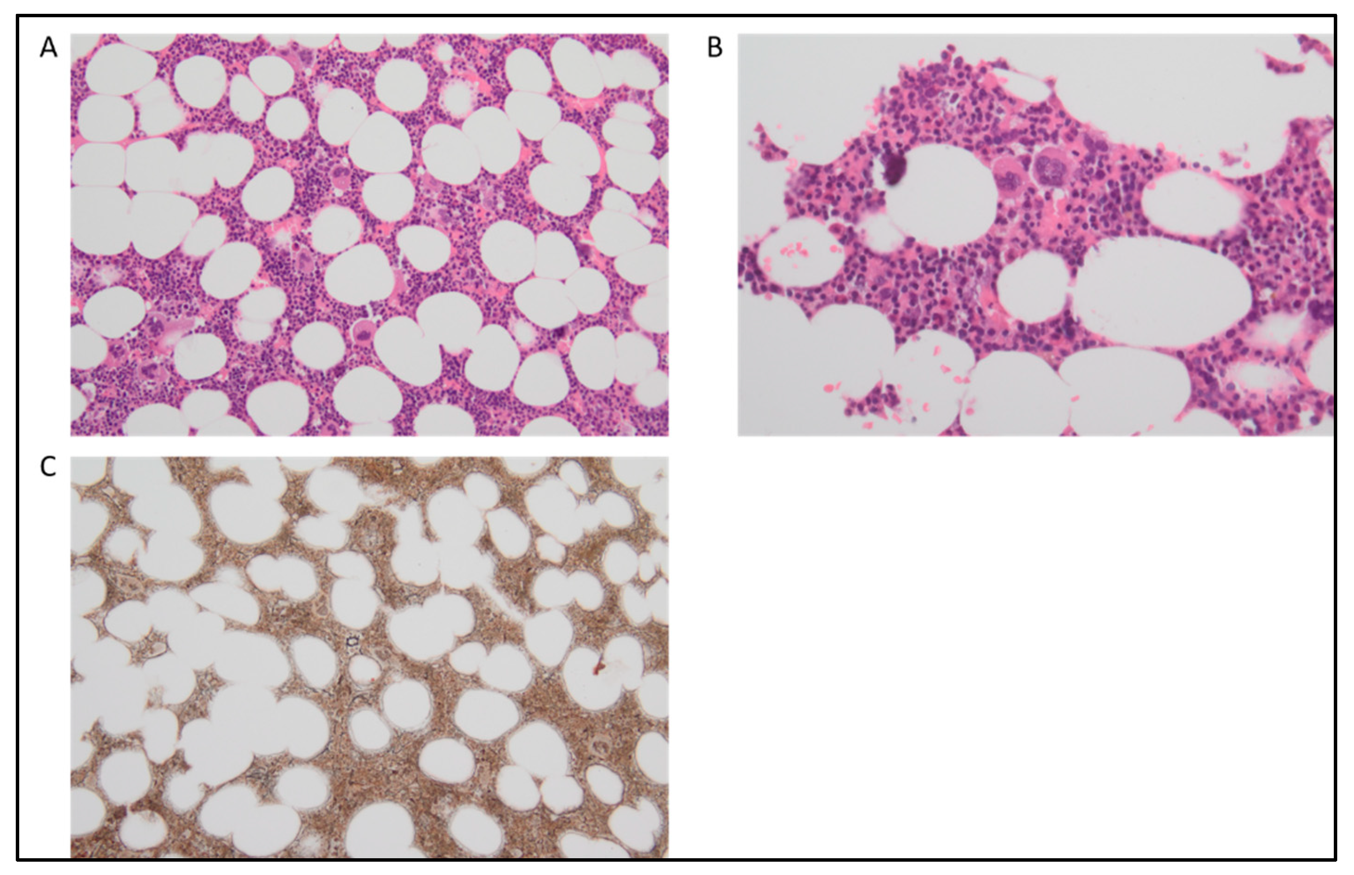
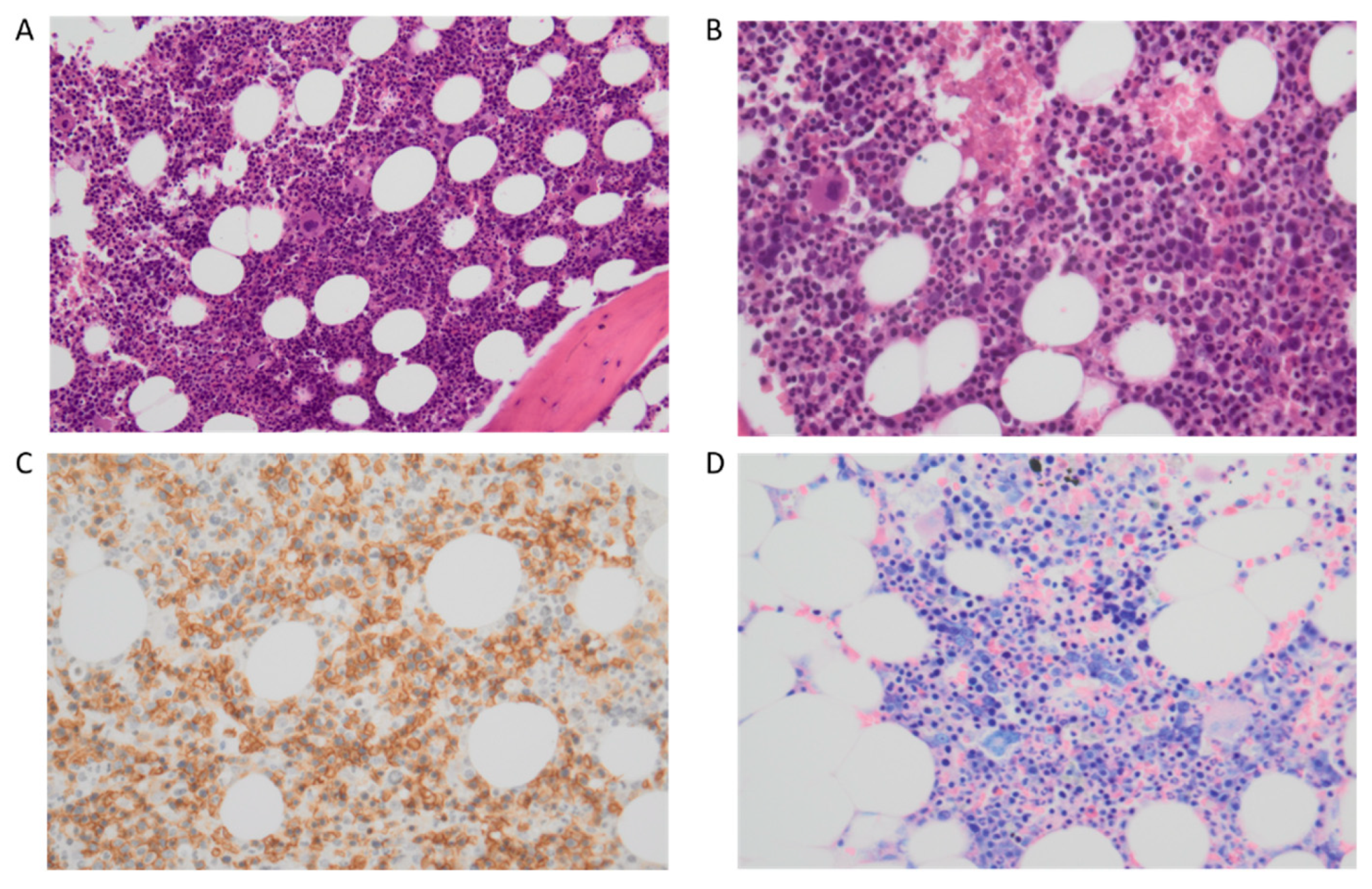
4. Chronic Myelomonocytic Leukemia

CMML is the most common (incidence 0.6 × 100,000 people in the United States) MDS/MPN disease. It is a clonal stem cell disorder that is characterized by sustained peripheral blood (PB) monocytosis (≥0.5 × 10⁹/L and ≥10% of white blood cells [WBC] differential count) and an inherent tendency for transformation to acute myeloid leukemia (AML; 15% over 3 to 5 years) [18].
The median age at diagnosis for CMML is 73 to 75 years, with a male preponderance. CMML diagnosis remains largely based on morphology. Histologically, CMML was classified as CMML-0 (<2% PB blasts and <5% bone marrow [BM] blasts), CMML-1 (2–4% PB blasts and/or 5–9% BM blasts), and CMML-2 (5–19% PB blasts and/or 10–19% BM blasts or when Auer rods are present) [48]. However, recent studies have shown that the prognostic impact of CMML-0 and CMML-1 is virtually identical. In addition, the classification of CMML patients as CMML-0 or CMML-1 requires accurate counting of a low number of blast cells. This is particularly challenging in the case of CMML, in which blasts include promonocytes, whose distinction from abnormal monocytes can at times be problematic. For these reasons, the WHO 2022 classification reverted to the fourth edition 2-tiered system of CMML-1 (<5% blasts in PB, <10% in BM) and CMML-2 (5–19% blasts PB, 10–19% in BM, or Auer rods), therefore incorporating CMML-0 cases into the CMML-1 group [2].
Based on the presenting WBC count, the French–American–British (FAB) classification distinguished CMML into two subtypes: myeloproliferative CMML (MP-CMML; WBC count ≥ 13 × 109/L) and myelodysplastic CMML (MD-CMML; WBC count < 13 × 109/L), where the former has a poor outcome and a higher rate of AML transformation [49]. In CMML, the proliferative component is manifested as monocytosis often in association with splenomegaly and/or leukocytosis [50]. Several prognostic models exist for CMML; however, the CMML-specific prognostic scoring system–molecular model (CPSS molecular score) is the principal one [49]. This includes the percentage of BM blasts, the FAB subtype of CMML (WBC > 13 × 109/L), the need for transfusion support, and the presence of genetic markers (ASXL1, RUNX1, NRAS, SETBP1, and cytogenetic abnormalities) as risk factors.

4.1. Cytogenetics and Molecular Genetics

CMML is a clonal disorder predominantly arising in the context of age-related clonal hematopoiesis [51]. Clonal cytogenetic abnormalities occur in ~30% of patients, with a variability largely due to small numbers, inclusion criteria, and referral patterns. CMML is not characterized by specific cytogenetic aberrations; however, trisomy 8 and monosomy 7 are the most frequent, while complex karyotypes are infrequent [52]. A strong association between specific cytogenetic abnormalities (trisomy 8, chromosome 7 abnormalities, or complex karyotype) and the risk of AML evolution and overall survival (OS) is described [53].
In the coding regions of CMML patients’ genome, an average of 10 to 15 somatic mutations can be found [54]. Compared to MDS and AML the mutational spectrum of CMML is more homogeneous [50]. The most common somatic mutations involve splicing genes and epigenetic modifiers (e.g., SRSF2, TET2, and/or ASXL1), which occur in about 80% of cases [18]. Other mutated genes include SETBP1, NRAS/KRAS, RUNX1, CBL, and EZH2. The prototypical molecular fingerprint combines a mutation in a gene encoding an epigenetic regulator (mainly TET2 and ASXL1) with a mutation affecting the spliceosome machinery (SRSF2, less often SF3B1, ZRSR2) with or without a mutation in the RAS/MAPK signaling pathway [50]. Overall, >90% of patients with CMML would be expected to show at least one of these mutations with modern next generation sequencing (NGS) techniques [55][56]. Mutations involving TET2 (epigenetic) and SRSF2 (splicing) often skew hematopoiesis toward monocytosis, with subsequent mutations in epigenetic regulators (ASXL1) or signaling pathways (NRAS, CBL, KRAS, PTPN11, and JAK2), giving rise to a full-blown disease [48].
MPN-CMML variant is enriched in active RAS/MAPK signaling, with ~70% of patients demonstrating RAS pathway mutations (e.g., NRAS, KRAS, CBL), with NRAS being the most commonly involved RAS-family gene. RAS pathway mutations, along with epigenetic events, also play a role in CMML transformation to AML. Of note, NPM1 mutation is seen in a rare subset of CMML (3–5%) [57]. The development of an NPM1 mutation in the setting of a known CMML should be noted, as it bears prognostic relevance. It is important to note that, although the new WHO classification states that the presence of the NPM1 variant alone leads to a diagnosis of AML independently from the blast percentage [2], such a finding in the ICC does not automatically define de novo AML in the setting of known CMML, as a bone marrow blast count of at least 10% is also required [2]. Regardless of these differences in the WHO classification and the ICC, CMML NPM1 mutated appears to herald a particularly aggressive clinical course with a median overall survival of 12.5 months compared to 20.5 months for NPM1 wild-type patients [57]. Therefore, this subgroup of CMML NPM1 mutated probably deserves an AML-like therapeutic approach.
A recommended minimal NGS panel for CMML [50] is represented by the following genes: TET2 (frequency: 29–61%), ASXL1 (frequency: 32–44%), DNMT3A (frequency: 2–12%), EZH2 (frequency: 5–13%), IDH1 (frequency: 1–2%), IDH2 (frequency: 6–7%), BCOR (frequency: 6–7%), SRSF2 (frequency: 29–52%), U2AF1 (frequency: 4–10%), SF3B1 (frequency: 6–10%), ZRSR2 (frequency: 4–8%), CBL (frequency: 8–22%), KRAS (frequency: 7–16%), NRAS (frequency: 4–22%), NF1 (frequency: 6–7%), JAK2 (frequency: 1–10%), RUNX1 (frequency: 8–23%), SETBP1 (frequency: 4–18%), NPM1 (frequency: 1–3%), and FLT3 (frequency: 1–3%) [49][50][51][54][58][59][60][61][62][63][64][65][66][67][68][69][70].

4.2. Flow Cytometry Immunophenotyping

CMML diagnosis can benefit from flow cytometry analyses of BM and PB cells. Indeed, the detection of subtle changes in the surface antigen expression of myelomonocytic cells and the erythroid lineage can potentially facilitate the monitoring of the disease [50]. In particular, flow cytometry analysis of monocyte subsets readily distinguishes CMML from benign reactive monocytosis in patients with PB monocytosis ≥ 1 × 10⁹/L. As reported by the current nomenclature of normal human monocyte subsets, the fraction of classical monocytes (CD14+/CD16), also known as MO1, can be distinguished from intermediate, MO2 monocytes expressing CD14+ and CD16+, and from nonclassical, MO3 monocytes, typically expressing CD16 and low levels of CD14 [71]. This is important in the context of CMML, as the proportion of MO1 is increased in CMML patients, while it is decreased in those affected by a reactive disorder [72]. In particular, a MO1 ≥ 94% provides a very high level of specificity and sensitivity to distinguish CMML from reactive monocytosis [71][72].

4.3. Diagnosis and Differential Diagnosis of Chronic Myelomonocytic Leukemia 

The revised diagnostic criteria according to the WHO 2022 classification include prerequisite and supporting criteria. The first prerequisite criterion is persistent absolute (≥0.5 × 10⁹/L) and relative (≥10%) peripheral blood monocytosis, in the absence of reactive etiologies. Namely, the cutoff for absolute monocytosis is lowered from 1.0 × 10⁹/L to 0.5 × 10⁹/L, to incorporate cases formerly referred to as oligomonocytic CMML. The second prerequisite is to have a PB and BM blast count of less than 20%, with or without BM dysplasia. The third and fourth prerequisites require not meeting the diagnostic criteria of chronic myeloid leukemia or other myeloproliferative neoplasms, and of myeloid/lymphoid neoplasms with tyrosine kinase fusions, respectively. In line with these exclusion criteria, CMML diagnosis also requires the absence of molecular aberrations, such as PDGFRA, PDGFRB, and PCM1::JAK2, that can be associated with clonal disorders potentially characterized by the presence of monocytosis.
Supporting criteria are instead represented (1) by the presence of dysplasia in one or more than one lineage (Figure 3A), (2) by the evidence of clonality, typically demonstrated by the identification of somatic mutations or cytogenetic abnormalities, and (3) by the evidence of abnormal partitioning of peripheral blood monocyte subsets (Figure 3B). The latter is introduced as a new supporting criterion, with most CMML patients demonstrating an expansion (>94%) of classical monocytes (MO1CD14+/CD16). This modality has also shown promise in distinguishing CMML from other causes of monocytosis, including MPN with monocytosis, and it is useful in cases of CMML without evidence of clonality [71][72]. In the presence of clear evidence of absolute monocytosis, i.e., with monocytes ≥ 1.0 × 10⁹/L, all prerequisite criteria must be present and further strengthened by at least one of the supporting criteria. To enhance diagnostic accuracy when absolute monocytosis is low, i.e., between 0.5 × 10⁹/L and 1.0 × 10⁹/L, the detection of one or more clonal cytogenetic or molecular abnormalities and the documentation of dysplasia in at least one lineage are required.
In the new WHO classification, the presence of somatic mutations as a supporting criterion is felt to be at least as critical as the presence of dysplasia for confirming a diagnosis of CMML. Indeed, having established the need for clonality as one of the supporting criteria, the evidence of dysplasia becomes necessary only for those rare patients who lack the presence of CMML-associated mutations or when monocytosis is low, as already discussed. The rationale for this approach is based on three considerations: (1) the frequent evidence of clonal variants in CMML, which makes the complete lack of mutations highly unlikely; (2) the availability of myeloid NGS panels able to reliably detect subclonal mutations down to the 1–5% variant allele frequency (VAF) range; (3) the challenges associated with the objective demonstration of dysplasia in bone marrow specimens. In addition, mutations in CMML have prognostic implications; hence, the implementation of mutation detection in the diagnostic routine of CMML allows for the killing of two birds with one stone, on the one hand, and confirming the diagnosis on the other, to refine the prognosis.
An absolute monocytosis is uncommon in classical CML; however, it may be present in a subset of patients with MPN and can be detected at the time of primary diagnosis or during the disease progression. Monocytosis is also associated with a more unfavorable outcome in patients affected by polycythemia vera (PV) [73]. Monocytosis is also a powerful and independent predictor of inferior survival in primary myelofibrosis (PMF) [74]. Among classical driver mutations, in 10% of CMML patients, JAK2V617F can occur, while mutations in MPL and CALR genes are extremely infrequent and their detection should raise questions with regard to a bona fide CMML diagnosis. In addition, the multiparametric flow cytometry (discussed below) can help differentiate CMML from MPN with monocytosis [75].
Triple-negative primary myelofibrosis (TN-PMF) and myelodysplastic syndromes with fibrosis (F-MDS) are rare entities, which are often difficult to distinguish and occasionally associated with a CMML-like phenotype. Currently, no specific molecular markers are available, and the integration of clinical data, such as BM morphology and blood counts, remains essential for diagnosis. Of note, neutrophil granulopoiesis, age-related cellularity, and changes in erythropoiesis, together with the severity of BM fibrosis should be considered in addition to megakaryocytic atypia (Figure 3C). NGS tests might be useful to distinguish between both entities and to refine prognosis. TN-PMF and F-MDS show a high rate of mutations in myeloid genes, with TET2, U2AF1, SETBP1, TP53, or RUNX1 being more frequently mutated in F-MDS. TN MF presents poor outcomes and a high risk of leukemic transformation [76].
Mutations involving SRSF2, SETBP1, IDH2, CBL, and GNAS are significantly enriched in TN disease.
Reactive monocytosis is very common in clinical practice and the principal causes are represented by viral infections, recovery from injury, drugs or chemotherapy, bacterial subacute endocarditis, tuberculosis, brucellosis, leishmaniosis, and autoimmune diseases such as systemic lupus erythematosus, sarcoidosis, and mixed connective tissue disorder. Reactive monocytosis can also be seen in the context of metastatic solid neoplasms [50]
Figure 3. Chronic myelomonocytic leukemia. (A) H&E staining (20×) multilineage dysplasia with irregular megakaryocytes characterized by hypolobulated and hyperchromatic nuclei. (B) A predominant monocyte differentiation is highlighted by CD68 pgm1 immunohistochemical staining (20×). (C) At silver staining (20×) large bands of extensive fibrosis (MF2, WHO score 2018).

5. Clonal Monocytosis of Undetermined Significance and Clonal Cytopenia with Monocytosis of Undetermined Significance

The ICC recognizes the clonal monocytosis of undetermined significance (CMUS), a CMML precursor condition, as an independent entity [2]. The CMUS was proposed in order to classify a particular disorder that does not yet fulfill the criteria of CMML [77]. Indeed, the CMUS is characterized by persistent monocytosis (monocytes ≥ 10% and≥ 0.5 × 10⁹/L of the WBC), the presence of myeloid neoplasm-associated mutation(s), and the absence of BM morphologic findings of CMML. In cases of cytopenia, the disorder is renamed clonal cytopenia and monocytosis of undetermined significance (CCMUS) [2].

References

  1. Khoury, J.D.; Solary, E.; Abla, O.; Akkari, Y.; Alaggio, R.; Apperley, J.F.; Bejar, R.; Berti, E.; Busque, L.; Chan, J.K.C.; et al. The 5th edition of the World Health Organization Classification of Haematolymphoid Tumours: Myeloid and Histiocytic/Dendritic Neoplasms. Leukemia 2022, 36, 1703–1719.
  2. Arber, D.A.; Orazi, A.; Hasserjian, R.P.; Borowitz, M.J.; Calvo, K.R.; Kvasnicka, H.M.; Wang, S.A.; Bagg, A.; Barbui, T.; Branford, S.; et al. International Consensus Classification of Myeloid Neoplasms and Acute Leukemias: Integrating morphologic, clinical, and genomic data. Blood 2022, 140, 1200–1228.
  3. Patnaik, M.M.; Barraco, D.; Lasho, T.L.; Finke, C.M.; Reichard, K.; Hoversten, K.P.; Ketterling, R.P.; Gangat, N.; Tefferi, A. Targeted next generation sequencing and identification of risk factors in World Health Organization defined atypical chronic myeloid leukemia. Am. J. Hematol. 2017, 92, 542–548.
  4. Vardiman, J.W.; Bain, B.; Inbert, M.; Brunning, R.D.; Pierre, R.V.; Flandrin, G. Atypical chronic myeloid leukemia. In WHO Classification of Tumours: Pathology and Genetics of Tumours of Haematopoietic and Lymphoid Tissues; Jaffe, E., Harris, N.L., Stein, H., Vardiman, J., Eds.; IARC Press: Lyon, France, 2001; pp. 53–57.
  5. Breccia, M.; Biondo, F.; Latagliata, R.; Carmosino, I.; Mandelli, F.; Alimena, G. Identification of risk factors in atypical chronic myeloid leukemia. Haematologica 2006, 91, 1566–1568.
  6. Oscier, D. Atypical chronic myeloid leukemias. Pathol. Biol. 1997, 45, 587–593.
  7. Cazzola, M.; Malcovati, L.; Invernizzi, R. Myelodysplastic/myeloproliferative neoplasms. Hematol. Am. Soc. Hematol. Educ. Program 2011, 1, 264–272.
  8. Hernandez, J.; Villaescusa, T.; Arefi, M.; López, L.; Garcia, J. Atypical Chronic Myeloid Leukemia (aCML). Atlas Genet. Cytogenet. Oncol. Haematol. 2009, 13, 432–433.
  9. Zhang, H.; Wilmot, B.; Bottomly, D.; Dao, K.T.; Stevens, E.; Eide, C.A.; Khanna, V.; Rofelty, A.; Savage, S.; Reister Schultz, A.; et al. Genomic landscape of neutrophilic leukemias of ambiguous diagnosis. Blood 2019, 134, 867–879.
  10. Fontana, D.; Ramazzotti, D.; Aroldi, A.; Redaelli, S.; Magistroni, V.; Pirola, A.; Niro, A.; Massimino, L.; Mastini, C.; Brambilla, V.; et al. Integrated Genomic, Functional, and Prognostic Characterization of Atypical Chronic Myeloid Leukemia. Hemasphere 2020, 4, e497.
  11. Giri, S.; Pathak, R.; Martin, M.G.; Bhatt, V.R. Characteristics and survival of BCR/ABL negative chronic myeloid leukemia: A retrospective analysis of the Surveillance, Epidemiology and End Results database. Ther. Adv. Hematol. 2015, 6, 308–312.
  12. Orazi, A.; Bennet, J.M.; Bain, B.J.; Brunning, R.D.; Thiele, J. Atypical chronic myeloid leukaemia. In WHO Classification of Tumours of Haematopoietic and Lymphoid Tissues, 4th ed.; Swerdlow, S.H., Campo, E., Harris, N.L., Jaffe, E.S., Pileri, S.A., Stein, H., Thiele, J., Eds.; IARC Press: Lyon, France, 2017; pp. 87–89.
  13. Vardiman, J.W.; Thiele, J.; Arber, D.A.; Brunning, R.D.; Borowitz, M.J.; Porwit, A.; Harris, N.L.; Le Beau, M.M.; Hellström-Lindberg, E.; Tefferi, A.; et al. The 2008 revision of the World Health Organization (WHO) classification of myeloid neoplasms and acute leukemia: Rationale and important changes. Blood 2009, 114, 937–951.
  14. Crisà, E.; Nicolosi, M.; Ferri, V.; Favini, C.; Gaidano, G.; Patriarca, A. Atypical Chronic Myeloid Leukemia: Where Are We Now? Int. J. Mol. Sci. 2020, 21, 6862.
  15. Fontana, D.; Gambacorti-Passerini, C.; Piazza, R. Molecular Pathogenesis of BCR-ABL-Negative Atypical Chronic Myeloid Leukemia. Front. Oncol. 2021, 11, 756348.
  16. Arber, D.A.; Orazi, A.; Hasserjian, R.; Thiele, J.; Borowitz, M.J.; Le Beau, M.M.; Bloomfield, C.D.; Cazzola, M.; Vardiman, J.W. The 2016 revision to the World Health Organization classification of myeloid neoplasms and acute leukemia. Blood 2016, 127, 2391–2405.
  17. Kurzrock, R.; Bueso-Ramos, C.E.; Kantarjian, H.; Freireich, E.; Tucker, S.L.; Siciliano, M.; Pilat, S.; Talpaz, M. BCR rearrangement-negative chronic myelogenous leukemia revisited. J. Clin. Oncol. 2001, 19, 2915–2926.
  18. Patnaik, M.M.; Tefferi, A. Atypical chronic myeloid leukemia and myelodysplastic/myeloproliferative neoplasm, not otherwise specified: 2023 update on diagnosis, risk stratification, and management. Am. J. Hematol. 2023, 98, 681–689.
  19. Gotlib, J.; Maxson, J.E.; George, T.I.; Tyner, J.W. The new genetics of chronic neutrophilic leukemia and atypical CML: Implications for diagnosis and treatment. Blood 2013, 122, 1707–1711.
  20. Orazi, A.; Germing, U. The myelodysplastic/myeloproliferative neoplasms: Myeloproliferative diseases with dysplastic features. Leukemia 2008, 22, 1308–1319.
  21. Maxson, J.E.; Tyner, J.W. Genomics of chronic neutrophilic leukemia. Blood 2017, 129, 715–722.
  22. Dao, K.H.; Tyner, J.W. What’s different about atypical CML and chronic neutrophilic leukemia? Hematol. Am. Soc. Hematol. Educ. Program 2015, 2015, 264–271.
  23. Maxson, J.E.; Gotlib, J.; Pollyea, D.A.; Fleischman, A.G.; Agarwal, A.; Eide, C.A.; Bottomly, D.; Wilmot, B.; McWeeney, S.K.; Tognon, C.E.; et al. Oncogenic CSF3R Mutations in Chronic Neutrophilic Leukemia and Atypical CML. N. Engl. J. Med. 2013, 368, 1781–1790.
  24. Schwartz, L.C.; Mascarenhas, J. Current and evolving understanding of atypical chronic myeloid leukemia. Blood Rev. 2019, 33, 74–81.
  25. Carreño-Tarragona, G.; Alvarez-Larran, A.; Harrison, C.N.; Martínez-Ávila, J.C.; Hernandez-Boluda, J.C.; Ferrer-Marin, F.; Radia, D.H.; Mora Casterá, E.; Francis, S.; González-Martínez, T.; et al. CNL and aCML should be considered as single entity based on molecular profiles and outcomes. Blood Adv. 2022, 9, 1672–1681.
  26. Thomopoulos, T.P.; Symeonidis, A.; Kourakli, A.; Papageorgiou, S.G.; Pappa, V. Chronic Neutrophilic Leukemia: A Comprehensive Review of Clinical Characteristics, Genetic Landscape and Management. Front. Oncol. 2022, 12, 891961.
  27. Thakral, B.; Anastasi, J.; Wang, S.A. 17-Myeloproliferative and “Overlap” Myelodysplastic/Myeloproliferative Neoplasms. Found. Diagn. Pathol. 2018, 488–538.e484.
  28. Wang, S.A.; Hasserjian, R.P.; Fox, P.S.; Rogers, H.J.; Geyer, J.T.; Chabot-Richards, D.; Weinzierl, E.; Hatem, J.; Jaso, J.; Kanagal-Shamanna, R.; et al. Atypical chronic myeloid leukemia is clinically distinct from unclassifiable myelodysplastic/myeloproliferative neoplasms. Blood 2014, 123, 2645–2651.
  29. Wang, S.A.; Hasserjian, R.P.; Tam, W.; Tsai, A.G.; Geyer, J.T.; George, T.I.; Foucar, K.; Rogers, H.J.; Hsi, E.D.; Rea, B.A.; et al. Bone marrow morphology is a strong discriminator between chronic eosinophilic leukemia, not otherwise specified and reactive idiopathic hypereosinophilic syndrome. Haematologica 2017, 102, 1352–1360.
  30. Fend, F.; Horn, T.; Koch, I.; Vela, T.; Orazi, A. Atypical chronic myeloid leukemia as defined in the WHO classification is a JAK2 V617F negative neoplasm. Leuk. Res. 2008, 32, 1931–1935.
  31. Gambacorti-Passerini, C.B.; Donadoni, C.; Parmiani, A.; Pirola, A.; Redaelli, S.; Signore, G.; Piazza, V.; Malcovati, L.; Fontana, D.; Spinelli, R.; et al. Recurrent ETNK1 mutations in atypical chronic myeloid leukemia. Blood 2015, 125, 499–503.
  32. Fontana, D.; Mauri, M.; Renso, R.; Docci, M.; Crespiatico, I.; Røst, L.M.; Jang, M.; Niro, A.; D’Aliberti, D.; Massimino, L.; et al. ETNK1 mutations induce a mutator phenotype that can be reverted with phosphoethanolamine. Nat. Commun. 2020, 11, 5938.
  33. Fontana, D.; Gambacorti-Passerini, C.; Piazza, R. Impact of ETNK1 somatic mutations on phosphoethanolamine synthesis, ROS production and DNA damage. Mol. Cell. Oncol. 2021, 8, 1877598.
  34. Piazza, R.; Valletta, S.; Winkelmann, N.; Redaelli, S.; Spinelli, R.; Pirola, A.; Antolini, L.; Mologni, L.; Donadoni, C.; Papaemmanuil, E.; et al. Recurrent SETBP1 mutations in atypical chronic myeloid leukemia. Nat. Genet. 2013, 45, 18–24.
  35. Piazza, R.; Magistroni, V.; Redaelli, S.; Mauri, M.; Massimino, L.; Sessa, A.; Peronaci, M.; Lalowski, M.; Soliymani, R.; Mezzatesta, C.; et al. SETBP1 induces transcription of a network of development genes by acting as an epigenetic hub. Nat. Commun. 2018, 9, 2192.
  36. Meggendorfer, M.; Bacher, U.; Alpermann, T.; Haferlach, C.; Kern, W.; Gambacorti-Passerini, C.; Haferlach, T.; Schnittger, S. SETBP1 mutations occur in 9% of MDS/MPN and in 4% of MPN cases and are strongly associated with atypical CML, monosomy 7, isochromosome i(17)(q10), ASXL1 and CBL mutations. Leukemia 2013, 27, 1852–1860.
  37. Dhakal, P.; Gundabolu, K.; Amador, C.; Rayamajhi, S.; Bhatt, V.R. Atypical chronic myeloid leukemia: A rare entity with management challenges. Future Oncol. 2018, 14, 177–185.
  38. Gotlib, J. How I treat atypical chronic myeloid leukemia. Blood 2017, 129, 838–845.
  39. Carulli, G.; Fabiani, O.; Azzara, A. The syndrome of abnormal chromatin clumping in leukocytes. Haematologica 1997, 82, 635–636.
  40. Tefferi, A.; Elliot, M.A.; Pardanani, A. Atypical myeloproliferative disorders: Diagnosis and management. Mayo Clin. Proc. 2006, 81, 553–563.
  41. Jones, A.V.; Kreil, S.; Zoi, K.; Waghorn, K.; Curtis, C.; Zhang, L.Y.; Score, J.; Seear, R.; Chase, A.J.; Grand, F.H.; et al. Widespread occurrence of the JAK2 V617F mutation in chronic myeloproliferative disorders. Blood 2005, 106, 2162–2168.
  42. Mughal, T.I.; Cross, N.C.; Padron, E.; Tiu, R.V.; Savona, M.; Malcovati, L.; Tibes, R.; Komrokji, R.S.; Kiladjian, J.J.; Garcia-Manero, G.; et al. An International MDS/MPN Working Group’s perspective and recommendations on molecular pathogenesis, diagnosis and clinical characterization of myelodysplastic/myeloproliferative neoplasms. Haematologica 2015, 100, 1117–1130.
  43. Tiu, R.V.; Sekeres, M.A. Making sense of the myelodysplastic/myeloproliferative neoplasms overlap syndromes. Curr. Opin. Hematol. 2014, 21, 131–140.
  44. Zoi, K.; Cross, N.C. Molecular pathogenesis of atypical CML, CMML and MDS/MPN-unclassifiable. Int. J. Hematol. 2015, 101, 229–242.
  45. Meggendorfer, M.; Haferlach, T.; Alpermann, T.; Jeromin, S.; Haferlach, C.; Kern, W.; Schnittger, S. Specific molecular mutation patterns delineate chronic neutrophilic leukemia, atypical chronic myeloid leukemia, and chronic myelomonocytic leukemia. Haematologica 2014, 99, e244–e246.
  46. Makishima, H.; Yoshida, K.; Nguyen, N.; Przychodzen, B.; Sanada, M.; Okuno, Y.; Ng, K.P.; Gudmundsson, K.O.; Vishwakarma, B.A.; Jerez, A.; et al. Somatic SETBP1 mutations in myeloid malignancies. Nat. Genet. 2013, 45, 942–946.
  47. Abdel-Wahab, O.; Mullally, A.; Hedvat, C.; Garcia-Manero, G.; Patel, J.; Wadleigh, M.; Malinge, S.; Yao, J.; Kilpivaara, O.; Bhat, R.; et al. Genetic characterization of TET1, TET2, and TET3 alterations in myeloid malignancies. Blood 2009, 114, 144–147.
  48. Patnaik, M.M.; Lasho, T. Myelodysplastic syndrome/myeloproliferative neoplasm overlap syndromes: A focused review. Hematol. Am. Soc. Hematol. Educ. Program 2020, 2020, 460–464.
  49. Elena, C.; Gallì, A.; Such, E.; Meggendorfer, M.; Germing, U.; Rizzo, E.; Cervera, J.; Molteni, E.; Fasan, A.; Schuler, E.; et al. Integrating clinical features and genetic lesions in the risk assessment of patients with chronic myelomonocytic leukemia. Blood 2016, 128, 1408–1417.
  50. Itzykson, R.; Fenaux, P.; Bowen, D.; Cross, N.C.P.; Cortes, J.; De Witte, T.; Germing, U.; Onida, F.; Padron, E.; Platzbecker, U.; et al. Diagnosis and Treatment of Chronic Myelomonocytic Leukemias in Adults: Recommendations From the European Hematology Association and the European LeukemiaNet. Hemasphere 2018, 2, e150.
  51. Mason, C.C.; Khorashad, J.S.; Tantravahi, S.K.; Kelley, T.W.; Zabriskie, M.S.; Yan, D.; Pomicter, A.D.; Reynolds, K.R.; Eiring, A.M.; Kronenberg, Z.; et al. Age-related mutations and chronic myelomonocytic leukemia. Leukemia 2016, 30, 906–913.
  52. Palomo, L.; Ibáñez, M.; Abáigar, M.; Vázquez, I.; Álvarez, S.; Cabezón, M.; Tazón-Vega, B.; Rapado, I.; Fuster-Tormo, F.; Cervera, J.; et al. Spanish Guidelines for the use of targeted deep sequencing in myelodysplastic syndromes and chronic myelomonocytic leukaemia. Br. J. Haematol. 2020, 188, 605–622.
  53. Woo, J.; Choi, D.R.; Storer, B.E.; Yeung, C.; Halpern, A.B.; Salit, R.B.; Sorror, M.L.; Woolston, D.W.; Monahan, T.; Scott, B.L.; et al. Impact of clinical, cytogenetic, and molecular profiles on long-term survival after transplantation in patients with chronic myelomonocytic leukemia. Haematologica 2020, 105, 652–660.
  54. Merlevede, J.; Droin, N.; Qin, T.; Meldi, K.; Yoshida, K.; Morabito, M.; Chautard, E.; Auboeuf, D.; Fenaux, P.; Braun, T.; et al. Mutation allele burden remains unchanged in chronic myelomonocytic leukaemia responding to hypomethylating agents. Nat. Commun. 2016, 7, 10767.
  55. Cargo, C.; Cullen, M.; Taylor, J.; Short, M.; Glover, P.; Van Hoppe, S.; Smith, A.; Evans, P.; Crouch, S. The use of targeted sequencing and flow cytometry to identify patients with a clinically significant monocytosis. Blood 2019, 133, 1325–1334.
  56. Palomo, L.; Meggendorfer, M.; Hutter, S.; Twardziok, S.; Ademà, V.; Fuhrmann, I.; Fuster-Tormo, F.; Xicoy, B.; Zamora, L.; Acha, P.; et al. Molecular landscape and clonal architecture of adult myelodysplastic/myeloproliferative neoplasms. Blood 2020, 136, 1851–1862.
  57. Vallapureddy, R.; Lasho, T.L.; Hoversten, K.; Finke, C.M.; Ketterling, R.; Hanson, C.; Gangat, N.; Tefferi, A.; Patnaik, M.M. Nucleophosmin 1 (NPM1) mutations in chronic myelomonocytic leukemia and their prognostic relevance. Am. J. Hematol. 2017, 92, E614–E618.
  58. Ball, M.; List, A.F.; Padron, E. When clinical heterogeneity exceeds genetic heterogeneity: Thinking outside the genomic box in chronic myelomonocytic leukemia. Blood 2016, 128, 2381–2387.
  59. Meggendorfer, M.; Roller, A.; Haferlach, T.; Eder, C.; Dicker, F.; Grossmann, V.; Kohlmann, A.; Alpermann, T.; Yoshida, K.; Ogawa, S.; et al. SRSF2 mutations in 275 cases with chronic myelomonocytic leukemia (CMML). Blood 2012, 120, 3080–3088.
  60. Itzykson, R.; Kosmider, O.; Renneville, A.; Gelsi-Boyer, V.; Meggendorfer, M.; Morabito, M.; Berthon, C.; Adès, L.; Fenaux, P.; Beyne-Rauzy, O.; et al. Prognostic score including gene mutations in chronic myelomonocytic leukemia. J. Clin. Oncol. 2013, 31, 2428–2436.
  61. Malcovati, L.; Papaemmanuil, E.; Ambaglio, I.; Elena, C.; Gallì, A.; Della Porta, M.G.; Travaglino, E.; Pietra, D.; Pascutto, C.; Ubezio, M.; et al. Driver somatic mutations identify distinct disease entities within myeloid neoplasms with myelodysplasia. Blood 2014, 124, 1513–1521.
  62. Ricci, C.; Fermo, E.; Corti, S.; Molteni, M.; Faricciotti, A.; Cortelezzi, A.; Lambertenghi Deliliers, G.; Beran, M.; Onida, F. RAS mutations contribute to evolution of chronic myelomonocytic leukemia to the proliferative variant. Clin. Cancer Res. 2010, 16, 2246–2256.
  63. Kohlmann, A.; Grossmann, V.; Klein, H.U.; Schindela, S.; Weiss, T.; Kazak, B.; Dicker, F.; Schnittger, S.; Dugas, M.; Kern, W.; et al. Next-generation sequencing technology reveals a characteristic pattern of molecular mutations in 72.8% of chronic myelomonocytic leukemia by detecting frequent alterations in TET2, CBL, RAS, and RUNX1. J. Clin. Oncol. 2010, 28, 3858–3865.
  64. Patnaik, M.M.; Lasho, T.L.; Vijayvargiya, P.; Finke, C.M.; Hanson, C.A.; Ketterling, R.P.; Gangat, N.; Tefferi, A. Prognostic interaction between ASXL1 and TET2 mutations in chronic myelomonocytic leukemia. Blood Cancer J. 2016, 6, e385.
  65. Steensma, D.P.; Dewald, G.W.; Lasho, T.L.; Powell, H.L.; McClure, R.F.; Levine, R.L.; Gilliland, D.G.; Tefferi, A. The JAK2 V617F activating tyrosine kinase mutation is an infrequent event in both “atypical” myeloproliferative disorders and myelodysplastic syndromes. Blood 2005, 106, 1207–1209.
  66. Itzykson, R.; Kosmider, O.; Renneville, A.; Morabito, M.; Preudhomme, C.; Berthon, C.; Adès, L.; Fenaux, P.; Platzbecker, U.; Gagey, O.; et al. Clonal architecture of chronic myelomonocytic leukemias. Blood 2013, 121, 2186–2198.
  67. Beran, M.; Shen, Y.; Onida, F.; Wen, S.; Kantarjian, H.; Estey, E. Prognostic significance of monocytosis in patients with myeloproliferative disorders. Leuk Lymphoma 2006, 47, 417–423.
  68. Meggendorfer, M.; Jeromin, S.; Haferlach, C.; Kern, W.; Haferlach, T. The mutational landscape of 18 investigated genes clearly separates four subtypes of myelodysplastic/myeloproliferative neoplasms. Haematologica 2018, 103, e192–e195.
  69. Peng, J.; Zuo, Z.; Fu, B.; Oki, Y.; Tang, G.; Goswami, M.; Priyanka, P.; Muzzafar, T.; Medeiros, L.J.; Luthra, R.; et al. Chronic myelomonocytic leukemia with nucleophosmin (NPM1) mutation. Eur. J. Haematol. 2016, 96, 65–71.
  70. Ernst, T.; Chase, A.; Zoi, K.; Waghorn, K.; Hidalgo-Curtis, C.; Score, J.; Jones, A.; Grand, F.; Reiter, A.; Hochhaus, A.; et al. Transcription factor mutations in myelodysplastic/myeloproliferative neoplasms. Haematologica 2010, 95, 1473–1480.
  71. Ziegler-Heitbrock, L.; Ancuta, P.; Crowe, S.; Dalod, M.; Grau, V.; Hart, D.N.; Leenen, P.J.; Liu, Y.J.; MacPherson, G.; Randolph, G.J.; et al. Nomenclature of monocytes and dendritic cells in blood. Blood 2010, 116, e74–e80.
  72. Selimoglu-Buet, D.; Wagner-Ballon, O.; Saada, V.; Bardet, V.; Itzykson, R.; Bencheikh, L.; Morabito, M.; Met, E.; Debord, C.; Benayoun, E.; et al. Characteristic repartition of monocyte subsets as a diagnostic signature of chronic myelomonocytic leukemia. Blood 2015, 125, 3618–3626.
  73. Barraco, D.; Cerquozzi, S.; Gangat, N.; Patnaik, M.M.; Lasho, T.; Finke, C.; Hanson, C.A.; Ketterling, R.P.; Pardanani, A.; Tefferi, A. Monocytosis in polycythemia vera: Clinical and molecular correlates. Am. J. Hematol. 2017, 92, 640–645.
  74. Tefferi, A.; Shah, S.; Mudireddy, M.; Lasho, T.L.; Barraco, D.; Hanson, C.A.; Ketterling, R.P.; Elliott, M.A.; Patnaik, M.S.; Pardanani, A.; et al. Monocytosis is a powerful and independent predictor of inferior survival in primary myelofibrosis. Br. J. Haematol. 2018, 183, 835–838.
  75. Patnaik, M.M. How I diagnose and treat chronic myelomonocytic leukemia. Haematologica 2022, 107, 1503–1517.
  76. Wan, Z.; Han, B. Comparison and Implications of Mutational Profiles of Myelodysplastic Syndromes, Myeloproliferative Neoplasms, and Myelodysplastic/Myeloproliferative Neoplasms: A Meta-Analysis. Front. Oncol. 2020, 10, 579221.
  77. Valent, P.; Orazi, A.; Savona, M.R.; Patnaik, M.M.; Onida, F.; van de Loosdrecht, A.A.; Haase, D.; Haferlach, T.; Elena, C.; Pleyer, L.; et al. Proposed diagnostic criteria for classical chronic myelomonocytic leukemia (CMML), CMML variants and pre-CMML conditions. Haematologica 2019, 104, 1935–1949.
More
Information
Subjects: Hematology
Contributors MDPI registered users' name will be linked to their SciProfiles pages. To register with us, please refer to https://encyclopedia.pub/register : , , ,
View Times: 328
Revisions: 2 times (View History)
Update Date: 21 Jun 2023
1000/1000
Video Production Service