Your browser does not fully support modern features. Please upgrade for a smoother experience.
Submitted Successfully!
 +1 credit
Thank you for your contribution! You can also upload a video entry or images related to this topic.
For video creation, please contact our Academic Video Service.
| Version | Summary | Created by | Modification | Content Size | Created at | Operation |
|---|---|---|---|---|---|---|
| 1 | Qaisar Abbas | -- | 1626 | 2023-05-27 12:44:11 | | | |
| 2 | Camila Xu | Meta information modification | 1626 | 2023-05-29 02:24:42 | | | | |
| 3 | Camila Xu | Meta information modification | 1626 | 2023-05-29 05:06:58 | | |
Video Upload Options
We provide professional Academic Video Service to translate complex research into visually appealing presentations. Would you like to try it?
Cite
If you have any further questions, please contact Encyclopedia Editorial Office.
Sajid, M.Z.; Qureshi, I.; Abbas, Q.; Albathan, M.; Shaheed, K.; Youssef, A.; Ferdous, S.; Hussain, A. Mobile-Hypertensive Retinopathy. Encyclopedia. Available online: https://encyclopedia.pub/entry/44916 (accessed on 21 July 2026).
Sajid MZ, Qureshi I, Abbas Q, Albathan M, Shaheed K, Youssef A, et al. Mobile-Hypertensive Retinopathy. Encyclopedia. Available at: https://encyclopedia.pub/entry/44916. Accessed July 21, 2026.
Sajid, Muhammad Zaheer, Imran Qureshi, Qaisar Abbas, Mubarak Albathan, Kashif Shaheed, Ayman Youssef, Sehrish Ferdous, Ayyaz Hussain. "Mobile-Hypertensive Retinopathy" Encyclopedia, https://encyclopedia.pub/entry/44916 (accessed July 21, 2026).
Sajid, M.Z., Qureshi, I., Abbas, Q., Albathan, M., Shaheed, K., Youssef, A., Ferdous, S., & Hussain, A. (2023, May 27). Mobile-Hypertensive Retinopathy. In Encyclopedia. https://encyclopedia.pub/entry/44916
Sajid, Muhammad Zaheer, et al. "Mobile-Hypertensive Retinopathy." Encyclopedia. Web. 27 May, 2023.
Copy Citation
Hypertensive retinopathy (HR) is a serious eye disease that causes the retinal arteries to change. This change is mainly due to the fact of high blood pressure. Cotton wool patches, bleeding in the retina, and retinal artery constriction are affected lesions of HR symptoms.
computer-aided diagnosis
hypertensive retinopathy
retinal fundus images
1. Introduction
Hypertensive retinopathy is the most common cause of retinal disease (HR). According to reports from the World Health Organization (WHO), most of the increase in the number of people with hypertension has occurred in low- and middle-income countries, increasing from 594 million in 1975 to 1.13 billion in 2015. The prevalence of existing conditions that enhance the possibility of developing hypertension is primarily responsible for this trend. A total of 1.56 billion people are expected to have hypertension by the year 2025. In addition, over 66% of people with hypertension reside in poor or underdeveloped countries, which makes the condition worse due to the lack of resources for healthcare that may be used in its diagnosis, monitoring, and treatment [1].
In practice, the ophthalmoscopic features of an HR examination show the effects on arteriolar constriction, arteriovenous nicking, vascular wall changes, flame-shaped hemorrhages, cotton wool spots, yellow hard exudates, and optic disk edema. Hypertension-induced ocular damage mainly includes choroidopathy, optic neuropathy, and hypertensive retinopathy [2]. Hypertensive retinopathy (HR) is an important disease to classify because it can cause vision loss. HR might also cause heart disease, which can be fatal. Thus, it has been recognized as posing a severe threat to general human health worldwide. If hypertension is detected and treated early, the risk of HR may be decreased. The early stages of HR are difficult to identify, since there are not enough experienced ophthalmologists or advanced imaging technologies that can classify the disease at this stage [3]. HR symptoms induce nicking of the retina, arteriolar narrowing, and arteriovenous narrowing [4]. Cotton wool spots, hemorrhages, papilledema, microaneurysms, and optical nerve and retinal edema are further notable signs of an eye condition associated with HR. Previous literature suggests that approximately 10% of adults without diabetes have modest signs of HR [5]. Fundus images captured by an optical device can reveal retinal microvascular abnormalities, according to recent studies. Due to the fact of its low cost, ease of use, and ability to accurately portray many clinical lesion structures in its fundus images, this fundus camera is frequently used to safely evaluate HR patients [6]. According to several research studies [7], mobile-based AI can assist in the detection of HR. Because mobile devices have lower memory and processing capacities, most of the significant research effort focuses on employing designs that are bulky and computationally costly. A dense block may create fewer convolution kernels and more feature maps, fully utilize the output feature maps of the preceding convolution layers, and realize the recurrent usage of features. To make MobileNet’s parameters and calculations even more manageable for mobile devices with limited memory, a modest growth rate option is used. HR can cause damage to certain eye areas. If these damaged areas are not recognized at an early stage, hypertensive retinopathy develops. Figure 1 displays images of a clean retinal fundus and images showing symptoms of an eye disease brought on by HR.
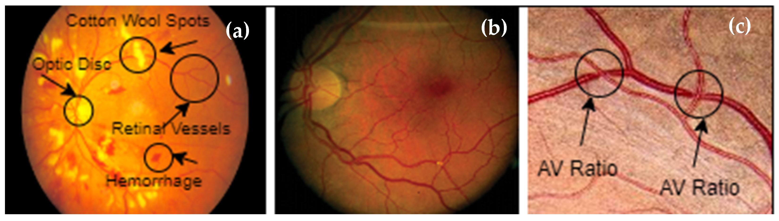
Figure 1. Illustration of the vascular system: (a) optic disk, cotton wool spots, and hemorrhages; (b) tortuosity; (c) A/V ratio.
Ophthalmologists can find different retinal diseases, such as those linked to HR, with the help of computerized methods [8]. These technologies aid academics and the global medical profession by enabling self-diagnosis. Optometrists use these technologies to treat and diagnose eye-related illnesses, particularly those that are HR-related. Hypertensive retinopathy (HR) can be identified by segmenting the retina’s structural features, such as the macular, optic nerves, arteries, and vasculature, as illustrated in Figure 1. There are two ways of extracting features from eye images: using either deep-learning models or handcrafted feature extraction techniques. In the case of using deep-learning techniques for feature extraction, these features can be statistically evaluated to determine the best features to identify HR or non-HR illness. Deep learning techniques have been applied for many applications, such as computer vision algorithms [9], biological behavior analysis, and many other different applications. A visual diagram of (a) normal and (b) hypertensive retinopathy (HR) is displayed in Figure 2.
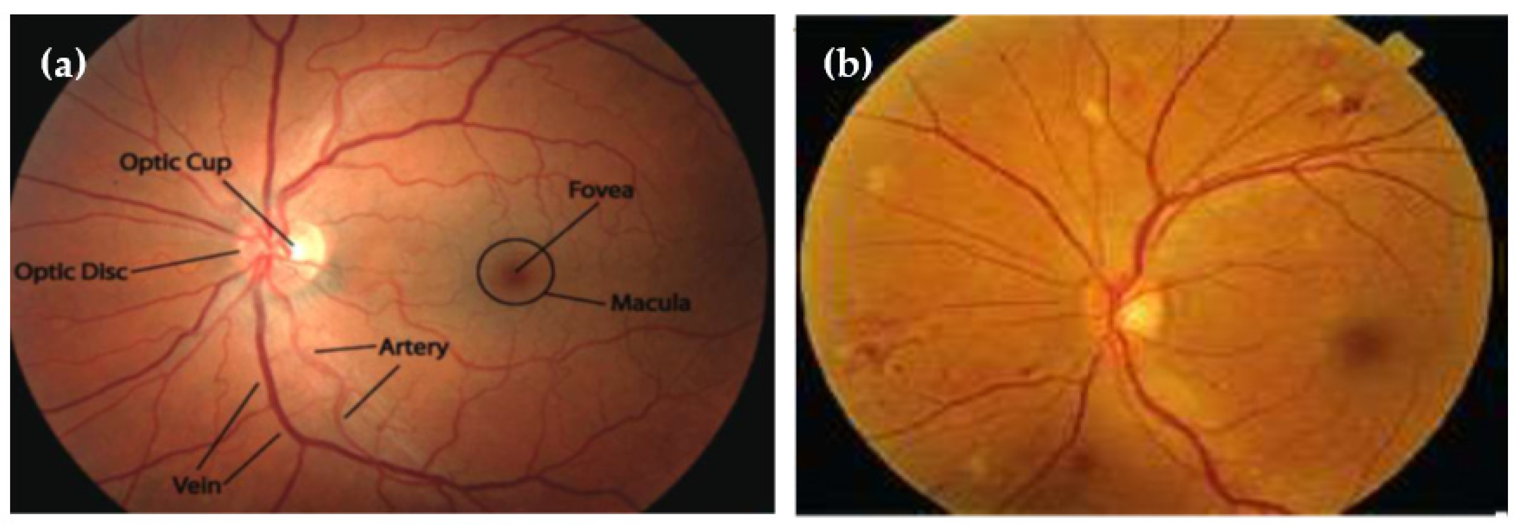
Figure 2. A visual diagram of (a) normal and (b) hypertensive retinopathy via diagnosis by retinal fundus images.
2. Mobile-HR: An Ophthalmologic-Based Classification System for Diagnosis of Hypertensive Retinopathy Using Optimized MobileNet Architecture
DL-based image diagnosis methods were developed in the past to help doctors make more accurate HR diagnoses. Before 2016, most studies used standard methods for preprocessing, segmentation, feature extraction, and classification. Public access to various skin lesion databases is now possible. To distinguish between HRs, researchers have developed DL algorithms [10]. Over time, it became clear that the CNNs retrieved more useful characteristics than the handcrafted techniques.
Many research articles [11][12][13][14][15][16][17][18][19][20][21][22][23][24][25][26][27][28][29][30][31] have focused on how to make small, effective networks that can be used for many different things. Researchers have tried out a wide range of methods, such as training network models and making older models smaller. Andrew et al. [12] came up with an efficient way to design lightweight neural networks that can be used in a number of computer vision applications, such as fine-grained classification and object identification, to achieve the benefits of small, low-latency models. Some authors have used the fuzzy inference method to determine how likely it is that diabetes will cause serious problems.
After looking at data with the VGG-19, MobileNet, and Resnet models [14], Wu and Hu [13] came up with a transfer learning classification method for hypertensive retinopathy. When the transfer learning approach is applied to the Kaggle dataset, the experimental accuracy is 60%, which is better than the model’s initial learning. Sun and Zhang published a model to detect hypertensive retinopathy [15]. Five distinct algorithms, including decision tree, random forest, support vector machine, logistic regression, and naive Bayesian, were utilized in electronic health records from 201 institutions [16] to improve diagnosis. They compared these models, and out of the five models, the random forest model had the highest accuracy (92%). In using the retinal vasculature as a crucial indicator for intelligent, DL-based screening and analysis of the diagnosis of diabetic and hypertensive retinopathy [17], when compared to cutting-edge techniques for automated vessel detection for diagnostic purposes, the authors’ accuracy results demonstrated the suggested method’s remarkable segmentation capability.
Mukesh et al. [18] proposed a system to detect HR lesions by suggesting a regional IoT-enabled federated learning-based categorization strategy (IoT-FHR) that integrates both global and local features. To improve the effectiveness of the classification of IoT-FHR, the local feature arterial and venous nicking (AVN) classification model was fused with the general IoT-FHR classification model. When evaluated on a private dataset, the recommended fusion model’s accuracy, sensitivity, and specificity were 93.50%, 69.83%, and 98.33%, respectively. Joseph et al. [19] proposed a machine-learning-based automated method for HR detection using fundus photographs. The study emphasizes how an early diagnosis of some medical issues using only a photograph acquired from the fundus image of the eye is more effective when conducted using computer-automated methods rather than manual observation techniques. In their study, Arslan et al. [20] suggest the dual-stream fusion network (DSF-Net) and the dual-stream aggregation network (DSA-Net) as two new shallow-DL architectures that can be used to recognize retinal vasculature. Semantic segmentation is used to find diabetic and hypertensive retinopathies in raw color fundus images. The effectiveness of the suggested strategy was evaluated using three publicly accessible metrics. The results of the experiments further demonstrate that the DSA-Net offers greater SE in comparison to the current methods. This study developed a system that is not as deep as standard semantic segmentation networks but delivers acceptable segmentation with limited trainable parameters and layers. Compared to existing methods, the proposed approach outperformed them on three publicly accessible datasets in terms of sensitivity, specificity, area under the curve, and accuracy metrics.
The study in [21] stated that deep learning ideas could be used to find HR and showed that the validation sensitivity was 95%. The most important thing that this study adds is the preprocessing step of adaptive histogram equalization. Some other interesting work in HR detection using machine learning models can be found in [22][23][24][25][26]. Qureshi et al. [27] developed a way to find HR using fundus images based on a depthwise separable CNN network. Their work reported a 95% accuracy and a 0.96 AUC. In [28], the authors propose using a fundus image as input for a CNN to classify images into HR and non-HR images. The proposed system was tested using the DRIVE dataset and based on experiments; the accuracy of the proposed model reached 98.6%.
Abbas et al. [29] came up with a new way to find hypertensive retinopathy (called DenseHyper) in retinal fundus images. Through ten-fold cross-validation, the proposed work performed much better than other algorithms, with an average accuracy of 95%. Recently, Arsalan et al. [30] came up with a new way for computers to help diagnose diabetes and high blood pressure retinopathy. They used shallow neural networks that are easy on memory, pool-less segmentation networks (PLS-net), and pool-less residual segmentation networks (PLRS-net). The DRIVE, CHASE-DB1, and STARE databases, which are all publicly accessible, were used for the research. The PLRS-net outperformed PLS-net by averaging an 82% sensitivity across all three datasets, which was better than PLS-net’s performance. The authors of [31] also created a five-step HR recognition system using semantic and instance segmentation in the DenseNet architecture.
There are three ways to classify segmented images in the literature [32]: Efficient-Net, VGG-16, and ResNet-152. Using the ensemble method, the generated feature vectors were combined, and the SoftMax classifier was used to accurately classify eleven different types of retinal disorders. The suggested method for recognizing HR had an accuracy of 99.71%, a precision of 98.63%, a recall of 98.25%, and an F measure of 99.22%. In [33], on the other hand, the authors used a segmentation method for blood vessels without considering any other HR properties.
References
- Mozaffarian, D.; Benjamin, E.J.; Go, A.S.; Arnett, D.K.; Blaha, M.J.; Cushman, M.; Das, S.R.; De Ferranti, S.; Després, J.P.; Fullerton, H.J.; et al. Executive summary: Heart disease and stroke statistics-2016 update: A report from the American heart association. Circulation 2016, 133, 447–454.
- Modi, P.; Arsiwalla, T. Hypertensive retinopathy. In StatPearls; StatPearls Publishing: Treasure Island, FL, USA, 2022.
- Rosendorff, C.; Lackland, D.T.; Allison, M.; Aronow, W.S.; Black, H.R.; Blumenthal, R.S.; Gersh, B.J. Treatment of hypertension in patients with coronary artery disease: A scientific statement from the American heart association, American college of cardiology, and american society of hypertension. J. Am. Coll. Cardiol. 2015, 65, 1998–2038.
- Qureshi, I.; Ma, J.; Abbas, Q. Diabetic retinopathy detection and stage classification in eye fundus images using active deep learning. Multimed. Tools Appl. 2021, 80, 11691–11721.
- Bhargava, M.; Ikram, M.K.; Wong, T.Y. How does hypertension affect your eyes? J. Hum. Hypertens. 2012, 26, 71–83.
- Wiharto; Suryani, E. The review of computer aided diagnostic hypertensive retinopathy based on the retinal image processing. IOP Conf. Ser. Mater. Sci. Eng. 2019, 620, 012099.
- Rajalakshmi, R.; Subashini, R.; Anjana, R.M.; Mohan, V. Automated diabetic retinopathy detec- tion in smartphone-based fundus photography using artificial intelligence. Eye 2018, 32, 1138–1144.
- Asiri, N.; Hussain, M.; Aboalsamh, H.A. Deep learning based computer-aided diagnosis Systems for Diabetic Retinopathy: A survey. Artif. Intell. Med. 2018, 99, 101701.
- Abbas, Q.; Ibrahim, M.E.; Jaffar, M.A. A comprehensive review of recent advances on deep vision systems. Artif. Intell. Rev. 2018, 52, 39–76.
- Jin, J.; Dundar, A.; Culurciello, E. Flattened convolutional neural networks for feed- forward acceleration. arXiv 2016, arXiv:1412.5474.
- Wang, M.; Liu, B.; Foroosh, H. Factorized convolutional neural networks. arXiv 2016, arXiv:1608.04337.
- Howard, A.G.; Zhu, M.; Chen, B.; Kalenichenko, D.; Wang, W.; Weyand, T.; Andreetto, M.; Adam, H. MobileNets: Efficient Convolutional Neural Networks for Mobile Vision Applications. arXiv 2017, arXiv:1704.04861v1.
- Pavate, A.; Nerurkar, P.; Ansari, N.; Bansode, R. Early prediction of five major complications ascends in diabetes mellitus using fuzzy logic. In Soft Computing in Data Analytics: Proceedings of the International Conference on SCDA, Singapore, 15–16 December 2019; Springer: Singapore; Volume 758, pp. 759–768.
- Wu, Y.; Hu, Z. Recognition of Diabetic Retinopathy Basedon Transfer Learning. In Proceedings of the 2019 IEEE 4th International Conference on Cloud Computing and Big Data Analysis (ICCCBDA), Chengdu, China, 12–15 April 2019; pp. 398–401.
- Sun, Y.; Zhang, D. Diagnosis and Analysis of Diabetic Retinopathy Based on Electronic Health Records. IEEE Access 2019, 7, 86115–86120.
- Sun, Y. The Neural Network of One-Dimensional Convolution—An Example of the Diagnosis of Diabetic Retinopathy. IEEE Access 2019, 7, 69657–69666.
- Arsalan, M.; Haider, A.; Lee, Y.W.; Park, K.R. Detecting retinal vasculature as a key biomarker for deep Learning-based intelligent screening and analysis of diabetic and hypertensive retinopathy. Expert Syst. Appl. 2022, 200, 117009.
- Soni, M.; Singh, N.K.; Das, P.; Shabaz, M.; Shukla, P.K.; Sarkar, P.; Singh, S.; Keshta, I.; Rizwan, A. IoT-Based Federated Learning Model for Hypertensive Retinopathy Lesions Classification. IEEE Trans. Comput. Soc. Systems 2022, 1–10.
- Joseph, R.; Chauhan, S.; Chichria, K.; Bhatia, T.; Thakur, H. Detection of Hypertension Retinopathy and Diabetes Using Machine Learning. In Proceedings of the International Conference on Recent Advances in Computational Techniques (IC-RACT), Mumbai, India, 9 October 2020; pp. 1–6.
- Arsalan, M.; Haider, A.; Choi, J.; Park, K.R. Diabetic and hypertensive retinopathy screening in fundus images using artificially intelligent shallow architectures. J. Pers. Med. 2022, 12, 7.
- Xu, K.; Feng, D.; Mi, H. Deep convolutional neural network-based early automated detection of diabetic retinopathy using fundus image. Molecules 2017, 22, 2054.
- Carson, L.; Yi, D.; Guo, M.; Lindsey, T. Automated detection of diabetic retinopathy using deep learning. AMIA Summits Transl. Sci. Proc. 2018, 2018, 147.
- Narayanan, B.N.; Hardie, R.C.; De Silva, M.S.; Kueterman, N.K. Hybrid machine learning architecture for automated detection and grading of retinal images for diabetic retinopathy. J. Med. Imaging 2020, 7, 034501.
- Hacisoftaoglu, R.E.; Karakaya, M.; Sallam, A.B. Deep learning frameworks for diabetic retinopathy detection with smartphone-based retinal imaging systems. Pattern Recognit. Lett. 2020, 135, 409–417.
- Riaz, H.; Park, J.; Choi, H.; Kim, H.; Kim, J. Deep and densely connected networks for classification of diabetic retinopathy. Diagnostics 2020, 10, 24.
- Pavate, A.; Mistry, J.; Palve, R.; Gami, N. Diabetic retinopathy detection-MobileNet binary classifier. Acta. Sci. Med. Sci. 2020, 4, 86–91.
- Qureshi, I.; Abbas, Q.; Yan, J.; Hussain, A.; Shaheed, K.; Baig, A.R. Computer-Aided Detection of Hypertensive Retinopathy Using Depth-Wise Separable CNN. Appl. Sci. 2022, 12, 12086.
- Triwijoyo, B.K.; Budiharto, W.; Abdurachman, E. The classification of hypertensive retinopathy using convolutional neural network. Procedia Comput. Sci. 2017, 116, 166–173.
- Abbas, Q.; Ibrahim, M.E. DenseHyper: An automatic recognition system for detection of hypertensive retinopathy using dense features transform and deep-residual learning. Multimed. Tools Appl. 2020, 20, 31595–31623.
- Arsalan, M.; Owais, M.; Mahmood, T.; Cho, S.W.; Park, K.R. Aiding the diagnosis of diabetic and hypertensive retinopathy using artificial intelligence-based semantic segmentation. J. Clin. Med. 2019, 8, 1446.
- Abbas, Q.; Qureshi, I.; Ibrahim, M.E. An Automatic Detection and Classification System of Five Stages for Hypertensive Retinopathy Using Semantic and Instance Segmentation in DenseNet Architecture. Sensors 2021, 21, 6936.
- Kumar, K.S.; Singh, N.P. Retinal disease prediction through blood vessel segmentation and classification using ensemble-based deep learning approaches. Neural Comput. Appl. 2023, 1–17.
- Sathananthavathi, V.; Indumathi, G. Deep learning approaches for the retinal vasculature segmentation in fundus images. In Computational Methods and Deep Learning for Ophthalmology; Academic Press: Cambridge, MA, USA, 2023; pp. 139–155.
More
Information
Contributors
MDPI registered users' name will be linked to their SciProfiles pages. To register with us, please refer to https://encyclopedia.pub/register
:
View Times:
1.1K
Revisions:
3 times
(View History)
Update Date:
29 May 2023
Table of Contents


Notice
You are not a member of the advisory board for this topic. If you want to update advisory board member profile, please contact office@encyclopedia.pub.
OK
Confirm
Only members of the Encyclopedia advisory board for this topic are allowed to note entries. Would you like to become an advisory board member of the Encyclopedia?
Yes
No
${ textCharacter }/${ maxCharacter }
Submit
Cancel
Back
Comments
${ item }
|
${ item.createdUser.fullName }
${ item.createdAt }
${ item.vote }
${ item.reply }
Delete
${ reply.createdUser.fullName }
${ reply.createdAt }
${ reply.vote }
Delete
There is no reply to this comment~
${ item.replyTextCharacter }/${ item.replyMaxCharacter }
Submit
Cancel
More
No more~
There is no comment~
${ textCharacter }/${ maxCharacter }
Submit
Cancel
${ selectedItem.replyTextCharacter }/${ selectedItem.replyMaxCharacter }
Submit
Cancel
Confirm
Are you sure to Delete?
Yes
No