Submitted Successfully!
To reward your contribution, here is a gift for you: A free trial for our video production service.
Thank you for your contribution! You can also upload a video entry or images related to this topic.
Version Summary Created by Modification Content Size Created at Operation
1 -- 2854 2023-05-25 11:09:20 |
2 Format correct Meta information modification 2854 2023-05-26 07:39:44 |

Video Upload Options

Do you have a full video?

Confirm

Are you sure to Delete?
Cite
If you have any further questions, please contact Encyclopedia Editorial Office.
Banger, A.; Gautam, S.; Jadoun, S.; Jangid, N.K.; Srivastava, A.; Pulidindi, I.N.; Dwivedi, J.; Srivastava, M. Applications of Carbon Nanodots. Encyclopedia. Available online: https://encyclopedia.pub/entry/44826 (accessed on 15 May 2024).
Banger A, Gautam S, Jadoun S, Jangid NK, Srivastava A, Pulidindi IN, et al. Applications of Carbon Nanodots. Encyclopedia. Available at: https://encyclopedia.pub/entry/44826. Accessed May 15, 2024.
Banger, Anjali, Sakshi Gautam, Sapana Jadoun, Nirmala Kumari Jangid, Anamika Srivastava, Indra Neel Pulidindi, Jaya Dwivedi, Manish Srivastava. "Applications of Carbon Nanodots" Encyclopedia, https://encyclopedia.pub/entry/44826 (accessed May 15, 2024).
Banger, A., Gautam, S., Jadoun, S., Jangid, N.K., Srivastava, A., Pulidindi, I.N., Dwivedi, J., & Srivastava, M. (2023, May 25). Applications of Carbon Nanodots. In Encyclopedia. https://encyclopedia.pub/entry/44826
Banger, Anjali, et al. "Applications of Carbon Nanodots." Encyclopedia. Web. 25 May, 2023.
Applications of Carbon Nanodots
Edit

Carbon dots have drawn immense attention and prompted intense investigation. The latest form of nanocarbon, the carbon nanodot, is attracting intensive research efforts, similar to its earlier analogues, namely, fullerene, carbon nanotube, and graphene. One outstanding feature that distinguishes carbon nanodots from other known forms of carbon materials is its water solubility owing to extensive surface functionalization (the presence of polar surface functional groups). These carbonaceous quantum dots, or carbon nanodots, have several advantages over traditional semiconductor-based quantum dots. They possess outstanding photoluminescence, fluorescence, biocompatibility, biosensing and bioimaging, photostability, feedstock sustainability, extensive surface functionalization and bio-conjugation, excellent colloidal stability, eco-friendly synthesis (from organic matter such as glucose, coffee, tea, and grass to biomass waste-derived sources), low toxicity, and cost-effectiveness.

carbon nanodots applications nanoparticles

1. Introduction

Nanoparticles are microscopic particles with a size range of 1–100 nm. During the past decade, considerable research was conducted on the fabrication and application of nanoparticles in many fields. Based on their unique properties, nanoparticles have a substantial impact in various industries, including health, cosmetics, energy, pharmaceuticals, and food.
Enormous work was completed in recent years to design nanostructured materials with specific characteristics that will ultimately influence their function and application. In this era of carbon nanotechnology, special emphasis is laid on the organic functionality of nanomaterials or organic nanomaterials, including graphene, carbon nanotubes, and fullerenes. Because of their biocompatibility, ease of fabrication, and fascinating features, especially their water solubility fluorescence emission, carbon nanodots with a size in the range of 1–10 nm have taken the central stage of materials research. Carbon nanodots (CDs) are known to have zero dimension with almost spherical geometry. This material has become a rising star in the field of luminescent nanomaterials [1]. Due to their desirable qualities, such as hydrophilicity, ease of functionalization, outstanding biocompatibility, bright luminescence, good solubility, high chemical inertness, and low toxicity, they are potent candidates for various applications in solar cells, biosensors [2][3][4][5][6][7][8][9][10], bioimaging, and optoelectronic devices, etc. CNDs exhibit many remarkable properties including outstanding photoinduced electron transfer, stable chemical inertness, low cytotoxicity [11][12][13][14][15], good biocompatibility, and efficient light harvesting. Dots made of carbon, such as carbon nanodots (CDs) and graphene quantum dots (GQDs), are a brand-new carbonaceous nanomaterial with zero dimensions [16][17][18][19][20][21][22]. Until now, a lot of work has been carried out, and substantial advances have been made in the synthesis and uses of carbon-based dots [23][24][25][26][27].
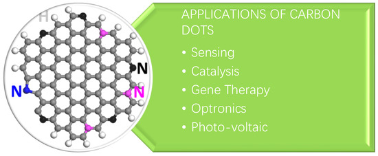
There are various applications which are associated with carbon dots. CDs also show a number of biomedical applications. The application of CDs is shown in Figure 1.
Figure 1. Application of Carbon dots.

2. Sensing

One of the most common and potentially significant uses of CDs is sensing [28][29][30]. Due to their superior optical qualities, high fluorescence sensitivity to the surrounding environment [31][32], and ability to function as effective electron donors [33][34][35], CDs are frequently suggested as detectors for a variety of harmful substances, including heavy metals such as mercury [36][37][38], copper, and iron [39][40][41][42]. To make CDs more sensitive to one or more of these analytes, persistent work is being conducted in this direction. Only a handful of studies, however, have attempted to examine the interactions of CDs with metal ions at a more fundamental level; for example, Goncalves and colleagues demonstrated that the fluorescence emissions of both CQD solution and CQDs immobilized in sol–gel are sensitive to the presence of Hg2+ [43]. In their study, laser-ablated and NH2-PEG200 and N-acetyl-L-cysteine-passivated CQDs were used as fluorescent probes. It was observed that the fluorescence intensity of the CQDs is efficiently quenched by micro molar amounts of Hg2+ with a Stern–Volmer constant of 1.3 × 10⁵ M−1. Therefore, judging from the relatively large magnitude of the Stern–Volmer constant [44], the quenching provoked by Hg2+ is probably due to the static quenching arising from the formation of a stable non-fluorescent complex between CQD and Hg2+. A substantial improvement in the sensitivity down to nanomolars was later realized by replacing the laser-ablated CQDs with N-CQDs. Again, static quenching is thought to be responsible for the quenching of fluorescence, but with a much larger Stern–Volmer constant of 1.4 × 10⁷ M−1, two orders of magnitude higher than that of the previous system [45]. It was suggested that the presence of the nitrogen element in the N-CQDs, most probably -CN groups on the N-CQD surface, is responsible for the much-improved performance of Hg2+ sensing.

3. Bio Imaging Probes

An intriguing application of C dots is their use as a potential agent for in vivo and in vitro bioimaging of cells and species due to their photoluminescence, which is an important property of C dots [46][47][48]. The bioimaging of cells and tissues is an important part of the diagnosis of many diseases, particularly cancer. Various fluorescent systems for diagnostic purposes have been reported, ranging from organic and inorganic dyes to the most recent nanoparticle-based systems.
To be considered suitable for use as an imaging probe, a bioimaging agent must have excellent biocompatibility, a tunable emission spectrum, and be free of cytotoxicity. Rapid progress in implementing a new class of nanoparticles has resulted in a material that meets these criteria and can be used for both diagnostic and therapeutic purposes. Chemical functionalization is used to successfully conjugate the required drug molecule to the fluorescent nanoprobes for these theranostic applications. Sahu et al. [49] reported the synthesis of C dots from orange juice hydrothermal treatment. This was one of the first examples of making fluorescent C dots from readily available natural resources [50][51]. The C dots were non-cytotoxic and efficiently taken up by MG-63 human osteosarcoma cells for cellular imaging.
The “central dogma” states that genetic information flows from DNA to RNA to proteins. Researchers investigated the physiological activity of RNA during cancer research by using RNA dynamics in cellular functions and the real-time monitoring of their temporo-spatial distribution. The experiments were carried out using fluorescent carbon dots created by the one-pot hydrothermal treatment of o-, m-, or p-phenylenediamines with triethylenetetramine by Chen et al. [52]. Because carbon has excellent biocompatibility and negligible cytotoxicity, there has been a lot of interest in using carbon nanodots as bioimaging probes instead of other types of nanoparticles. C dots are ideal candidates for theranostic applications due to their ease of synthesis, acceptable emission spectra, high photostability, and lack of cytotoxicity.
Tao et al. [53] used a mixed acid treatment to create C dots from carbon nanotubes (CNTs) and graphite. Under UV light, the C dots emit a strong yellow fluorescence with no cellular toxicity. They also demonstrated in vivo bioimaging in the near-infrared region using a rat model, and this experiment exemplified the possibilities for the development of fluorescent imaging probes in both the ultraviolet (UV) and infrared (IR) range spectra.

4. Photodynamic Therapy

Photodynamic therapy is a relatively new advancement in biomedical nanotechnology that uses energy transfer to destroy damaged cells and tissues. This method is useful in dealing with cancer cells because it effectively targets and destroys malignant tissue while leaving normal, healthy tissue alone. This targeted destruction in photodynamic therapy can be accomplished with fluorescent C dots that have adequate photostability [54].
Shi et al. [55] used the hydrothermal method to create N-doped C dots from rapeseed flowers and bee pollen. The authors demonstrated that C dots had no cytotoxic effect up to a limiting concentration of 0.5 mg/mL after this successful large-scale synthesis. Human colon carcinoma cells were imaged successfully in this study, and the C dots were found to have good photostability and biocompatibility.
Wang et al. [56] reported C dot synthesis from the condensation carbonization of linear polyethylenic amine (PEA) analogues and citric acid (CA) of different ratios. The authors successfully demonstrated that the extent of conjugated π-domains with CN in the carbon backbone was correlated with their photoluminescence quantum yield. The main conclusion from this study is that the emission arises not only from the sp2/sp3 carbon core and surface passivation of C nanodots, but also from the molecular fluorophores integrated into the C dot framework. This work provided an insight into the excellent biocompatibility, low cytotoxicity, and enhanced bioimaging properties of N-doped C dots, which opens the possibilities for new bioimaging applications.
Bankoti et al. [57] fabricated C dots from onion peel powder waste using the microwave method and studied cell imaging and wound healing aspects. The C dots exhibited stable fluorescence at an excitation wavelength of 450 nm and an emission wavelength of 520 nm at variable pH, along with the ability to scavenge free radicals, which can be further explored for antioxidant activity. The radical scavenging ability leads to an enhanced wound healing ability in a full-thickness wound in a rat model.

5. Photocatalysis

There has been significant research interest in photocatalysts over the past decade due to the scenario of environmental safety and sustainable energy. The applications of nanomaterials for the efficient fabrication of photocatalysts made the journey fast and effective.
Ming et al. [58] successfully developed C dots using a one-pot electrochemical method that only used water as the main reagent. This is an extremely promising synthetic methodology because it is a green protocol that is also cost-effective, with good photocatalytic activity of C dots for methyl orange degradation.
Song et al. [59] devised a two-step hydrothermal method for the creation of a C dot–WO2 photocatalyst. The authors used this system to photocatalytically degrade rhodamine B. It is worth noting that the reaction rate constant reported in this study is 0.01942 min−1, which is approximately 7.7 times higher than the catalytic rate using WO2 alone.
For photocatalytic hydrogen generation, a C-dot/g-C3N4 system was used. The authors created C dots from rapeseed flower pollen and hydrothermally incorporated them into g-C3N4. Under visible light irradiation, this system was able to photocatalytically generate hydrogen via sound with an output greater than that of bulk g-C3N4.

6. Biological Sensors and Chemical Sensors

There is great interest in using nanoparticles as biochemical sensors because C dots have been found to be useful in sensing chemical compounds or elements. Based on the properties of C dots, particularly their fluorescence properties and surface-functionalized chemical groups, various sensors for biological and chemical applications have been developed.
Qu et al. [60] developed ratiometric fluorescent nano-sensors using C dots in a single step of microwave-assisted synthesis. This research is significant in C-dot sensor research because the developed nanosensors are multi-sensory and can detect temperature, pH, and metal ions such as Fe (III). Because it can detect and estimate multiple metabolic parameters at the same time, this exciting feature is proving to be widely applicable in the biological environment. The sensory mechanism is non-cytotoxic and based on ratiometric fluorescence, which is a promising feature for future research.
Vedamalai et al. [61] developed C dots that are highly sensitive to copper (II) ions in cancer cells. They used a relatively simple hydrothermal synthesis method based on ortho-phenylenediamine (OPD). The orange color was caused by the formation of the Cu(OPD)2 complex on the surface of the C dots. Further investigation revealed that the C dots were highly water dispersible, photostable, chemically stable, and biocompatible.
Shi et al. [62] used C dots to detect Cu(II) ions in living cells as well. The hydrothermal pyrolysis of leeks resulted in blue and green fluorescent C dots. In a single step of hydrothermal carbonization, the C dots were modified with boronic acid using phenylboronic acid as the precursor. This C-dot-based sensor successfully detected blood sugar levels and demonstrated good selectivity with minimal chemical interference from other species [63].
Nie et al. [64] used a novel bottom-up method to develop a pH sensor out of C dots. This method yielded C dots with high crystallinity and stability. The procedure involved a one-pot synthesis with high reproducibility using chloroform and diethylamine. The authors were able to use the technique for cancer diagnosis after successfully implementing the pH detection of two C dots with different emission wavelengths.
Wang et al. [65] described an intriguing C-dot sensor for hemoglobin detection (Hb). The C dots were developed from glycine using an electrochemical method that included multiple steps, such as electro-oxidation, electro-polymerization, carbonization, and passivation. The authors successfully validated the sensitivity of Hb detection and discovered that the luminescence intensity varied inversely with Hb concentration in the 0.05–250 nM range.

7. Drug Delivery

Carbon dots’ excellent biocompatibility and clearance from the body meet the requirements for in vivo applications. Carbon dots with rich and tunable function groups, such as amino, carboxyl, or hydroxyl, can carry therapeutic agents, resulting in theranosticnanomedicines [66][67][68][69][70][71]. The bright emission of carbon dots allows for the dynamic and real-time monitoring of drug distribution and response. Zheng et al. [72] used carbon dots synthesized through the thermal pyrolysis of citric acid and polyene polyamine to transport oxaliplatin, a platinum-based drug, because platinum-based drugs are the most effective anticancer drugs and are used in more than 50% of clinical cancer patients’ chemotherapeutic treatments.

8. Micro-Fluidic Marker

The study of fluidic physics at the micro-scale is now best conducted using microfluidic systems. Because of their considerably higher surface-to-volume ratio, surface tension and viscosity dominate those of inertia, making the fluid easier to control. Static laminar flows and dynamic droplet formation are typical microfluidic situations. Both exhibit many advantages, including minimal reagent use, high sensitivity, and high output, which leads to a wide range of applications in bioassays, chemical reactions, drug delivery, etc. The majority of applications rely on the microfluidic circuit’s ability to visualize fluid flow. However, the biocompatibility and cheap cost of the fluorescent materials currently in use cannot be balanced, which is a critical issue for microfluidic applications, particularly for bio-applications. Sun’s colleagues used carbon dots, synthesized by heating glucose and urea in a microwave, to visualize microfluid flows for the first time to address this problem [73][74][75]. The scientists used carbon dots dissolved in the deionized water as a fluorescent marker to investigate the dynamics of the mixture of glycerol and deionized water. When the interface is ruptured by an electric field above a threshold, fast mixing occurs at the microscale. In addition to laminar flow, the authors also synthesized mono-dispersed droplets in a flow focusing system, where the continuous phase was mineral oil while the aqueous solution of carbon dots appeared as the dispersed phase. The diameter of the droplets will shrink because a higher capillary number results in a greater interfacial shear force. Additionally, the authors successfully demonstrated the multiple component droplet, merged droplet, and double emulsion, each of which has a distinct core-shell structure. To more accurately determine the speed of the flow field, luminescent seeding carbon dots were made via a mixture of carbon dots (liquid state) and polystyrene microparticles [76][77][78].

9. Bioimaging

Carbon dots have significant advantages over fluorescent organic dyes and genetically engineered fluorescent proteins, such as high PL quantum yield, photostability, and resistance to metabolic degradation, which endows them with enormous potential for use in bioapplications. While the toxicity testing of carbon dots is required before exploring their bioapplications, Yang et al. [79] used human breast cancer MCF-7 cells and human colorectal adenocarcinoma HT-29 cells (previously reported by other scientists, Yang modified and used it) to assess the in vitro toxicity of carbon dots synthesized by the laser ablation of graphite powder and cement with PEG1500N [80][81][82] as a surface passivation agent. All the observations of cell proliferation, mortality, and viability from both cell lines indicated that the carbon dots exhibited superior biocompatibility, even at concentrations as high as 50 mg/mL, which is much higher than the practical application demand, for example, in living cell imaging.

10. Carbon Dots Chiral Photonics

Chirality is essential in a number of practical application fields, such as chiral drug recognition, chiral molecular biology, and chiral chemistry [83][84][85]. As a result, as previously proposed by M. Va’zquez-Nakagawa et al. [86], chirality and carbon dots can be combined to form intriguing chiral optics based on carbon dots. The carbon dots used in their groundbreaking research were created by chemically exfoliating graphite with strong sulfuric and nitric acids. The carbon dots’ surface carboxylic acid groups were subsequently converted to acid chlorides using thionyl chloride. When the acid chlorides and the (R) or (S)-2-phenyl-1-propanol reacted simultaneously, enantiomerically pure esters and chiral carbon dots were created (chiral molecular). Enantiomerically esters and chiral carbon dots were formed, and their formation was verified using 13C-NMR and FTIR spectroscopy. The presence of phenyl substituents was suggested by the appearance of peaks in the 13C–NMR. The recent work in this field is the most notable development in the chiral regulation of bioreactions for chiral carbon dots. Xin et al. [87] described the destruction of the cell walls of gram-positive and gram-negative bacteria via carbon dots in the presence of D-glutamic acid, which resulted in the fatality of bacteria. In contrast, the carbon dots formed in the presence of L-glutamic acid demonstrated an insignificant effect on bacterial cells. This implied that antimicrobial nanoagents with chirality can be synthesized from carbon dots. The D-form and L-form of cysteine-based carbon dots were used to regulate the chirality of the enzyme. For instance, L-form cysteine carbon dots reduce the enzymatic activity while D-form cysteine carbon dots enhance the enzymatic activity of the enzyme. According to Li et al. [88], these cysteine-based carbon nanodots have the capacity to affect cellular energy metabolism. We anticipate that other chiral carbon dots-based applications will be investigated in the future [89], and that carbon dots with chirality will emerge as a novel but exciting topic because of their wide applications.

References

  1. Xu, X.Y.; Ray, R.; Gu, Y.L.; Ploehn, H.J.; Gearheart, L.; Raker, K.; Scrivens, W.A. Electrophoretic Analysis and Purification of Fluorescent Single-Walled Carbon Nanotube Fragments. J. Am. Chem. Soc. 2004, 126, 12736–12737.
  2. Sun, Y.P.; Zhou, B.; Lin, Y.; Wang, W.; Fernando, K.A.S.; Pathak, P.; Meziani, M.J.; Harruff, B.A.; Wang, X.; Wang, H.; et al. QuantumSized Carbon Dots for Bright and Colorful Photoluminescence. J. Am. Chem. Soc. 2006, 128, 7756–7757.
  3. Zheng, L.; Chi, Y.; Dong, Y.; Lin, J.; Wang, B. Electrochemiluminescence of Water-Soluble Carbon Nanocrystals Released Electrochemically from Graphite. J. Am. Chem. Soc. 2009, 131, 4564–4565.
  4. Baker, S.N.; Baker, G.A. Luminescent Carbon Nanodots: Emergent Nanolights. Angew. Chem. Int. Ed. 2010, 49, 6726–6744.
  5. Li, H.T.; He, X.D.; Kang, Z.H.; Huang, H.; Liu, Y.; Liu, J.L.; Lian, S.Y.; Tsang, C.C.A.; Yang, X.B.; Lee, S.T. Water-Soluble Fluorescent Carbon Quantum Dots and Catalyst Design. Angew. Chem. Int. Ed. 2010, 49, 4430–4434.
  6. Ma, Z.; Zhang, Y.L.; Wang, L.; Ming, H.; Li, H.T.; Zhang, X.; Wang, F.; Liu, Y.; Kang, Z.H.; Lee, S.T. Bioinspired Photoelectric Conversion System Based on Carbon Quantum Dot Doped Dye—Semiconductor Complex. ACS Appl. Mater. Interfaces 2013, 5, 5080–5084.
  7. Mu, X.W.; Wu, M.X.; Zhang, B.; Liu, X.; Xu, S.M.; Huang, Y.B.; Wang, X.H.; Sun, Q. A sensitive “off-on” carbon dots-Ag nanoparticles fluorescent probe for cysteamine detection via the inner filter effect. Talanta 2021, 221, 121463.
  8. Huang, H.L.; Ge, H.; Ren, Z.P.; Huang, Z.J.; Xu, M.; Wang, X.H. Controllable Synthesis of Biocompatible Fluorescent Carbon Dots from Cellulose Hydrogel for the Specific Detection of Hg2+. Front. Bioeng. Biotechnol. 2021, 9, 617097.
  9. Wang, Y.; Zhuang, Q.; Ni, Y. Facile Microwave Assisted Solid Phase Synthesis of Highly Fluorescent Nitrogen Sulfur Co-doped Carbon Quantum Dots for Cellular Imaging Applications. Chem. Eur. J. 2015, 21, 13004–13011.
  10. Li, D.; Li, W.; Zhang, H.; Zhang, X.; Zhuang, J.; Liu, Y.; Hu, C.; Lei, B. Far-Red Carbon Dotsas Efficient Light-Harvesting Agents for Enhanced Photosynthesis. ACS Appl. Mater. Interfaces 2020, 12, 21009–21019.
  11. Wang, J.; Li, R.S.; Zhang, H.Z.; Wang, N.; Zhang, Z.; Huang, C.Z. Highly fluorescent carbon dots as selective and visual probes for sensing copper ions in living cells via an electron transfer process. Biosens. Bioelectron. 2017, 97, 157–163.
  12. Jana, J.; Lee, H.J.; Chung, J.S.; Kim, M.H.; Hur, S.H. Blue emitting nitrogen-doped carbon dots as a fluorescent probe for nitrite ion sensing and cell-imaging. Anal. Chim. Acta 2019, 1079, 212–219.
  13. Hu, J.; Tang, F.; Jiang, Y.; Liu, C. Rapid screening and quantitative detection of Salmonella using a quantum dot nanobead-based biosensor. Analyst 2020, 145, 2184–2190.
  14. Liu, M.L.; Chen, B.B.; Li, C.M.; Huang, C.Z. Carbon Dots: Synthesis, Formation Mechanism, Fluorescence Origin and Sensing Applications. Green Chem. 2019, 21, 449–471.
  15. Qin, K.H.; Zhang, D.F.; Ding, Y.F.; Zheng, X.D.; Xiang, Y.Y.; Hua, J.H.; Zhang, Q.; Ji, X.L.; Li, B.; Wei, Y.L. Applications of hydrothermal synthesis of Escherichia coli derived carbon dots in in vitro and in vivo imaging and p-nitrophenol detection. Analyst 2020, 145, 177–183.
  16. Cui, L.; Wang, J.; Sun, M.T. Graphene plasmon for optoelectronics. Rev. Phys. 2021, 6, 100054.
  17. Yuan, F.; Wang, Y.K.; Sharma, G.; Dong, Y.; Zheng, X.; Li, P.; Johnston, A.; Bappi, G.; Fan, J.Z.; Kung, H. Bright High-Colour Purity Deep-Blue Carbon Dot Light-Emitting Diodes via Efficient Edge Amination. Nat. Photonics 2020, 14, 171–176.
  18. Hu, C.; Li, M.Y.; Qiu, J.S.; Sun, Y.P. Design and fabrication of carbon dots for energy conversion and storage. Chem. Soc. Rev. 2019, 48, 2315–2337.
  19. Cao, Y.; Cheng, Y.; Sun, M.T. Graphene-based SERS for sensor and catalysis. Appl. Spectrosco. Rev. 2021, 58, 1–38.
  20. Hsu, P.C.; Shih, Z.Y.; Lee, C.H.; Chang, H.T. Synthesis and analytical applications of photoluminescent carbon nanodots. Green Chem. 2012, 14, 917–920.
  21. Chandra, S.; Pathan, S.H.; Mitra, S.; Modha, B.H.; Goswami, A.; Pramanik, P. Tuning of photoluminescence on different surface functionalized carbon quantum dots. RSC Adv. 2012, 2, 3602–3606.
  22. Liu, M.; Chen, W. Green synthesis of silver nanoclusters supported on carbon nanodots: Enhanced photoluminescence and high catalytic activity for oxygen reduction reaction. Nanoscale 2013, 5, 12558–12564.
  23. Kottam, N.; Smrithi, S.P. Luminescent carbon nanodots: Current prospects on synthesis, properties and sensing applications. Methods Appl. Fluoresc. 2021, 9, 012001.
  24. Xu, A.; Wang, G.; Li, Y.; Dong, H.; Yang, S.; He, P.; Ding, G. Carbon-based quantum dots with solid-state photoluminescent: Mechanism, implementation, and application. Small 2020, 16, 2004621.
  25. Roy, P.; Chen, P.C.; Periasamy, A.P.; Chen, Y.N.; Chang, H.T. Photoluminescent carbon nanodots: Synthesis, physicochemical properties and analytical applications. Mater. Today 2015, 18, 447–458.
  26. Li, H.; Kang, Z.; Liu, Y.; Lee, S.T. Carbon nanodots: Synthesis, properties and applications. J. Mat. Chem. 2012, 22, 24230–24253.
  27. Philippidis, A.; Stefanakis, D.; Anglos, D.; Ghanotakis, D. Microwave heating of arginine yields highly fluorescent nanoparticles. J. Nanopart. Res. 2013, 15, 1–9.
  28. Luo, X.; Huang, G.; Bai, C.; Wang, C.; Yu, Y.; Tan, Y.; Tang, C.; Kong, J.; Huang, J.; Li, Z. A versatile platform for colorimetric, fluorescence and photothermal multi-mode glyphosate sensing by carbon dots anchoring ferrocene metal-organic framework nanosheet. J. Hazard. Mat. 2023, 443, 130277.
  29. Wang, C.; Xu, Z.; Zhang, C. Polyethyleneimine-functionalized fluorescent carbon dots: Water stability, pH sensing, and cellular imaging. ChemNanoMat 2015, 1, 122–127.
  30. Mandal, P.; Sahoo, D.; Sarkar, P.; Chakraborty, K.; Das, S. Fluorescence turn-on andturn-off sensing of pesticides by carbon dot-based sensor. New J. Chem. 2019, 43, 12137–12151.
  31. Papaioannou, N.; Marinovic, A.; Yoshizawa, N.; Goode, A.E.; Fay, M.; Khlobystov, A.; Titirici, M.M.; Sapelkin, A. Structure and solvents effects on the optical properties of sugar-derivedcarbon nanodots. Sci. Rep. 2018, 8, 6559.
  32. Wang, J.C.; Violette, K.; Ogunsolu, O.O.; Hanson, K. Metal ion mediated electrontransfer at dye–semiconductor interfaces. Phys. Chem. Chem. Phys. 2017, 19, 2679–2682.
  33. Zheng, H.; Wang, Q.; Long, Y.; Zhang, H.; Huang, X.; Zhu, R. Enhancing theluminescence of carbon dots with a reduction pathway. Chem. Commun. 2011, 47, 10650–10652.
  34. Deshmukh, S.; Deore, A.; Mondal, S. Ultrafast dynamics in carbon dots as photosensitizers: A review. ACS Appl. Nano Mater. 2021, 4, 7587–7606.
  35. Sciortino, A.; Madonia, A.; Gazzetto, M.; Sciortino, L.; Rohwer, E.J.; Feurer, T.; Gelardi, F.M.; Cannas, M.; Cannizzo, A.; Messina, F. The interaction of photoexcited carbon nanodotswith metal ions disclosed down to the femtosecond scale. Nanoscale 2017, 9, 11902–11911.
  36. Liu, R.; Li, H.; Kong, W.; Liu, J.; Liu, Y.; Tong, C.; Zhang, X.; Kang, Z. Ultra-sensitiveand selective Hg2+ detection based on fluorescent carbon dots. Mater. Res. Bull. 2013, 48, 2529–2534.
  37. Huang, H.; Lv, J.J.; Zhou, D.L.; Bao, N.; Xu, Y.; Wang, A.J.; Feng, J.J. One-pot greensynthesis of nitrogen-doped carbon nanoparticles as fluorescent probes for mercury ions. RSC Adv. 2013, 3, 21691–21696.
  38. Wang, X.; Zhang, J.; Zou, W.; Wang, R. Facile synthesis of polyaniline/carbon dotnanocomposites and their application as a fluorescent probe to detect mercury. RSC Adv. 2015, 5, 41914–41919.
  39. Liu, C.; Tang, B.; Zhang, S.; Zhou, M.; Yang, M.; Liu, Y.; Zhang, Z.L.; Zhang, B.; Pang, D.W. Photoinduced electron transfer mediated by coordination between carboxyl on carbon nanodotsand Cu2+ quenching photoluminescence. J. Phy. Chem. C 2018, 122, 3662–3668.
  40. Zhu, A.; Qu, Q.; Shao, X.; Kong, B.; Tian, Y. Carbon-dot-based dual-emissionnanohybrid produces a ratiometric fluorescent sensor for in vivo imaging of cellular copperions. Angew. Chem. Int. Ed. 2012, 51, 7185–7189.
  41. Mohammed, L.J.; Omer, K.M. Dual functional highly luminescence B, N Co-dopedcarbon nanodots as nanothermometer and Fe3+/Fe2+ sensor. Sci. Rep. 2020, 10, 3028.
  42. Sekar, A.; Yadav, R.; Basavaraj, N. Fluorescence quenching mechanism and the application of green carbon nanodots in the detection of heavy metal ions: A review. New J. Chem. 2021, 45, 2326–2360.
  43. Goncalves, H.M.; Duarte, A.J.; da Silva, J.C.E. Optical fiber sensor for Hg (II) based on carbon dots. Biosen. Bioelect. 2010, 26, 1302–1306.
  44. Gharat, P.M.; Pal, H.; Choudhury, S.D. Photophysics and luminescence quenching ofcarbon dots derived from lemon juice and glycerol. Spectrochim. Acta Part A Mol. Biomol. Spectro. 2019, 209, 14–21.
  45. Karali, K.K.; Sygellou, L.; Stalikas, C.D. Highly fluorescent N-doped carbon nanodotsas an effective multi-probe quenching system for the determination of nitrite, nitrate and ferric ions infood matrices. Talanta 2018, 189, 480–488.
  46. Guo, J.; Liu, D.; Filpponen, I.; Johansson, L.S.; Malho, J.M.; Quraishi, S.; Liebner, F.; Santos, H.A.; Rojas, O.J. Photoluminescent hybrids of cellulose nanocrystals and carbon quantum dots as cytocompatible probes for in vitro bioimaging. Biomacromolecules 2017, 18, 2045–2055.
  47. Cronican, J.J.; Thompson, D.B.; Beier, K.T.; McNaughton, B.R.; Cepko, C.L.; Liu, D.R. Potent delivery of functional proteins into Mammalian cells in vitro and in vivo using a superchargedprotein. ACS Chem. Biol. 2010, 5, 747–752.
  48. Pan, L.; Sun, S.; Zhang, L.; Jiang, K.; Lin, H. Near-infrared emissive carbon dots for two-photon fluorescence bioimaging. Nanoscale 2016, 8, 17350–17356.
  49. Sahu, S.; Behera, B.; Maiti, T.K.; Mohapatra, S. Simple one-step synthesis of highly luminescent carbon dots from orange juice: Application as excellent bio-imaging agents. Chem. Commun. 2012, 48, 8835–8837.
  50. Sivasankarapillai, V.S.; Jose, J.; Shanavas, M.S.; Marathakam, A.; Uddin, M.; Mathew, B. Silicon quantum dots: Promising theranostic probes for the future. Curr. Drug Targets 2019, 20, 1255–1263.
  51. Liu, C.; Zhang, P.; Tian, F.; Li, W.; Li, F.; Liu, W. One-step synthesis of surfacepassivated carbon nanodots by microwave assisted pyrolysis for enhanced multicolour photoluminescence and bioimaging. J. Mater. Chem. 2011, 21, 13163–13167.
  52. Chen, B.B.; Wang, X.Y.; Qian, R.C. Rolling “wool-balls”: Rapid live-cell mapping of membrane sialic acids via poly-p-benzoquinone/ethylenediamine nanoclusters. Chem. Commun. 2019, 55, 9681–9684.
  53. Tao, H.; Yang, K.; Ma, Z.; Wan, J.; Zhang, Y.; Kang, Z.; Liu, Z. In vivo NIR fluorescenceimaging, biodistribution, and toxicology of photoluminescent carbon dots produced from carbonnanotubes and graphite. Small 2012, 8, 281–290.
  54. Choi, Y.; Kim, S.; Choi, M.H.; Ryoo, S.R.; Park, J.; Min, D.H.; Kim, B.S. Highly biocompatible carbon nanodots for simultaneous bioimaging and targeted photodynamic therapy invitro and in vivo. Adv. Functional Mater. 2014, 24, 5781–5789.
  55. Shi, Q.Q.; Li, Y.H.; Xu, Y.; Wang, Y.; Yin, X.B.; He, X.W.; Zhang, Y.K. High-yield and high-solubility nitrogen-doped carbon dots: Formation, fluorescence mechanism and imaging application. RSC Adv. 2014, 4, 1563–1566.
  56. Wang, J.; Zhang, P.; Huang, C.; Liu, G.; Leung, K.C.F.; Wáng, Y.X.J. High performancephotoluminescent carbon dots for in vitro and in vivo bioimaging: Effect of nitrogen dopingratios. Langmuir 2015, 31, 8063–8073.
  57. Bankoti, K.; Rameshbabu, A.P.; Datta, S.; Das, B.; Mitra, A.; Dhara, S. Onion derived carbon nanodots for live cell imaging and accelerated skin wound healing. J. Mater. Chem. B 2017, 5, 6579–6592.
  58. Ming, H.; Ma, Z.; Liu, Y.; Pan, K.; Yu, H.; Wang, F.; Kang, Z. Large scaleelectrochemical synthesis of high-quality carbon nanodots and their photocatalytic property. Dalton Trans. 2012, 41, 9526–9531.
  59. Song, B.; Wang, T.; Sun, H.; Shao, Q.; Zhao, J.; Song, K.; Hao, L.; Wang, L.; Guo, Z. Two-step hydrothermally synthesized carbon nanodots/WO3 photocatalysts with enhanced photocatalytic performance. Dalton Trans. 2017, 46, 15769–15777.
  60. Qu, S.; Chen, H.; Zheng, X.; Cao, J.; Liu, X. Ratiometric fluorescent nanosensor based on water soluble carbon nanodots with multiple sensing capacities. Nanoscale 2013, 5, 5514–5518.
  61. Vedamalai, M.; Periasamy, A.P.; Wang, C.W.; Tseng, Y.T.; Ho, L.C.; Shih, C.C.; Chang, H.T. Carbon nanodots prepared from o-phenylenediamine for sensing of Cu2+ ions in cells. Nanoscale 2014, 6, 13119–13125.
  62. Shi, L.; Li, Y.; Li, X.; Zhao, B.; Wen, X.; Zhang, G.; Dong, C.; Shuang, S. Controllable synthesis of green and blue fluorescent carbon nanodots for pH and Cu2+ sensing in living cells. Biosen. Bioelect. 2016, 77, 598–602.
  63. Xu, B.; Zhao, C.; Wei, W.; Ren, J.; Miyoshi, D.; Sugimoto, N.; Qu, X. Aptamer carbon-nanodot sandwich used for fluorescent detection of protein. Analyst 2012, 137, 5483–5486.
  64. Nie, H.; Li, M.; Li, Q.; Liang, S.; Tan, Y.; Sheng, L.; Shi, W.; Zhang, S.X.A. Carbon dots with continuously tunable full-color emission and their application in ratiometric pH sensing. Chem. Mater. 2014, 26, 3104–3112.
  65. Wang, C.I.; Wu, W.C.; Periasamy, A.P.; Chang, H.T. Electrochemical synthesis of photoluminescent carbon nanodots from glycine for highly sensitive detection of hemoglobin. Green Chem. 2014, 16, 2509–2514.
  66. Gogoi, N.; Chowdhury, D. Novel carbon dot coated alginate beads with superior stability, swelling and pH responsive drug delivery. J. Mater. Chem. B 2014, 2, 4089–4099.
  67. Karthik, S.; Saha, B.; Ghosh, S.K.; Singh, N.P. Photoresponsivequinoline tethered fluorescent carbon dots for regulated anticancer drug delivery. Chem. Commun. 2013, 49, 10471–10473.
  68. Chen, B.; Yang, Z.; Zhu, Y.; Xia, Y. Zeolitic imidazolate framework materials: Recent progress in synthesis and applications. J. Mater. Chem. A 2014, 2, 16811–16831.
  69. He, L.; Wang, T.; An, J.; Li, X.; Zhang, L.; Li, L.; Li, G.; Wu, X.; Su, Z.; Wang, C. Carbon zeolitic imidazolate framework-8 nanoparticles for simultaneous pH-responsive drug delivery and fluorescence imaging. Cryst. Eng. Commun. 2014, 16, 3259–3263.
  70. Kim, Y.; Jang, G.; Lee, T.S. New fluorescent metal-ion detection using a paper-based sensor strip containing tethered rhodamine carbon nanodots. ACS Appl. Mater. Interfaces 2015, 7, 15649–15657.
  71. Wang, J.; Zhang, Z.; Zha, S.; Zhu, Y.; Wu, P.; Ehrenberg, B.; Chen, J.Y. Carbon nanodots featuring efficient FRET for two-photon photodynamic cancer therapy with a low fs laser power density. Biomaterials 2014, 35, 9372–9381.
  72. Zheng, M.; Liu, S.; Li, J.; Qu, D.; Zhao, H.; Guan, X.; Hu, X.; Xie, Z.; Jing, X.; Sun, Z. Integrating oxaliplatin with highly luminescent carbon dots: An unprecedented theranostic agent for personalized medicine. Adv. Mat. 2014, 26, 3554–3560.
  73. Huang, Y.; Chen, J.; Wong, T.; Liow, J.L. Experimental and theoretical investigations of non-Newtonian electro-osmotic driven flow in rectangular microchannels. Soft Matter 2016, 12, 6206–6213.
  74. Atencia, J.; Beebe, D.J. Controlled microfluidic interfaces. Nature 2005, 437, 648–655.
  75. Stewart, M.P.; Sharei, A.; Ding, X.; Sahay, G.; Langer, R.; Jensen, K.F. In vitro and ex vivo strategies for intracellular delivery. Nature 2016, 538, 183–192.
  76. Song, H.; Chen, D.L.; Ismagilov, R.F. Reactions in droplets in microfluidic channels. Angew. Chem. Int. Ed. 2006, 45, 7336–7356.
  77. Huang, Y.; Xiao, L.; An, T.; Lim, W.; Wong, T.; Sun, H. Fast dynamic visualizations in microfluidics enabled by fluorescent carbon nanodots. Small 2017, 13, 1700869.
  78. Lin, H.; Storey, B.D.; Oddy, M.H.; Chen, C.H.; Santiago, J.G. Instability of electrokinetic microchannel flows with conductivity gradients. Phys. Fluids 2004, 16, 1922–1935.
  79. Yang, S.T.; Wang, X.; Wang, H.; Lu, F.; Luo, P.G.; Cao, L.; Meziani, M.J.; Liu, J.H.; Liu, Y.; Chen, M.; et al. Carbon Dots as Nontoxic and High-Performance Fluorescence Imaging Agents. J. Phys. Chem. C 2009, 113, 18110–18114.
  80. Zhu, A.; Luo, Z.; Ding, C.; Li, B.; Zhou, S.; Wang, R.; Tian, Y. A two-photon “turn-on” fluorescent probe based on carbon nanodots for imaging and selective biosensing of hydrogen sulfide in live cells and tissues. Analyst 2014, 139, 1945–1952.
  81. Chandra, A.; Deshpande, S.; Shinde, D.B.; Pillai, V.K.; Singh, N. Mitigating the cytotoxicity of graphene quantum dots and enhancing their applications in bioimaging and drug delivery. ACS Macro Lett. 2014, 3, 1064–1068.
  82. Song, Y.; Shi, W.; Chen, W.; Li, X.; Ma, H. Fluorescent carbon nanodots conjugated with folic acid for distinguishing folate-receptor-positive cancer cells from normal cells. J. Mater. Chem. 2012, 22, 12568–12573.
  83. Milton, F.P.; Govan, J.; Mukhina, M.V.; Gun’ko, Y.K. The chiral nano-world: Chiroptically active quantum nanostructures. Nano. Hori. 2016, 1, 14–26.
  84. Liu, X.L.; Tsunega, S.; Jin, R.H. Self-directing chiral information in solid–solid transformation: Unusual chiral-transfer without racemization from amorphous silica to crystalline silicon. Nano. Hori. 2017, 2, 147–155.
  85. Walker, R.; Pociecha, D.; Abberley, J.P.; Martinez-Felipe, A.; Paterson, D.A.; Forsyth, E.; Lawrence, G.B.; Henderson, P.A.; Storey, J.M.; Gorecka, E.; et al. Spontaneous chirality through mixing achiral components: A twist-bend nematic phase driven by hydrogen-bonding between unlike components. Chem. Commun. 2018, 54, 3383–3386.
  86. Vázquez-Nakagawa, M.; Rodríguez-Pérez, L.; Herranz, M.A.; Martín, N. Chirality transfer from graphene quantum dots. Chem. Commun. 2016, 52, 665–668.
  87. Xin, Q.; Liu, Q.; Geng, L.; Fang, Q.; Gong, J.R. Chiral nanoparticles as a new efficient antimicrobial nanoagent. Adv. Healthcare Mater. 2017, 6, 1601011.
  88. Li, F.; Li, Y.; Yang, X.; Han, X.; Jiao, Y.; Wei, T.; Yang, D.; Xu, H.; Nie, G. Highly fluorescent chiral N-S-doped carbon dots from cysteine: Affecting cellular energy metabolism. Angew. Chem. 2018, 130, 2401–2406.
  89. Malishev, R.; Arad, E.; Bhunia, S.K.; Shaham-Niv, S.; Kolusheva, S.; Gazit, E.; Jelinek, R. Chiral modulation of amyloid beta fibrillation and cytotoxicity by enantiomeric carbon dots. Chem. Commun. 2018, 54, 7762–7765.
More
Information
Contributors MDPI registered users' name will be linked to their SciProfiles pages. To register with us, please refer to https://encyclopedia.pub/register : , , , , , , ,
View Times: 345
Revisions: 2 times (View History)
Update Date: 30 May 2023
1000/1000