Submitted Successfully!
To reward your contribution, here is a gift for you: A free trial for our video production service.
Thank you for your contribution! You can also upload a video entry or images related to this topic.
Version Summary Created by Modification Content Size Created at Operation
1 -- 2286 2023-05-04 11:46:39 |
2 format correct Meta information modification 2286 2023-05-06 04:29:41 | |
3 format correct Meta information modification 2286 2023-05-08 10:58:30 |

Video Upload Options

Do you have a full video?

Confirm

Are you sure to Delete?
Cite
If you have any further questions, please contact Encyclopedia Editorial Office.
Patano, A.; Malcangi, G.; Inchingolo, A.D.; Garofoli, G.; De Leonardis, N.; Azzollini, D.; Latini, G.; Mancini, A.; Carpentiere, V.; Laudadio, C.; et al. Diagnosis and Management of Mandibular Crowding. Encyclopedia. Available online: https://encyclopedia.pub/entry/43760 (accessed on 04 July 2024).
Patano A, Malcangi G, Inchingolo AD, Garofoli G, De Leonardis N, Azzollini D, et al. Diagnosis and Management of Mandibular Crowding. Encyclopedia. Available at: https://encyclopedia.pub/entry/43760. Accessed July 04, 2024.
Patano, Assunta, Giuseppina Malcangi, Alessio Danilo Inchingolo, Grazia Garofoli, Nicole De Leonardis, Daniela Azzollini, Giulia Latini, Antonio Mancini, Vincenzo Carpentiere, Claudia Laudadio, et al. "Diagnosis and Management of Mandibular Crowding" Encyclopedia, https://encyclopedia.pub/entry/43760 (accessed July 04, 2024).
Patano, A., Malcangi, G., Inchingolo, A.D., Garofoli, G., De Leonardis, N., Azzollini, D., Latini, G., Mancini, A., Carpentiere, V., Laudadio, C., Inchingolo, F., D’agostino, S., Di Venere, D., Tartaglia, G.M., Dolci, M., Dipalma, G., & Inchingolo, A.M. (2023, May 04). Diagnosis and Management of Mandibular Crowding. In Encyclopedia. https://encyclopedia.pub/entry/43760
Patano, Assunta, et al. "Diagnosis and Management of Mandibular Crowding." Encyclopedia. Web. 04 May, 2023.
Diagnosis and Management of Mandibular Crowding
Edit

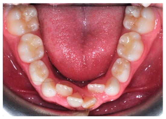
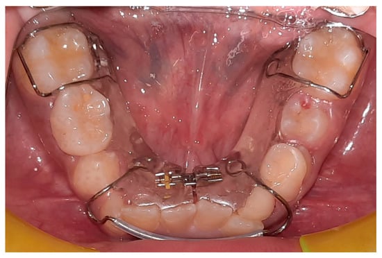
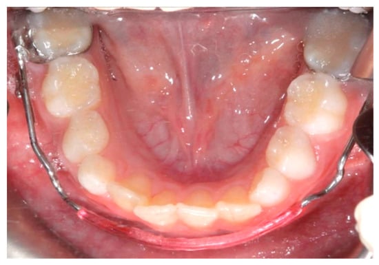
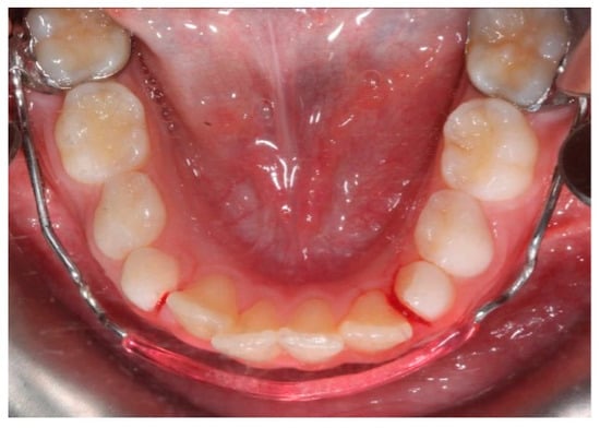
Crowding is the most frequent malocclusion in orthodontics, with a strong hereditary tendency. It already occurs in pediatric age and is mainly hereditary. It is a sign of a lack of space in the arches, and is not self-correcting, but can worsen over time. The main cause of the worsening of this malocclusion is a progressive and physiological decrease in the arch perimeter.  An orthodontic treatment cannot ignore the concept of “guide arch”, which concerns the lower arch, because of the objective difficulty in increasing its perimeter; the bone structure of the lower jaw is more compact than that of the upper one. Its expansion, in fact, is limited to a slight vestibularization of the incisors and lateral sectors that may be associated with a limited distalization of the molars. There are various therapeutic solutions available to the orthodontist, and a correct diagnosis through clinical examination, radiographs and model analysis are essential. The decision of how to deal with crowding cannot be separated from an overall assessment of the malocclusion to be treated.

mandibular crowding mixed dentition space recovery serial extractions slicing orthodontic expansion leeway space

1. Introduction

Crowding is a common orthodontic malocclusion with a strong hereditary tendency. It is caused by a variety of factors, including the impact of environmental and genetic factors on dental arch dimensions [1]. Dental crowding is defined as an inconsistency between tooth size and arch dimension that results in malocclusion; it occurs because of a lack of coordination between tooth size and arch dimensions [2][3][4][5]. The lower incisors are the teeth most frequently involved [6][7][8] (Figure 1).
Figure 1. Crowding of the permanent mandibular incisors.
Studies show that 46 per cent of children between 6 and 12 years old and 85% of children between 12 and 17 years old have crowding. This is because, if action is not taken, leeway space is lost, and consequently, a mild and easily resolved crowding in mixed dentition becomes stable and significant in permanent dentition [9][10][11][12]. During the third phase of the permutation, when deciduous canines and molars are replaced by permanent canines and premolars, there is a decrease in the length of the dental arch perimeter. The maximum increase in size of both dental arches occurs in the first 2 years of life; after that, the arch length increases up to 13 years in the maxilla and up to 8 years in the mandible; after this age, length decreases in both arches [13]. Crowding is thus considered a malocclusion that never self-corrects and instead worsens over time [14][15][16][17]. If present in deciduous teeth, it will worsen over the next two dental stages [18][19][20]. It generally involves 50% of individuals who were exempt in the first decade of life [21]. An orthodontic treatment cannot ignore the concept of the “guide arch”, which concerns the lower one, because of the objective difficulty in increasing its perimeter; the bone structure of the lower jaw is more compact than that of the upper one. The identification of factors that contribute to mandibular tooth crowding in mixed dentition is critical for treatment planning [22]. Several factors can be assumed to affect the development and severity of crowding, such as the direction of mandibular growth, the early loss of deciduous molars, mesiodistal tooth and arch dimensions, oral and perioral musculature and incisor and molar inclination [23]. The correction of severe mandibular crowding in mixed dentition could be carried out by extraction, distalization and surgical and non-surgical expansion of the mandible [18][24]. It is useful to emphasize that the clinician is confronted not only with crowding but also with clinical situations that are predictive of it or represent a different aspect, such as the lack of diastemas in deciduous dentition, the early loss of deciduous canines, the loss of arch length due to destructive caries, the early loss of the deciduous second molar, the appearance of teeth in ectopic position and protrusion or the accentuated retrusion of the incisors [25]. The early detection of mandibular teeth crowding is critical for interceptive orthodontic treatment planning.

2. Diagnostic Methods

Regarding the diagnosis, the lack of space in the primary dentition is a predictor to crowding. The inability to accommodate the size difference between the primary and permanent incisors is also due to a “closed” primary dentition, which prevents the mesial shift in the erupting permanent molars into a class I molar relationship during the closing of the private space [26]. Measurements for diagnosing crowding are usually taken on the plaster model with a digital caliper, both before and after treatment. Tooth size is the sum of the mesiodistal diameters of all teeth [26]. Arch length is calculated as the perpendicular distance between a line that connects the medial contact point of the first permanent molars and the most vestibular point between the lower central incisors. Crowding is measured as the difference between tooth size and arch length [26]. As result, crowding is associated with both bigger teeth and a smaller dental arch. The degree of crowding is influenced by the direction of mandibular development, early loss of primary molars, arch size, oral musculature and incisor and molar inclination [22]. Many studies have found a correlation between crowding and the direction of mandibular rotation. Extreme mandibular rotation has been linked to increased crowding, and crowding is also brought on by some growth/skeletal patterns at the start of adolescence [27]. Other factors that should not be underestimated for the diagnosis of crowding are the changes in facial morphology brought on by growth or orthodontic treatment [28]. Dental crowding is also caused by several reasons, including the impact of environmental and genetic variables on dental arch measurements such arch width, arch length and arch perimeter. Further factors influencing mesiodistal tooth width include racial characteristics, sex and inherited traits [18]. Lateral cephalograms can be used for skeletal parameters contributing to dental crowding such as effective maxillary and mandibular length, mandibular plane angle, Y axis, lower anterior face height and dental parameters such as axial inclination of the lower incisor, inclination of the lower incisor to the mandibular plane and interincisal angle [29]. Some authors have found that the use of 2D lateral cephalograms or profile photos for orthodontic measurements may not be adequately accurate. Three-dimensional computed tomography provides better frontal and three-quarter profile data for diagnosis, allowing structures to be precisely measured, aiding in analysis and diagnosis [18]. A template applied for the measurement of crowding is Little’s Irregularity Index. It denotes the total linear displacement of the six mandibular anterior teeth’s anatomical contact locations, represented in millimeters: 0 indicates perfect alignment, 1–3 mm indicates the least amount of irregularity, 4–6 mm indicates moderate abnormality, 7–9 mm indicates severe irregularity and 10 mm or more indicates very severe misalignment [30]. For treatment planning, it is crucial to identify the causes of anterior mandibular tooth crowding in mixed dentition [22]. If malocclusion is characterized by a deep bite, the cause could be skeletal, dental or both [31].
Two clinical disorders can coexist as a result of altered tooth eruption, which is the failure of a tooth to erupt into the proper location in the arch: transmigration and inclusion [32].

3. Prophylaxis

Primary dentition is essential for influencing the eruption of permanent teeth. Early primary tooth loss can result in undesired tooth motions and space loss in the permanent dentition [2].
It is recommended to maintain the primary dentition in the arch until exfoliation; nevertheless, if early loss is inevitable, it should be managed to minimize the negative consequences on the developing occlusion [23].
Space maintainers can be utilized for this purpose.
Early primary molar loss might result in a reduction in arch length, increasing the severity of crowding/malocclusion; therefore, in the affected patient, every effort should be made to preserve the natural leeway space [33]. The unilateral loss of a primary canine or first molar can result in a significant centerline disparity and mesial migration of the buccal segments, which is another critical clinical condition to preserve space [29]. Space maintainers are classified into three types: fixed unilateral appliances, fixed bilateral appliances and removable partial dentures. The band and loop space maintainer is one of the most prevalent permanent unilateral appliances [34]. The appliance has a band that cements to the primary second molar. It also has a loop that contacts the distal surface of the primary canine [2].
The distal shoe is another permanent unilateral appliance. A stainless-steel wire extends in front of the unerupted permanent first molar to guide it into position as it grows in. Distal shoes can only be placed on one tooth [2].
Bilateral space maintainers are used once teeth on both sides of the mouth are lost. Common types include Lingual Holding Arch, Nance Arch and Transpalatal Arch.
Removable dentures are often used for cosmetic purposes rather than to avoid space loss, particularly when anterior (front) teeth are lost [2].

4. Treatment

The lower jaw is considered the guiding arch in crowding therapy because it is difficult to modify its perimeter due to the more compact bone structure and the continuity with the mandibular branch, which does not allow for distalization [28][35]. In addition, the symphysis cartilage ossifies in the first year of life, so it is not possible to perform an orthopedic bone expansion, as in the upper jaw, working at the level of the median palatine suture [35][36]. The modalities of space recovery in mixed dentition are: arch perimeter increment, reduction in mesiodistal widths of teeth and serial extractions [2][37][38]. In the upper arch, the expansions are quite stable, but inferiorly, it is universally recognized that the expansion of the intercanine diameter always recurs, whereas expansion at the molar level is quite stable, which should be considered [26][39][40][41]. The space that can be recovered in the lower arch depends on the type of sector: in general, in the posterior sector, utilizing a lip bumper-style device, it is possible to recover a maximum of 2 mm of space per molar distalization in the posterior regions [42]. At the molar level in the lateral sectors, the arch length could increase by about 0.4 mm; at the canine level, the arch length increases about 0.7 mm [43]. The vestibularization of one millimeter of the incisal margin in the anterior sector results in the gain of one millimeter of arch space, or roughly a ratio of one to one (changing the arch form) [41]. Schwarz’s appliance (Figure 2) and lip bumper (Figure 3) are two commonly used appliances for increasing lower dental arch dimensions [44]. In their study, Vincenzo Quinzi et al. compared the effects of these appliances on reducing mandibular crowding by increasing lower arch dimensions [26]. The study included twenty subjects (10 males and 10 females). Ten patients were treated with Schwarz’s appliance, and ten with lip bumper. The Schwarz appliance was more effective in increasing intercanine arch dimensions and arch perimeter, although the lip bumper reached a greater increase in arch length [26]. Since the 1970s, there have been reports of spontaneous changes in mandibular dentition caused by maxillary expansion [45][46]. Di Ventura et al. assessed the consequences of rapid maxillary expansion (RME) anchored to primary molars on the mandibular arch. A total of 54 patients were recruited for this study and divided into two groups: a test group (21 patients, 7.4 ± 1.2 years) who underwent RME, and a control group (17 patients, 7.3 ± 1.1 years old) who did not receive any treatment. The results of this study showed a significant increase in interdental width in the lower arch after 9 months of RME therapy [45]. Olivia Griswold et al. evaluated the changes in sagittal mandibular incisors’ position in response to lip bumper therapy using CBCT [47]. In this study, the authors compared a group that was treated only with rapid maxillary expansion (experimental group) and an RME + LB (lip bumper) group (control group) [47]. The CBCTs were placed in 3D on the mandibular structure, and the angular and linear alterations in the mandibular incisors throughout LB therapy were assessed. In the investigation, there was no statistically significant difference in the degree of mandibular incisor protrusion between the two groups; the lip bumper did not generate substantial proclination, protrusion or extrusion of the mandibular incisors. [13]. Air-rotor stripping (ARS) (Figure 4) is a technique for creating space during the mixed dentition period by reducing interproximal enamel thickness. Yahya B. Nakhjavani et al. assessed the efficacy of the mesial stripping of mandibular deciduous canines for the correction of rotated and lingually erupted lateral incisors in 42 patients with <3 mm mandibular crowding [48]. In this study, the mesial stripping of mandibular primary canines resulted in full crowding correction; in just few cases, the amount of crowding did not reach zero, and a small crowding in the range of 0.06 to 0.1 mm remained [48]. The extraction of all the first premolars with subsequent orthodontic treatment is the most used method to relieve dental crowding [49]. The importance and timing of extraction as a component of orthodontic therapy for late incisor crowding have been well investigated [50]. No difference in late incisor crowding is shown by the data, regardless of whether serial extraction or early or late premolar extraction is performed prior to orthodontic treatment. Additionally, selecting a non-extraction orthodontic procedure has been linked to post-retention crowding [50]. Maurits Persson et al. investigated changes in the mandibular incisor area from early adolescence to late adulthood in patients with a class I crowding malocclusion treated in the mixed dentition by the extraction of all first premolars without subsequent orthodontic treatment [50]. The extraction group included 24 subjects who had all their first premolars extracted at a mean age of about 11.5 years to treat a class I space deficiency malocclusion. The control group included 21 subjects who had normal occlusions at the age of 13 years [50]. The extraction group showed no improvements in lower incisor irregularity, and a significant increase in lower tooth space insufficiency into adulthood. Lower incisor irregularity and space shortage developed significantly in the control group throughout late adulthood [50]. Premolar extraction is the sole treatment option for severe crowding in a class I occlusion, allowing for spontaneous adjustments and more stable incisor alignment in late adulthood, according to the authors [50].
Figure 2. Schwarz’s appliance.
Figure 3. Lip bumper.
Figure 4. Air-rotor stripping (ARS).

References

  1. Normando, D.; Almeida, M.A.O.; Quintão, C.C.A. Dental Crowding. Angle Orthod. 2013, 83, 10–15.
  2. Jadhav, V.; Tiwari, M.; Seegavadi, V.; Kamble, R.; Daigavane, P. A Formula for Estimating the Mesiodistal Width of Permanent Mandibular Central Incisors. J. Datta Meghe Inst. Med. Sci. Univ. 2021, 16, 29–32.
  3. Rizzo, C.; Di Bartolo, I.; Santantonio, M.; Coscia, M.F.; Monno, R.; De Vito, D.; Ruggeri, F.M.; Rizzo, G. Epidemiological and Virological Investigation of a Norovirus Outbreak in a Resort in Puglia, Italy. BMC Infect. Dis. 2007, 7, 135.
  4. De Vito, D.; Monno, R.; Nuccio, F.; Legretto, M.; Oliva, M.; Coscia, M.F.; Dionisi, A.M.; Calia, C.; Capolongo, C.; Pazzani, C. Diffusion and Persistence of Multidrug Resistant Salmonella Typhimurium Strains Phage Type DT120 in Southern Italy. BioMed Res. Int. 2015, 2015, 1–8.
  5. Coscia, M.F.; Monno, R.; Ballini, A.; Mirgaldi, R.; Dipalma, G.; Pettini, F.; Cristallo, V.; Inchingolo, F.; Foti, C.; De Vito, D. Human Papilloma Virus (HPV) Genotypes Prevalence in a Region of South Italy (Apulia). Ann. Dell’istituto Super. Di Sanita 2015, 51, 248–251.
  6. Paul, S.; Garg, S.; Saraf, B.G.; Sheoran, N.; Chawla, M.; Saji, S.E. Arch Measurements, Bigonial Width, Dental Caries, and Their Effect on Occurrence of Mandibular Incisors Crowding in Early Mixed Dentition Period. Int. J. Clin. Pediatr. Dent. 2021, 14, S57–S62.
  7. Tartaglia, G.M.; Mapelli, A.; Maspero, C.; Santaniello, T.; Serafin, M.; Farronato, M.; Caprioglio, A. Direct 3D Printing of Clear Orthodontic Aligners: Current State and Future Possibilities. Materials 2021, 14, 1799.
  8. Maspero, C.; Fama, A.; Cavagnetto, D.; Abate, A.; Farronato, M. Treatment of Dental Dilacerations. J. Biol. Regul. Homeost. Agents 2019, 33, 1623–1627.
  9. Grainger, R.M. Orthodontic Treatment Priority Index; Vital Health Stat 2; United States Department of Health, Education, and Welfare: Washington, DC, USA, 1967; pp. 1–49.
  10. Cazzolla, A.P.; Zhurakivska, K.; Ciavarella, D.; Lacaita, M.G.; Favia, G.; Testa, N.F.; Marzo, G.; La Carbonara, V.; Troiano, G.; Lo Muzio, L. Primary Hyperoxaluria: Orthodontic Management in a Pediatric Patient: A Case Report. Spec. Care Dent. 2018, 38, 259–265.
  11. Lo Russo, L.; Ciavarella, D.; Salamini, A.; Guida, L. Alignment of Intraoral Scans and Registration of Maxillo-Mandibular Relationships for the Edentulous Maxillary Arch. J. Prosthet. Dent. 2019, 121, 737–740.
  12. Cassano, M.; Russo, G.; Granieri, C.; Ciavarella, D. Modification of Growth, Immunologic and Feeding Parameters in Children with OSAS after Adenotonsillectomy. Acta Otorhinolaryngol. Ital. 2018, 38, 124–130.
  13. Maspero, C.; Abate, A.; Inchingolo, F.; Dolci, C.; Cagetti, M.G.; Tartaglia, G.M. Incidental Finding in Pre-Orthodontic Treatment Radiographs of an Aural Foreign Body: A Case Report. Children 2022, 9, 421.
  14. Farronato, M.; Maspero, C.; Abate, A.; Grippaudo, C.; Connelly, S.T.; Tartaglia, G.M. 3D Cephalometry on Reduced FOV CBCT: Skeletal Class Assessment through AF-BF on Frankfurt Plane-Validity and Reliability through Comparison with 2D Measurements. Eur. Radiol. 2020, 30, 6295–6302.
  15. Ergoren, M.C.; Paolacci, S.; Manara, E.; Dautaj, A.; Dhuli, K.; Anpilogov, K.; Camilleri, G.; Suer, H.K.; Sayan, M.; Tuncel, G.; et al. A Pilot Study on the Preventative Potential of Alpha-Cyclodextrin and Hydroxytyrosol against SARS-CoV-2 Transmission. Acta Bio Med. Atenei Parm. 2020, 91, e2020022.
  16. Maspero, C. Operculectomy and Spontaneous Eruption of Impacted Second Molars: A Retrospective Study. J. Biol. Regul. Homeost. Agents 2019, 33, 1909–1912.
  17. Maspero, C.; Cappella, A.; Dolci, C.; Cagetti, M.G.; Inchingolo, F.; Sforza, C. Is Orthodontic Treatment with Microperforations Worth It? A Scoping Review. Children 2022, 9, 208.
  18. Das, P.J.; Dkhar, W.; Pradhan, A. An Evaluation of Dental Crowding in Relation to the Mesiodistal Crown Widths and Arch Dimensions in Southern Indian Population. J. Clin. Diagn. Res. 2017, 11, TC10–TC13.
  19. Ciavarella, D.; Monsurrò, A.; Padricelli, G.; Battista, G.; Laino, L.; Perillo, L. Unilateral Posterior Crossbite in Adolescents: Surface Electromyographic Evaluation. Eur. J. Paediatr. Dent. 2012, 13, 25–28.
  20. Inchingolo, A.M.; Fatone, M.C.; Malcangi, G.; Avantario, P.; Piras, F.; Patano, A.; Di Pede, C.; Netti, A.; Ciocia, A.M.; De Ruvo, E.; et al. Modifiable Risk Factors of Non-Syndromic Orofacial Clefts: A Systematic Review. Children 2022, 9, 1846.
  21. Brunelle, J.A.; Bhat, M.; Lipton, J.A. Prevalence and Distribution of Selected Occlusal Characteristics in the US Population, 1988–1991. J. Dent. Res. 1996, 75, 706–713.
  22. Jain, R. Prevalence of Mandibular Anterior Teeth Crowding in Mixed Dentition Subjects Reporting to a University Hospital in Chennai City. Int. J. Dent. Oral Sci. 2019, S3, 6–11.
  23. Grippaudo, M.M.; Quinzi, V.; Manai, A.; Paolantonio, E.G.; Valente, F.; La Torre, G.; Marzo, G. Orthodontic Treatment Need and Timing: Assessment of Evolutive Malocclusion Conditions and Associated Risk Factors. Eur. J. Pediatr. Dent. 2020, 21, 203–208.
  24. Farronato, M.; Boccalari, E.; Del Rosso, E.; Lanteri, V.; Mulder, R.; Maspero, C. A Scoping Review of Respirator Literature and a Survey among Dental Professionals. Int. J. Environ. Res. Public Health 2020, 17, 5968.
  25. Leighton, B.C. The Early Signs of Malocclusion. Eur. J. Orthod. 2007, 29, i89–i95.
  26. Jain, R. Association Of Mandibular Arch Crowding And Vertical Growth Pattern—A Retrospective Study. Int. J. Dent. Oral Sci. 2021, 8, 4096–4100.
  27. Satra, P.; Vichare, G.; Bhosale, V. Relationship of Maxillary and Mandibular Effective Base Length, Arch Length and Dental Crowding in Different Vertical Growth Pattern. APOS 2022, 12, 108–114.
  28. Singh, S.; Shivaprakash, G. To Evaluate the Correlation Between Skeletal and Dental Parameters to the Amount of Crowding in Class II Div. 1 Malocclusions. J. Clin. Diagn. Res. 2017, 11, ZC22–ZC27.
  29. Pasciuti, E.; Coloccia, G.; Inchingolo, A.D.; Patano, A.; Ceci, S.; Bordea, I.R.; Cardarelli, F.; Di Venere, D.; Inchingolo, F.; Dipalma, G. Deep Bite Treatment with Aligners: A New Protocol. Appl. Sci. 2022, 12, 6709.
  30. Di Ventura, A.; Lanteri, V.; Farronato, G.; Gaffuri, F.; Beretta, M.; Lanteri, C.; Cossellu, G. Three-Dimensional Evaluation of Rapid Maxillary Expansion Anchored to Primary Molars: Direct Effects on Maxillary Arch and Spontaneous Mandibular Response. Eur. J. Pediatr. Dent. 2019, 20, 38–42.
  31. Inchingolo, A.D.; Carpentiere, V.; Piras, F.; Netti, A.; Ferrara, I.; Campanelli, M.; Latini, G.; Viapiano, F.; Costa, S.; Malcangi, G.; et al. Orthodontic Surgical Treatment of Impacted Mandibular Canines: Systematic Review and Case Report. Appl. Sci. 2022, 12, 8008.
  32. Tepedino, M.; Iancu-Potrubacz, M.; Ciavarella, D.; Masedu, F.; Marchione, L.; Chimenti, C. Expansion of Permanent First Molars with Rapid Maxillary Expansion Appliance Anchored on Primary Second Molars. J. Clin. Exp. Dent. 2018, 10, e241–e247.
  33. Antoszewska-Smith, J.; Bohater, M.; Kawala, M.; Sarul, M.; Rzepecka-Skupień, M. Treatment of Adults with Anterior Mandibular Teeth Crowding: Reliability of Little’s Irregularity Index. Int. J. Dent. 2017, 2017, 5057941.
  34. Persson, M.; Al-Taai, N.; Pihlgren, K.; Westerlund, A. Early Extractions of Premolars Reduce Age-Related Crowding of Lower Incisors: 50 Years of Follow-Up. Clin. Oral Investig. 2022, 26, 4525–4535.
  35. Ciavarella, D.; Tepedino, M.; Chimenti, C.; Troiano, G.; Mazzotta, M.; Foschino Barbaro, M.P.; Lo Muzio, L.; Cassano, M. Correlation between Body Mass Index and Obstructive Sleep Apnea Severity Indexes—A Retrospective Study. Am. J. Otolaryngol. 2018, 39, 388–391.
  36. Troiano, G.; Dioguardi, M.; Cocco, A.; Zhurakivska, K.; Ciavarella, D.; Muzio, L.L. Increase the Glyde Path Diameter Improves the Centering Ability of F6 Skytaper. Eur. J. Dent. 2018, 12, 089–093.
  37. Inchingolo, A.D.; Patano, A.; Coloccia, G.; Ceci, S.; Inchingolo, A.M.; Marinelli, G.; Malcangi, G.; Montenegro, V.; Laudadio, C.; Pede, C.D.; et al. The Efficacy of a New AMCOP® Elastodontic Protocol for Orthodontic Interceptive Treatment: A Case Series and Literature Overview. Int. J. Environ. Res. Public Health 2022, 19, 988.
  38. Ciavarella, D.; Parziale, V.; Mastrovincenzo, M.; Palazzo, A.; Sabatucci, A.; Suriano, M.M.; Bossù, M.; Cazzolla, A.P.; Lo Muzio, L.; Chimenti, C. Condylar Position Indicator and T-Scan System II in Clinical Evaluation of Temporomandibular Intracapsular Disease. J. Cranio-Maxillofac. Surg. 2012, 40, 449–455.
  39. Cianci, C.; Pappalettera, G.; Renna, G.; Casavola, C.; Laurenziello, M.; Battista, G.; Pappalettere, C.; Ciavarella, D. Mechanical Behavior of PET-G Tooth Aligners Under Cyclic Loading. Front. Mater. 2020, 7, 104.
  40. Inchingolo, A.M.; Malcangi, G.; Costa, S.; Fatone, M.C.; Avantario, P.; Campanelli, M.; Piras, F.; Patano, A.; Ferrara, I.; Di Pede, C.; et al. Tooth Complications after Orthodontic Miniscrews Insertion. Int. J. Environ. Res. Public Health 2023, 20, 1562.
  41. De Vito, D.; Zullo, M.J.; Benincasa, C.; Aiello, E.; Giacomello, M.S.; Mortellaro, C.; Greco Lucchina, A. Facial Mask and Plexiglass Box: A Critical Overview on the Current Strategies to Protect Patients from COVID-19 Infection. J. Biol. Regul. Homeost. Agents 2021, 35, 139–145.
  42. De Vito, D.; Di Ciaula, A.; Palmieri, V.O.; Trerotoli, P.; Larocca, A.M.V.; Montagna, M.T.; Portincasa, P. Reduced COVID-19 Mortality Linked with Early Antibodies against SARS-CoV-2, Irrespective of Age. Eur. J. Intern. Med. 2022, 98, 77–82.
  43. Raucci, G.; Pachêco-Pereira, C.; Elyasi, M.; d’Apuzzo, F.; Flores-Mir, C.; Perillo, L. Predictors of Postretention Stability of Mandibular Dental Arch Dimensions in Patients Treated with a Lip Bumper during Mixed Dentition Followed by Fixed Appliances. Angle Orthod. 2017, 87, 209–214.
  44. Coloccia, G.; Inchingolo, A.D.; Inchingolo, A.M.; Malcangi, G.; Montenegro, V.; Patano, A.; Marinelli, G.; Laudadio, C.; Limongelli, L.; Di Venere, D.; et al. Effectiveness of Dental and Maxillary Transverse Changes in Tooth-Borne, Bone-Borne, and Hybrid Palatal Expansion through Cone-Beam Tomography: A Systematic Review of the Literature. Medicina 2021, 57, 288.
  45. Griswold, O.; Li, C.; Orr, J.; Boucher, N.; Shah, S.; Chung, C.-H. Lip Bumper Therapy Does Not Influence the Sagittal Mandibular Incisor Position in a Retrospective CBCT Study. J. Clin. Med. 2022, 11, 6032.
  46. Freitas, K.M.S.; Massaro, C.; Miranda, F.; de Freitas, M.R.; Janson, G.; Garib, D. Occlusal Changes in Orthodontically Treated Subjects 40 Years after Treatment and Comparison with Untreated Control Subjects. Am. J. Orthod. Dentofac. Orthop. 2021, 160, 671–685.
  47. Nakhjavani, Y.B.; Nakhjavani, F.B.; Jafari, A. Mesial Stripping of Mandibular Deciduous Canines for Correction of Permanent Lateral Incisors. Int. J. Clin. Pediatr. Dent. 2017, 10, 229–233.
  48. Verma Comparative Evaluation of Stability of Mandibular Anterior Crowding Correction Done with Two Different Treatment Protocols: A Retrospective Study. Available online: https://www.jioh.org/article.asp?issn=0976-7428;year=2022;volume=14;issue=2;spage=189;epage=194;aulast=Verma (accessed on 24 January 2023).
  49. Mahmoudzadeh, M.; Mirzaei, H.; Farhadian, M.; Mollabashi, V.; Khosravi, M. Comparison of Anterior Crowding Relapse Tendency in Patients Treated with Incisor Extraction, Premolar Extraction, and Nonextraction Treatment. J. World Fed. Orthod. 2018, 7, 61–65.
  50. Berbert, M.; Cotrin, P.; de Oliveira, R.G.; de Oliveira, R.G.; Valarelli, F.P.; de Freitas, M.R.; Freitas, K.M.S. The Influence of 3x3 Bonded Retainer on Anterior Crowding Relapse in Mandibular Incisor Extraction Cases. Dent. Press J. Orthod. 2021, 26, e212081.
  51. Freitas, K.M.S.; Massaro, C.; Miranda, F.; de Freitas, M.R.; Janson, G.; Garib, D. Occlusal Changes in Orthodontically Treated Subjects 40 Years after Treatment and Comparison with Untreated Control Subjects. Am. J. Orthod. Dentofac. Orthop. 2021, 160, 671–685.
  52. Mahmoudzadeh, M.; Mirzaei, H.; Farhadian, M.; Mollabashi, V.; Khosravi, M. Comparison of Anterior Crowding Relapse Tendency in Patients Treated with Incisor Extraction, Premolar Extraction, and Nonextraction Treatment. J. World Fed. Orthod. 2018, 7, 61–65.
More
Information
Contributors MDPI registered users' name will be linked to their SciProfiles pages. To register with us, please refer to https://encyclopedia.pub/register : , , , , , , , , , , , , , , , ,
View Times: 407
Revisions: 3 times (View History)
Update Date: 08 May 2023
1000/1000
Video Production Service