Submitted Successfully!
To reward your contribution, here is a gift for you: A free trial for our video production service.
Thank you for your contribution! You can also upload a video entry or images related to this topic.
Version Summary Created by Modification Content Size Created at Operation
1 -- 4630 2023-04-27 11:46:49 |
2 update references and layout Meta information modification 4630 2023-04-27 11:53:53 |

Video Upload Options

Do you have a full video?

Confirm

Are you sure to Delete?
Cite
If you have any further questions, please contact Encyclopedia Editorial Office.
Thangaleela, S.; Sivamaruthi, B.S.; Radha, A.; Kesika, P.; Chaiyasut, C. Molecular Mechanism of Neuromyelitis Optica Spectrum Disorders. Encyclopedia. Available online: https://encyclopedia.pub/entry/43566 (accessed on 17 May 2024).
Thangaleela S, Sivamaruthi BS, Radha A, Kesika P, Chaiyasut C. Molecular Mechanism of Neuromyelitis Optica Spectrum Disorders. Encyclopedia. Available at: https://encyclopedia.pub/entry/43566. Accessed May 17, 2024.
Thangaleela, Subramanian, Bhagavathi Sundaram Sivamaruthi, Arumugam Radha, Periyanaina Kesika, Chaiyavat Chaiyasut. "Molecular Mechanism of Neuromyelitis Optica Spectrum Disorders" Encyclopedia, https://encyclopedia.pub/entry/43566 (accessed May 17, 2024).
Thangaleela, S., Sivamaruthi, B.S., Radha, A., Kesika, P., & Chaiyasut, C. (2023, April 27). Molecular Mechanism of Neuromyelitis Optica Spectrum Disorders. In Encyclopedia. https://encyclopedia.pub/entry/43566
Thangaleela, Subramanian, et al. "Molecular Mechanism of Neuromyelitis Optica Spectrum Disorders." Encyclopedia. Web. 27 April, 2023.
Molecular Mechanism of Neuromyelitis Optica Spectrum Disorders
Edit

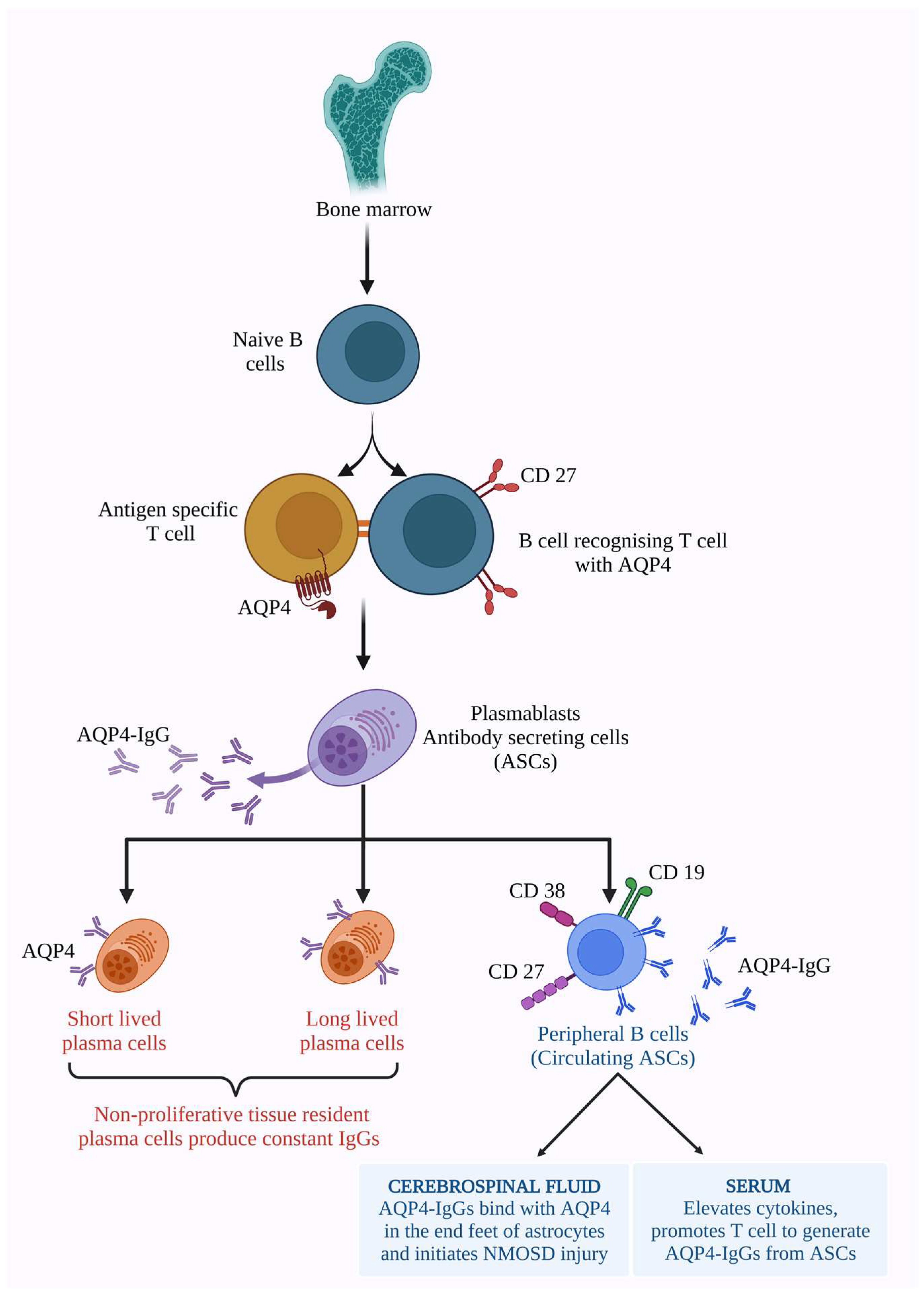
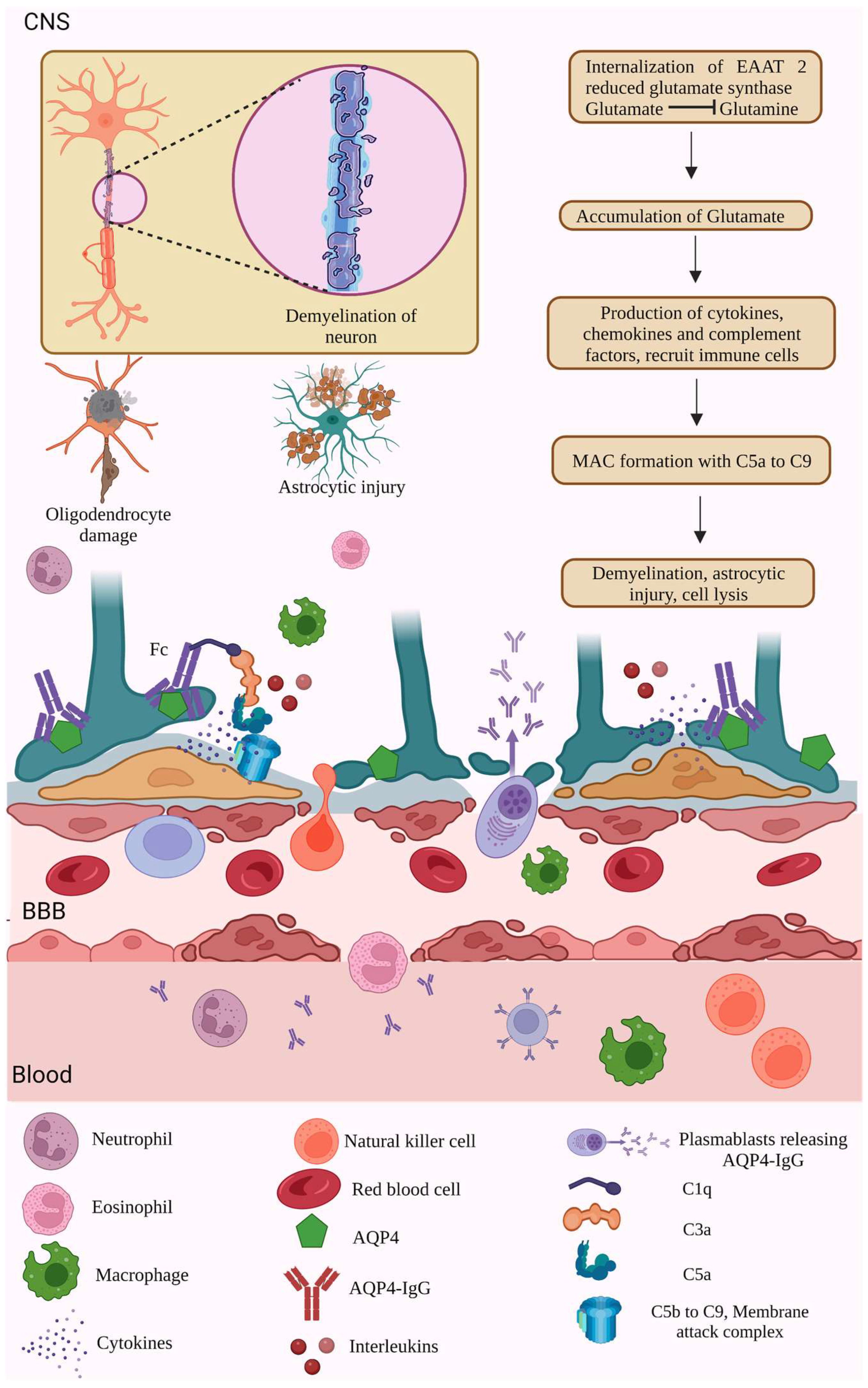
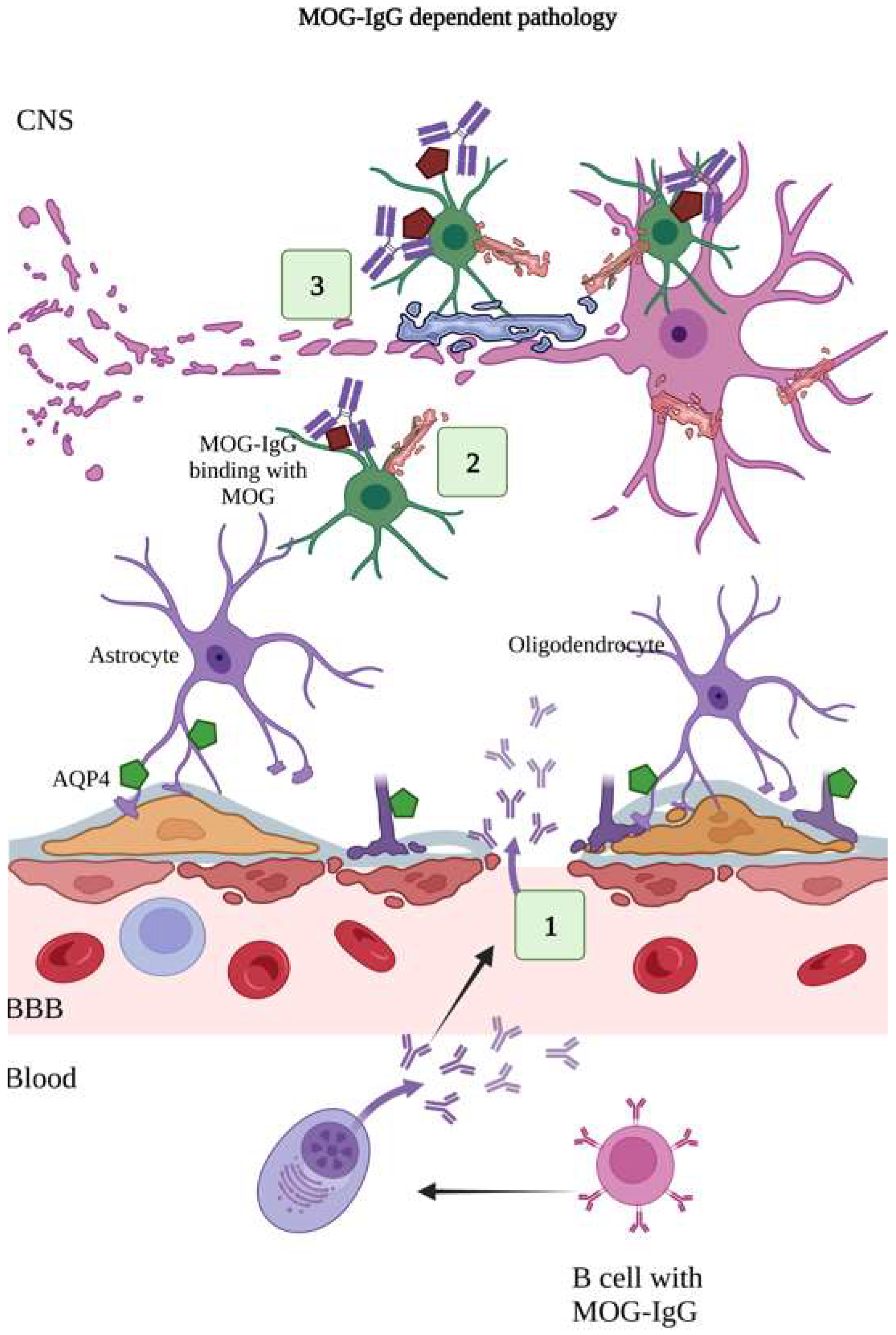
Neuromyelitis optica (NMO) is a rare autoimmune inflammatory disorder affecting the central nervous system (CNS), specifically the optic nerve and the spinal cord, with severe clinical manifestations, including optic neuritis (ON) and transverse myelitis. Initially, NMO was wrongly understood as a condition related to multiple sclerosis (MS), due to a few similar clinical and radiological features, until the discovery of the AQP4 antibody (NMO-IgG/AQP4-ab).NMO was expanded to NMO spectrum disorder (NMOSD) because of its varied clinical phenotypes. NMOSD is characterized by the activation of the complement cascade, granulocyte, eosinophil and lymphocyte infiltration, oligodendrocyte, and astrocyte injury, demyelination, and neuronal loss. The studies on gut microbiota and NMOSD explain the correlation between gut dysbiosis and NMOSD by signifying the abundance of pathogenic bacteria and reduction in commensal organisms, which cause abnormal metabolism and metabolic signals in the pathogenesis of autoimmune diseases such as NMOSD. 

neuromyelitis optica neuromyelitis optica spectrum disorders autoimmune diseases

1. Introduction

Autoimmune diseases (ADs) are chronic conditions initiated due to loss of immunological tolerance or immune responses toward self-antigens. ADs are a heterogeneous group of disorders, having multifactorial origins and affecting specific organs or multiple organs of the body [1][2]. Genetics, drugs, immune responses initiated by environmental agents and microbiota, air pollution, stress, low levels of vitamin D, medications, vaccination, sex hormones, and the interplay between gene-environment context are the possible risk factors of ADs, in addition to cigarette smoking, alcohol, and coffee consumption [1][3].
Neurological autoimmune disease (NAD) is an organ-specific AD that particularly affects the central nervous system (CNS) and peripheral nervous system (PNS). Certain autoimmune disorders, such as multiple sclerosis (MS), acute disseminated encephalomyelitis (ADEM), anti-NMDAR encephalitis, longitudinally extensive transverse myelitis, Guillain-Barré Syndrome (GBS), Myasthenia Gravis (MG), Sjogren’s syndrome (SS), immunoglobulin G4 related diseases, limbic encephalitis, opsoclonus-myoclonus, Miller-Fisher syndrome, Stiff-person syndrome, Morvan syndrome, sensory ganglionopathy, Lambert-Eaton myasthenic syndrome, and neuromyelitis optica (NMO), are categorized as NADs. Among these NADs, MS is the most prevalent worldwide, while other NADs are less common [4]. NMO is one of the immune-mediated inflammatory disorders of the CNS. NMO predominantly causes astrocyte loss and dysfunction resulting in secondary myelination and neurodegeneration, particularly affecting the optical nerves and spinal cord with concurrent inflammation and demyelination of ON and the spinal cord (myelitis) [5]. Astrocytic lesions, caused by the binding of IgG to aquaporin 4 (AQP4) channels, and subsequent deposition of complement factors resulting in lytic reactions in astrocytes lead to astrocyte damage. Human astrocytes are more complex than rodent astrocytes. Detailed immunohistochemical, molecular, and histopathological studies of astrocytes in optic nerves, brain, and spinal cord are obligatory in studying NMO patients to understand the neuropsychological symptoms in NMO cases [6].
NMO was expanded to NMO spectrum disorder (NMOSD) because of its varied clinical phenotypes. NMOSD is characterized by the activation of the complement cascade, granulocyte, eosinophil and lymphocyte infiltration, oligodendrocyte, and astrocyte injury, demyelination, and neuronal loss [7]. Most NMOSD patients exhibit acute inflammatory responses activated by autoantibodies of IgG against AQP4 in the optic nerve and spinal cord [8], CNS attack relapses, ON, and encephalitis of the diencephalon, area postrema, and brain stem [9]. Classically, about 30% of NMOSD patients have been observed with severe neurological disability, ON, worse clinical outcomes, and the presence of oligoclonal bands in the CSF [10].

2. Molecular Mechanism of NMOSD

2.1. AQP4 Dependent Pathology

AQP4 is a bidirectional transmembrane protein highly present in the astrocyte end feet of the BBB and the ependymal cells of the CNS [11]. AQP4 has been found to be necessary for the functioning of the BBB and important for maintaining CNS water homeostasis [11]. BBB disruption is the hallmark pathological sign of NMOSD, resulting in BBB breakdown and an influx of serum AQP4 IgG and complement factors into the CNS compartment, followed by the entry of B cells into the CNS and the formation of lesions [12]. Many histological studies in NMOSD patients have revealed the loss of AQP4 and CNS lesions, with or without astrocyte loss [13]. The presence of the circulating serum autoantibody against the AQP4 water channel (AQP4-IgG) induces neuroinflammation by breaching the BBB, releasing proinflammatory cytokines into the CNS, and producing acute CNS attacks, CNS pathologies, area postrema syndrome, astrocyte inactivation, and astrocyte loss through complement inactivation, BBB disruption, and neural injury [13][14][15].
Besides the astrocytes, the retinal muller cells are highly packed with AQP4 and inwardly rectifying potassium (Kir4.1) channels [16]. The binding of AQP4-ab with AQP4 in the astrocyte initiates immune complement cascade signals, which results in the infiltration of granulocytes, lymphocytes, and eosinophils, leading to astrocyte and oligodendrocyte injury and demyelination, myelin loss, neurodegeneration [5], and thickened hyalinization blood [17].
The high expression of AQP4 in the periventricular zone and particularly in the area postrema make these regions more prone to NMOSD pathology [18]. The international consensus diagnostic criteria for NMOSD suggest that AQP4-IgG plays a major causative role in NMOSD. AQP4-IgG seropositive individuals showed six affected regions in the CNS, including the optic nerves, spinal cord, area postrema, brain stem, and cerebrum. The spinal cord and optic nerves are affected in AQP4-IgG seronegative individuals [19]. The astrocyte-specific, glial fibrillary acidic protein (GFAP) necessary for maintaining the structural integrity of the BBB was significantly higher in the CSF of NMOSD patients [20]. A cohort study in North American patients with meningeal encephalomyelitis revealed that GFAP autoantibodies could act as the serum biomarker for CNS autoimmune diseases and potentially initiate complement-mediated immune responses and cause astrocyte damage [21][22].
Aquaporins are the transmembrane proteins essential for water transport and homeostasis in the CNS. Among the thirteen aquaporins in mammals, AQP1, AQP4, and AQP9 are in the brain, especially AQP4, which is present in the CNS–blood and CNS–CSF interfaces in the subarachnoid, subependymal, and pericapillary spaces [23][24]. The AQP4 protein is also expressed more in the retina, optic nerves, and optic tract [25]. AQP4 exists as two dominant isoforms, M1 and M23, due to the extra 22 amino acids in the intracellular N terminus of M1 isoforms. AQP4 monomers consist of six transmembrane helices and two helices with asparagine–proline–alanine motifs that form the pore for transporting water [26]. Both the AQP4 isoforms are highly selective, allowing only the water molecules and excluding other ions or molecules. AQP4 involves potassium reuptake, which is essential for maintaining BBB integrity, synaptic plasticity, and spatial memory [27][28]. The absence of AQP4 in the AQP4 −/− mice showed hyperpermeability of the BBB [7]. A study on autoimmune encephalitis AQP4 −/− mice revealed reduced demyelination, CNS inflammation, motor dysfunction, and cytokine production [29].
The antibodies of AQP4 (AQP4-ab), also known as NMO-IgG, are the prominent serum markers of autoimmune NMO. AQP4-mediated complement pathways and internalization of AQP4 are important reasons for NMO disease [30]. Most NMOSD patients are seropositive for circulating AQP4 immunoglobulin G (AQP4-IgG) antibodies, which target various extracellular epitopes of the AQP4 water channel [8][31]. AQP4-IgG autoantibodies possess a polyclonal ability that recognizes and binds to various epitopes and domains of the AQP4 channel [32]. Anti-AQP4-IgG produced by the plasma cells in peripheral blood enters the CNS through endothelial transcytosis and a permeable BBB and binds with the AQP4 channel. The binding of AQP-IgG with AQP4 activates the complement factors leading to astrocytic injuries through CDC and ADCC. The CDC and ADCC induce further inflammatory signals downstream and the migration of macrophages, neutrophils, and eosinophils to the inflammation site, resulting in BBB interruptions, loss of myelin, and neuronal injury [29][33]. Increased levels of IL-6 are correlated with increased anti-AQP4-IgG and glial fibrillary acidic protein and increased damage in NMOSD [34]. AQP4 crosses the BBB, and the Fc domain of AQP4-IgG binds to the AQP4 in the astrocyte end feet, thereby employing complement activation and causing complement-dependent cytotoxicity (CDC). CDC is activated by AQP4 binding with C1q complement protein. The formation of OAPs by clustering AQP4 results in adding more AQP4-IgGs, which enhances the multivalent binding of C1q onto the grouped AQP4-IgGs over the OAPs [35]. The binding of AQ4-IgG causes endocytosis of AQP4 and reduces water transport across the plasma membrane [34].
To develop memory for AQP4 antigen, B cells require AQP4-specific T cells [36]. AQP4-IgG-generating plasma cells were originally developed by the recognition of T cells with AQP4, which led to the expansion of B cell differentiation into plasmablasts [37]. Identifying the subsets of B cells that produce AQP4-IgG helps in targeted immunotherapies. Antibody-secreting cells (ASCs) are the proliferative plasmablasts that emerge from bone marrow. Initially they are naive B cells, later developing antigen memory, acquiring CD 27, and switching the IgD/IgM expression into IgG expression on its surface [33][38]. Wilson et al. studied the capacity of B cells to produce AQP4-IgGs. The circulating B cells produce AQP4 antibodies in the absence of antigens. Few other factors induce AQP4-IgGs without the cytokines. Hence, it was found that antigens are not necessary for AQP4-IgG production [37]. The production cycle and availability of AQP4-IgGs in the peripheral nervous system and the CNS, and their function in the serum and CNS, are described in Figure 1.
Figure 1. The AQP4-IgG antibodies are produced by antibody-secreting cells (ASCs). Precursors of ASCs secreted from bone marrow as naive B cells. Naive B cells acquire memory antigen CD 27 on their surface along with AQP4 antigen-specific T cells and switch their IgM/IgD expression into IgG. Thus, developed antibody-secreting plasma differentiates into short, long-lived (these are tissue-resident plasma cells that produce constant IgGs), and peripheral circulating B cells secreting AQP4-IgG antibodies with other cell surfaces markers such as CD 19, CD 27, and CD 38 on its surface. Thus, produced AQP4-IgGs are available in the serum and enter the CNS, thereby inducing cytokine production, developing the inflammation cycle, and promoting T cells to generate more ASCs. In the CNS, the AQP4-IgGs bind with AQP4 present in the end feet of astrocytes and initiate NMOSD injury and lesions.
The binding of AQP4-IgG to AQP4 induces endocytosis, which results in the loss or internalization of the excitatory amino acid transporter 2 (EAAT2) [39]. The internalization of glutamate receptors reduces the astrocytic enzyme glutamate synthase activity, causing glutamate accumulation [39]. More accretion of glutamate stimulates the production of various immunological factors, chemokines, cytokines, and complement factors [40]. The production of immunological components activates immune cells such as eosinophils, neutrophils, and macrophages, which damage neurons’ myelin [41][42]. The complement factors bind to the Fc region of the AQP4-IgG antibody; in that way, CDC and ADCC effector mechanisms are initiated by activating the multivalent complement factors such as C1q, C3a, C5a, and MAC over the AQP4-IgG [43][44]. Thus, the activated complement cascade recruits other factors such as C3a and C5a.
The binding of C5a attracts complement factors C5 b to 9, leading to the formation of a membrane attack complex (MAC), which further leads to cell lysis. In contrast, complement activation is modulated by regulatory proteins such as CD59. ADCC allows the binding of AQP4-IgG onto the Fc receptors and activates the ADCC pathway and degranulation. Thus, the inflammatory niche produced by the CDC and ADCC results in the infiltration of neutrophils, macrophages, and eosinophils, astrocytic and oligodendrocyte injury, BBB disruption and leakage, and demyelination and neuronal injury [43][44]. In addition to the neurons and oligodendrocytes, the bystander cells that do not express the AQP4 are also subjected to the inflammatory cytokine storm produced due to astrocytic injuries [43] (Figure 2).
Figure 2. AQP4- dependent NMOSD pathology. NMOSD pathogenesis is initiated through various etiological factors such as genetic reasons, environmental factors, genetic-environment context, low levels of vitamins, medications, and so on. NMOSD pathogenesis involves the synthesis of autoantibodies against AQP4, a water channel transmembrane protein present in the astrocyte end feet of the BBB. AQP4 antibodies moved from the plasma into the CSF through the BBB. The presence of more AQP4 autoantibodies circulating in the plasma results in inflammation and causes the production of pro-inflammatory cytokines, interleukins, and complement factors. Thus, produced cytokines, complement factors, and granulocytes breach into the BBB and CSF of NMOSD patients. The cytokines activate the complement cascade factors, activating NK cells and the MAC. The MAC targets the astrocytes’ AQP4 channels, leading to astrocyte damage and neuroinflammation.

2.2. MOG-IgG-Dependent Pathology

Myelin oligodendrocyte glycoprotein (MOG) is present on the outer surface of myelin sheaths [45] that consist of 218 amino acid residues spanning twice across the cell membrane and having IgG-like domains in the extracellular N-terminal-end [46]. In AQP4-ab seronegative NMO cases, autoantibodies against MOG (MOG-IgG) have been detected. The AQP4-IgG type of NMOSD primarily affects astrocytes and causes astrocytopathy, whereas MOG-IgG causes demyelination, as it expresses mostly in the oligodendrocytes and outermost myelin sheaths [47]. AQP4-ab and MOG-IgG are the two different target-mediated entities of NMOSD. The characteristics of this two-antibody-mediated NMOSD are different. AQP4-ab positive cases showed loss of AQP4 and dystrophic astrocytes. In the MOG-IgG type of NMOSD, the pathology is differentiated by the expression of AQP4, perivenous and primary demyelination, reactive gliosis in the white and grey matter, and numerous lesions in cortical regions [48]. The clinical features of MOGAD differ from AQP4-IgG seropositive NMOSD. Even though ON is a common feature in AQP4-IgG NMOSD and MOGAD, the phenotypic difference can be seen in the length of optic nerve involvement, optical disc morphology, and the ganglion cell inner plexiform layer pattern in OCT [49][50].
The clinical characteristics of MOGAD and AQP4+ NMOSD were compared with CSF analysis and peripheral T/B lymphocytes during active and chronic phases. The results showed that the protein level in MOGAD was significantly lower than in AQP4+ NMOSD. The level of myelin basic protein was significantly lower in MOGAD. Furthermore, the albumin and IgG reflecting BBB permeability and intrathecal IgG production were significantly lower in MOGAD patients compared to AQP4+ NMOSD patients. The increased permeability of the BBB in AQP4+ NMOSD was observed more than in MOGAD. Concurrently, the BBB allows AQP4-IgG to promote IL-6 production from AQP4-positive astrocytes. T/B lymphocyte subset plasmablasts are also normal in MOGAD and higher in AQP4+ NMOSD, and the B cell subsets differ in both conditions. Transitional B cells were higher in MOGAD than AQP4+ NMOSD and healthy controls, but there was no significant difference in memory B cells [51].
In MS, MOGAD, and AQP4+ NMOSD, the primary phenomenon is the neurodegeneration of myelin deficient retina. Analyzing the degeneration of retinal neurons is a frequently used diagnostic tool in MOGAD and NMOSD. The retinal nerve fibre layer was significantly reduced in NMOSD patients, and visual impairment was observed in MOGAD [50]. The inflammation and visual system degeneration were evaluated using spectral domain OCT and electroretinography in transgenic spontaneous opticospinal encephalomyelitis mice. The results revealed that the damage to the retina and optical nerve, which could affect the retinal functions, caused inflammation, complement factors activation, and degeneration (Figure 3). Further histological analysis of retinal and optic nerve degeneration is only capable in postmortem tissues [50].
Figure 3. MOG-IgG dependent pathology. MOG-IgG antibodies are specific for myelin oligodendrocyte glycoprotein present on the myelin-forming oligodendrocytes and myelin sheath, which insulates the neuronal cell. (1) The MOG-IgG antibodies produced by plasma cells cross the BBB. (2) MOG-IgG antibodies bind with MOG antigens. (3) Binding of MOG-IgG onto the MOG causes release of myelin basic proteins, resulting in demyelination, oligodendrocyte damage, and loss of neuronal cells.

2.3. IL-6 Pathophysiology

IL-6 is an important pathogenic factor of NMOSD, a pleiotropic cytokine and regulator of acute and chronic inflammation, and is responsible for the survival of plasmablasts, which is essential for AQP4-IgG production [52]. The survival of plasmablasts and B memory cell maturation are modulated by IL-6 signalling. Alternatively, IL-6 inhibits the differentiation of Th17 cells [53]. IL-6 initiates the pathophysiological processes and disease activity of NMOSD by stimulating AQP4-IgG secretion, disturbing BBB integrity, enhancing proinflammatory T cell differentiation and activation [54], and regulating the balance between Th-17 and regulatory T (Treg) cells. Studies have revealed that enhanced IL-6 levels inhibit regulatory T cell differentiation and induce B cell differentiation in NMOSD conditions. Levels of IL-6 and soluble IL-6 receptors are increased more in the CSF and serum of NMOSD patients during relapse than during the remission period [55][56].
IL-6 enhances BBB permeability, enhances the entry of antibodies into the CNS, and induces pro-inflammatory cell infiltration, leading to the binding of AQP4-IgG with AQP4 [54]. It was previously reported that NMO-IgG enhances the expression of the IL-6 gene [55]. The production of IL-6 in astrocytes depends on the Janus Kinase/Signal transducer and activator of the transcription 3 protein (JAK/STAT3) pathway and NFκB pathway [55][57]. Under usual circumstances, the IL-6 level is low in the CNS. CNS lesions could stimulate IL-6. T cells, B cells, endothelial cells, monocytes, and glial cells can produce IL-6 in different situations such as inflammation, antigen-specific immune responses, and during the use of host defence mechanisms [58]. In multiple neuroinflammatory diseases, the injured neurons, astrocytes, and microglial cells produce IL-6, which is helpful in differential diagnosis [59], oligodendrocyte and axonal injury, and demyelination [54].
The levels of IL-2 and interferon-γ (INF- γ) in the CSF remain unchanged in NMOSD [60][61]. The cytokines IL-1β, IL-6, IL-17, and TNF-α and other complement factors C1s, C3a, C4a, C4d, and C5 are elevated in NMOSD [62]. NMOSD pathogenesis can be promoted through IL-6-dependent Th-17 cells and Th-17-associated cytokines [63]. IL-6 induces the naive T cells and differentiates them into Th-17 inflammatory cells, which initiates further inflammation and disturbs BBB integrity. IL-6 also induces B cells to differentiate into plasmablasts and produce AQP4-IgG antibodies [64]. The increased IL-6 levels in the serum and CSF correlate with expanded the disability status scale (EDSS) score [56][58][59] and relate to the levels of AQP4-IgG and GFAP, which are directly proportional to the degree of astrocyte damage [60]. Thus, high levels of IL-6 in serum and the CSF can be marked as an indication of NMOSD relapse. Blocking the IL-6 receptors with the help of monoclonal antibodies might inhibit IL-6-associated humoral immune response mechanisms, T cell activation and pathways, BBB dysfunctions, recruitment and activation of the complement system, and macrophage activities [54].
Pro-inflammatory cytokine IL-6 levels remain high in the serum of NMOSD patients, which remains an important prognostic biomarker and a potential target for NMOSD therapy because the IL-6 initiates the production and maintenance of Th17, a kind of CD4+ T helper cell, which further induces other pro-inflammatory markers, neutrophil deposition, and organ-specific autoimmunity, enhancing AQP4-IgG secretion even more [54][56]. In addition to IL-6, the granulocyte-macrophage stimulating colony-forming factor (GM-CSF) could stimulate the function of Th17 cells [65]. Apart from IL-6, the other cytokines IL-1, IL-8, IL-10, IL-13, IL-17, IL-21, and IL-23 were found in higher levels in the serum and CSF of NMOSD patients [66].

2.4. Complement-Mediated Pathology

Other immune pathways, such as complement-dependent cytotoxicity (CDC), complement-dependent cell-mediated cytotoxicity (CDCC), and antibody-dependent cellular cytotoxicity (ADCC), also initiate astrocyte injuries. CDC produces a membrane attack complex (MAC) by triggering the complement pathway [43][44]. The complement cascade is activated by binding AQP4-IgG onto AQP4, which recruits the complement factors and forms the membrane attack complex formation that attacks the astrocytes, oligodendrocytes, and neurons, leading to demyelination and neuronal cell death [43]. The complement factor C3a is quite elevated in NMOSD patients and is found to be responsible for the disease activity, presence of neurologic disabilities, and AQP4-IgG [67]. ADCC mainly involves binding neutrophils, macrophages, and NK cells with the Fc region of the AQP4-IgG antibody [68]. CDC pathways induce membrane permeability, cause AQP4-IgG antibody influx into the BBB, and accelerate inflammation [69]. Another MAC inhibitory protein, CD59, constrains the accumulation of MACs on the AQP4-expressing tissues. A study demonstrated that rats lacking CD59 are highly prone to NMO pathology, and that CD59 is responsible for protecting AQP4 tissues. In CD59 −/− rats, the MAC gets activated, targets the AQP4 channels, and causes astrocyte damage, inflammation, and injury. CD59, the regulatory complement protein, protects AQP4 seropositive NMOSD cases [70].
The levels of complement factors C3 and C4 in the plasma of patients with MS, MOGAD, and AQP4-IgG+ NMOSD and healthy controls were examined. AQP4+ NMOSD and MOGAD patients have the lowest C3 and C4 levels compared to other participants, suggesting that C3 and C4 might be diagnostic markers for AQP4-IgG+, NMOSD, and MOGAD [63]. Another study demonstrated the association of the levels of complement factors C3 and C4 with AQP4-IgG titers, indicating the involvement of AQP4-IgG clones in activating the complement [69]. Purinergic receptors (P2Rs) were used to treat inflammatory diseases that protect against complement-mediated cell injury. P2Rs interact with AQP4-IgG and cause misfolding of antibodies, which disrupts the complement involvement and activation. The interaction of AQP4-IgG antibodies is of vital clinical importance because its interactions determine the complement activation and BBB permeability [71]. In particular, C5 initiates the terminal cascade reactions, and a C5 inhibitor (eculizumab) can neutralize C5 and inhibit MAC formation by blocking the CDC [72].
Complement activation requires aggregation of AQP4 monomers (OAPs). AQP4-IgGs bind to OAPs and C1q, a multiple interaction reaction in which a single C1q molecule binds to six AQP4-IgG molecules [35]. The binding of C1q to AQP4-IgG activates the CDC, recruits pro-inflammatory anaphylatoxins, forms a MAC, and induces cellular injury. In ADCC, the cascade gets activated by binding effector leukocytes onto the Fcγ region of AQP4-IgG. This can cause further cellular injury by degranulation and local cell death by infiltrating NK cells, neutrophils, eosinophils, and macrophages [68]. Thus, the injury creates an inflammatory response in astrocytes and produces other cytokines, disturbing the BBB and damaging oligodendrocytes, microglial cells, and nearby neurons [73][74]. The cell types involved in ADCC also participate in CDCC through enhanced phagocytosis and anaphylatoxin-induced degranulation of cells. The anaphylatoxins produced by NMO lesions act as chemoattractants for the nearby circulating eosinophils, neutrophils, monocytes, and macrophages [75]. Höftberger and her team histopathologically analysed autopsies and brain biopsies of patients with MOGAD. They observed that MOG-IgG activates the complement and complement deposition in all white matter lesions and that MOGAD pathology is dominated by perivenous and confluent white matter demyelination. MOG-IgG binds to the outer myelin surface, then the myelin is destroyed by complement and ADCC phagocytosis due to the endocytic internalization of MOG antigens into the cell [48].

2.5. Involvement of Gut Microbiome and NMOSD

Numerous microbial populations inhabit the gastrointestinal tract. They are involved in various functions in the host, such as digestion, absorption, protection, energy balance, immune response, intestinal permeability, enteric nervous system activity, and brain functions such as emotions, pain, behaviour, stress responses, etc. The gut is linked with the neural networks through vagus nerves and organizes the exchange of metabolites [76]. The gut microbiota (GM) is vital in developing neuroimmunological disorders. Recent studies signify the association between GM dysbiosis and NMOSD through intestinal mucosal barrier destruction and intestinal and peripheral immunity.
Any abnormality in GM functions may cause the release of endotoxins, microbial products, and proinflammatory cells and cytokines, which affects the intestinal mucosal barrier function, allowing the substances to enter the bloodstream, thereby damaging the BBB and producing demyelination, axonal loss, and CNS damage and initiating NMOSD pathology [77]. Th17 cells involved in organ-specific immunity originate from the gut and are abundant in the intestinal lamina propria. The differentiation or migration of Th17 cells to the lymphoid-rich sites depends on the gut commensals [78]. The magnitude and frequency of T cell proliferation are higher in NMOSD patients than in healthy controls in response to AQP4 determinants. The adenosine triphosphate-binding cassette transporter permease (ABC-TP) of Clostridium perfringens has 90% similarity to AQP4 p61–80 peptide. AQP4 p61–80 peptide binds to T cells, but T cells mimic the homologous sequence of C. perfringens, bind with the ABC-TP transporter peptide, and cause cross-reaction and Th17 polarization. Other AQP4 sequences, such as p63–76, also exhibit 60–70% homology with the ABC-TP of other commensal and pathogenic species of Clostridium, such as C. scindens, C. hylemonae and C. sporogenes. Thus, Clostridium species stimulate proinflammatory immune responses by influencing the balance between Th17 and Treg cells [79]. C. perfrigens has dual functions; i.e., promoting the polarization of proinflammatory Th17 cells and exposing ABC-TP homologous determinants that cross-react with AQP4-producing reactive T cells and regulate the balance between T cells and Th17 cells, eventually resulting in immunologic dysfunctions and NMOSD [80].
Cree and his team examined faecal microbiota in NMOSD patients for the first time. The study revealed that microbial diversity was reduced, and C. perfringens abundance was increased significantly in NMOSD patients, suggesting that C. perfringens could be involved in NMOSD pathogenesis [81]. C. perfringens is also implicated in MS and NMO. Other members, such as Fibrobacteres, were over-presented in NMO patients’ gut microbiota. Th-17 cells proliferate in response to the ABC peptide of C. perfringens, which shows the link between NMO and gut microbiota [81].
The abundance of butyrate-producing bacteria such as Faecalibacterium, Roseburia, Ruminococcus, and Coprococcus was reduced in AQP4+ and AQP4 groups. Bacteroides and Parabacteroides were predominant in AQP4 groups [82]. Investigation of possible foreign proteins that possess structural homology with AQP4 revealed that transmembrane proteins from Klebsiella pneumoniae were involved in cross-reactivity in place of AQP4 in the case of NMO [83]. A Chinese cohort study revealed the presence of a high abundance of Streptococcus in NMOSD patients. The high abundance of Streptococcus was associated with reduced short-chain fatty acids and increased CD4+ T cells promoting inflammatory reactions in NMOSD patients [84]. Another study in NMOSD patients showed that distributions of Streptococcus, Shigella, and Faecalibacterium were positively associated with NMOSD severity. Some species of Streptococcus, including S. oralis, S. salivarius, S. parasanguinis, S. pneumonia, and S. mitis, varied significantly between healthy subjects and NMOSD patients [85]. Studies showed that Bacteroides, Firmicutes, and Proteobacteria were more abundant, and other phyla such as Tenericutes and Actinobacteria were less abundant in NMOSD patients and healthy subjects [86]. Phyla Proteobacteria and Verrucomicrobia were significantly higher in AQP4+ and AQP4 NMOSD patients. Members of genera Clostridium, Streptococcus, Megamonas, Enterobacteriaceae, and Bilophila were predominant in AQP4+ cases, whereas members of Bacteroides, Megamonas, Phascolarctobacterium, and Akkermansia were prominent in AQP4 cases. At the species level, AQP4+ NMOSD patients had Clostridium boltae and Flavonifractor. In the case of AQP4 NMOSD patients, Megamonas funiformis and Clostridium ramosum were significantly abundant. Megamonas funiformis, Phascolarctobacterium faecium, Flavonifractor plautii, and Clostridium ramosum were described in both AQP4 seropositive and seronegative patients [86].
Like the C. perfringens ABC peptide, peptide 59–71 from C. boltae also showed homology with the AQP4 p91–110 peptide, was highly reactive to AQP4-specific T cells [86], and initiated disease pathogenesis. C. boltae caused immunopathogenesis in NMOSD patients through Th17, which induces inflammatory responses and influences the balance between Th17 and Treg cells [86]. NMOSD patients with high annual reoccurrence rates have a high abundance of Actinomyces and Sphingomonas and lower abundances of Veillomonas, Atopobium, and Haemophilus [87].
The studies on gut microbiota and NMOSD explain the correlation between gut dysbiosis and NMOSD by signifying the abundance of pathogenic bacteria and reduction in commensal organisms, which cause abnormal metabolism and metabolic signals in the pathogenesis of autoimmune diseases such as NMOSD. The exposure of intestinal epithelium to microbial pathogens provokes an inflammatory response. The transported pathogens and certain toxins are endocytosed in the lamina propria [88]. Still, failed defense mechanisms in the mucosal immune system damage gut integrity, which can elicit inflammatory responses [89] and disturb BBB integrity in NMOSD patients [90].
Streptococcus induces inflammatory factors, causes damage to the intestinal mucosal barrier, and affects immune function [77]. Once the tight junction in the intestinal barrier is affected, it increases gut leakage. The T helper1 cells and Th17 and Treg cells are stimulated by cytokines such as IL12 and IL18; later, this cascade induces interferon-γ production and results in inflammation and damage in the intestine [91]. A high abundance of Streptococcus might indicate the inducing of neuroinflammation and BBB degradation through Th17 activation [77]. Certain microbial antigens stimulate immunomodulation in autoimmune diseases of the CNS. A few bacterial species, such as Fusobacterium nucleatum and members of Clostridium, contain potent encephalitogenic peptides that closely mimic the myelin basic protein and myelin oligodendrocyte protein that trigger autoimmunity [92]. The immunomodulation of the CNS in autoimmune diseases needs to be further studied to reveal the involvement of the gut microbiome and its products.

References

  1. Anaya, J.M.; Ramirez-Santana, C.; Alzate, M.A.; Molano-Gonzalez, N.; Rojas-Villarraga, A. The Autoimmune Ecology. Front. Immunol. 2016, 7, 139.
  2. Acosta-Ampudia, Y.; Monsalve, D.M.; Ramírez-Santana, C. Identifying the culprits in neurological autoimmune diseases. J. Transl. Autoimmun. 2019, 2, 100015.
  3. Pierrot-Deseilligny, C.; Souberbielle, J.C. Vitamin D and multiple sclerosis: An update. Mult. Scler. Relat. Disord. 2017, 14, 35–45.
  4. Rubin, D.B.; Batra, A.; Vaitkevicius, H.; Vodopivec, I. Autoimmune Neurologic Disorders. Am. J. Med. 2018, 131, 226–236.
  5. Kawachi, I.; Lassmann, H. Neurodegeneration in multiple sclerosis and neuromyelitis optica. J. Neurol. Neurosurg. Psychiatry 2017, 88, 137–145.
  6. Guo, Y.; Lennon, V.A.; Parisi, J.E.; Popescu, B.; Vasquez, C.; Pittock, S.J.; Howe, C.L.; Lucchinetti, C.F. Spectrum of sub lytic astrocytopathy in neuromyelitis optica. Brain 2022, 145, 1379–1390.
  7. Carnero Contentti, E.; Correale, J. Neuromyelitis optica spectrum disorders: From pathophysiology to therapeutic strategies. J. Neuroinflamm. 2021, 18, 208.
  8. Papadopoulos, M.C.; Verkman, A.S. Aquaporin 4 and Neuromyelitis Optica. Lancet Neurol. 2012, 11, 535–544.
  9. Chan, K.H.; Lee, C.Y. Treatment of Neuromyelitis Optica Spectrum Disorders. Int. J. Mol. Sci. 2021, 22, 8638.
  10. Wingerchuk, D.M. Neuromyelitis optica: New findings on pathogenesis. Int. Rev. Neurobiol. 2007, 79, 665–688.
  11. Pereira, W.L.; Reiche, E.M.; Kallaur, A.P.; Kaimen-Maciel, D.R. Epidemiological, clinical, and immunological characteristics of neuromyelitis optica: A review. J. Neurol Sci. 2015, 355, 7–17.
  12. Kowarik, M.C.; Dzieciatkowska, M.; Wemlinger, S.; Ritchie, A.M.; Hemmer, B.; Owens, G.P.; Bennett, J.L. The cerebrospinal fluid immunoglobulin transcriptome and proteome in neuromyelitis optica reveals central nervous system-specific B cell populations. J. Neuroinflamm. 2015, 12, 19.
  13. Lucchinetti, C.F.; Guo, Y.; Popescu, B.F.G.; Fujihara, K.; Itoyama, Y.; Misu, T. The Pathology of an Autoimmune Astrocytopathy: Lessons Learned from Neuromyelitis Optica. Brain. Pathol. 2014, 24, 83–97.
  14. Pittock, S.J.; Lucchinetti, C.F. Neuromyelitis optica and the evolving spectrum of autoimmune aquaporin-4 channelopathies: A decade later. Ann. N. Y. Acad. Sci. 2016, 1366, 20–39.
  15. Popescu, B.; Lennon, V.A.; Parisi, J.E.; Howe, C.L.; Weigand, S.D.; Cabrera-Gomez, J.A.; Newell, K.; Mandler, R.N.; Pittock, S.J.; Weinshenker, B.G.; et al. Neuromyelitis optica unique area postrema lesions: Nausea, vomiting, and pathogenic implications. Neurology 2011, 76, 1229–1237.
  16. Katoozi, S.; Rao, S.B.; Skauli, N.; Froehner, S.C.; Ottersen, O.P.; Adams, M.E.; Amiry-Moghaddam, M. Functional specialization of retinal Müller cell endfeet depends on an interplay between two syntrophin isoforms. Mol. Brain 2020, 13, 40.
  17. Roemer, S.F.; Parisi, J.E.; Lennon, V.A.; Benarroch, E.E.; Lassmann, H.; Bruck, W.; Mandler, R.N.; Weinshenker, B.G.; Pittock, S.J.; Wingerchuk, D.M.; et al. Pattern-Specific Loss of Aquaporin-4 Immunoreactivity Distinguishes Neuromyelitis Optica from Multiple Sclerosis. Brain 2007, 130, 1194–1205.
  18. Whittam, D.; Wilson, M.; Hamid, S.; Keir, G.; Bhojak, M.; Jacob, A. What’s new in neuromyelitis optica? A short review for the clinical neurologist. J. Neurol. 2017, 264, 2330–2344.
  19. Wingerchuk, D.M.; Banwell, B.; Bennett, J.L.; Cabre, P.; Carroll, W.; Chitnis, T.; de Seze, J.; Fujihara, K.; Greenberg, B.; Jacob, A.; et al. International consensus diagnostic criteria for neuromyelitis optica spectrum disorder. Neurology 2015, 85, 177–189.
  20. Takano, R.; Misu, T.; Takahashi, T.; Sato, S.; Fujihara, K.; Itoyama, Y. Astrocytic damage is far more severe than demyelination in NMO: A clinical CSF biomarker study. Neurology 2010, 75, 208–216.
  21. Storoni, M.; Verbeek, M.M.; Illes, Z.; Marignier, R.; Teunissen, C.E.; Grabowska, M.; Confavreux, C.; Plant, G.T.; Petzold, A. Serum GFAP levels in optic neuropathies. J. Neurol. Sci. 2012, 317, 117–122.
  22. Fang, B.; McKeon, A.; Hinson, S.R.; Kryzer, T.J.; Pittock, S.J.; Aksamit, A.J.; Lennon, V.A. Autoimmune glial fibrillary acidic protein astrocytopathy: A novel meningo encephalomyelitis. JAMA Neurol. 2016, 73, 1297–1307.
  23. Jarius, S.; Aboul-Enein, F.; Waters, P.; Kuenz, B.; Hauser, A.; Berger, T.; Lang, W.; Reindl, M.; Vincent, A.; Kristoferitsch, W. Antibody to aquaporin-4 in the long-term course of neuromyelitis optica. Brain 2008, 131, 3072–3080.
  24. Ho, J.D.; Yeh, R.; Sandstrom, A.; Chorny, I.; Harries, W.E.; Robbins, R.A.; Miercke, L.J.; Stroud, R.M. Crystal structure of human aquaporin 4 at 1.8 a and its mechanism of conductance. Proc. Natl. Acad. Sci. USA 2009, 106, 7437–7442.
  25. Xu, M.; Xiao, M.; Li, S.; Yang, B. Aquaporins in Nervous System. Adv. Exp. Med. Biol. 2017, 969, 81–103.
  26. Jung, J.S.; Bhat, R.V.; Preston, G.M.; Guggino, W.B.; Baraban, J.M.; Agre, P. Molecular characterization of an aquaporin cDNA from brain: Candidate osmoreceptor and regulator of water balance. Proc. Natl. Acad. Sci. USA 1994, 91, 13052–13056.
  27. Zhang, S.; Shang, D.; Shi, H.; Teng, W.; Tian, L. Function of Astrocytes in Neuroprotection and Repair after Ischemic Stroke. Eur. Neurol. 2021, 84, 426–434.
  28. Szu, J.I.; Binder, D.K. The Role of Astrocytic Aquaporin-4 in Synaptic Plasticity and Learning and Memory. Front. Integr. Neurosci. 2016, 24, 10:18.
  29. Li, L.; Zhang, H.; Verkman, A.S. Greatly attenuated experimental autoimmune encephalomyelitis in aquaporin-4 knockout mice. BMC Neurosci. 2009, 10, 94.
  30. Netti, V.; Fernández, J.; Melamud, L.; Garcia-Miranda, P.; Di Giusto, G.; Ford, P.; Echevarría, M.; Capurro, C. Aquaporin-4 Removal from the Plasma Membrane of Human Müller Cells by AQP4-IgG from patients with neuromyelitis optica induces changes in cell volume homeostasis: The first step of retinal injury? Mol. Neurobiol. 2021, 58, 5178–5193.
  31. Lennon, V.A.; Kryzer, T.J.; Pittock, S.J.; Verkman, A.S. Hinson SR. IgG marker of optic-spinal multiple sclerosis binds to the aquaporin-4 water channel. J. Exp. Med. 2005, 202, 473–477.
  32. Jarius, S.; Wildemann, B.; Paul, F. Neuromyelitis optica: Clinical features, immunopathogenesis and treatment. Clin. Exp. Immunol. 2014, 176, 149–164.
  33. Dalakas, M.C. B cells as therapeutic targets in autoimmune neurological disorders. Nat. Clin. Pract. 2008, 4, 557–567.
  34. Chihara, N.; Aranami, T.; Sato, W.; Miyazaki, Y.; Miyake, S.; Okamoto, T.; Ogawa, M.; Toda, T.; Yamamura, T. Interleukin 6 signaling promotes anti-aquaporin 4 autoantibody production from plasmoblasts in neuromyelitis optica. Proc. Nat. Acad. Sci. USA 2011, 108, 3701–3706.
  35. Phuan, P.W.; Ratelade, J.; Rossi, A.; Tradtrantip, L.; Verkman, A.S. Complement-Dependent Cytotoxicity in Neuromyelitis Optica Requires Aquaporin-4 Protein Assembly in Orthogonal Arrays. J. Biol. Chem. 2012, 287, 13829–13839.
  36. Elsone, L.; Townsend, T.; Mutch, K.; Das, K.; Boggild, M.; Nurmikko, T.; Jacob, A. Neuropathic pruritis (itch) in neuromyelitis optica. Mult. Scler. 2012, 19, 475–479.
  37. Wilson, R.; Makuch, M.; Kienzler, A.K.; Varley, J.; Taylor, J.; Woodhall, M.; Palace, J.; Leite, M.I.; Waters, P.; Irani, S.R. Condition-dependent generation of aquaporin-4 antibodies from circulating B cells in neuromyelitis optica. Brain 2018, 141, 1063–1074.
  38. LeBien, T.W.; Tedder, T.F. B lymphocytes: How they develop and function. Blood 2008, 112, 1570.
  39. Hinson, S.R.; Roemer, S.F.; Lucchinetti, C.F.; Fryer, J.P.; Kryzer, T.J.; Chamberlain, J.L.; Howe, C.L.; Pittock, S.J.; Lennon, V.A. Aquaporin-4-binding autoantibodies in patients with neuromyelitis optica impair glutamate transport by down-regulating EAAT2. J. Exp. Med. 2008, 205, 2473–2481.
  40. Marignier, R.; Nicolle, A.; Watrin, C.; Touret, M.; Cavagna, S.; Varrin-Doyer, M.; Cavillon, G.; Rogemond, V.; Confavreux, C.; Honnorat, J.; et al. Oligodendrocytes are damaged by neuromyelitis optica immunoglobulin G via astrocyte injury. Brain 2010, 133, 2578–2591.
  41. Howe, C.L.; Kaptzan, T.; Magaña, S.M.; Ayers-Ringler, J.R.; LaFrance-Corey, R.G.; Lucchinetti, C.F. Neuromyelitis optica IgG stimulates an immunological response in rat astrocyte cultures. Glia 2014, 62, 692–708.
  42. Zhang, H.; Verkman, A.S. Eosinophil pathogenicity mechanisms and therapeutics in neuromyelitis optica. J. Clin. Investig. 2013, 123, 2306–2316.
  43. Tradtrantip, L.; Yao, X.; Su, T.; Smith, A.J.; Verkman, A.S. Bystander mechanism for complement-initiated early oligodendrocyte injury in neuromyelitis optica. Acta. Neuropathol. 2017, 134, 35–44.
  44. Duan, T.; Smith, A.J.; Verkman, A.S. Complement-dependent bystander injury to neurons in AQP4-IgG seropositive neuromyelitis optica. J. Neuroinflamm. 2018, 15, 294.
  45. Luppe, S.; Robertson, N.P. MOG-IgG in neuromyelitis optica. J. Neurol. 2014, 261, 640–642.
  46. John, T.G.; Bernard, C.A.C. The structure and function of myelin oligodendrocyte glycoprotein. J. Neurochem. 2002, 72, 1–9.
  47. Mader, S.; Gredler, V.; Schanda, K.; Rostasy, K.; Dujmovic, I.; Pfaller, K.; Lutterotti, A.; Jarius, S.; Di Pauli, F.; Kuenz, B.; et al. Complement activating antibodies to myelin oligodendrocyte glycoprotein in neuromyelitis optica and related disorders. J. Neuroinflamm. 2011, 8, 184.
  48. Höftberger, R.; Guo, Y.; Flanagan, E.P.; Lopez-Chiriboga, A.S.; Endmayr, V.; Hochmeister, S.; Joldic, D.; Pittock, S.J.; Tillema, J.M.; Gorman, M.; et al. The pathology of central nervous system inflammatory demyelinating disease accompanying myelin oligodendrocyte glycoprotein autoantibody. Acta Neuropathol. 2020, 139, 875–892.
  49. De Lott, L.B.; Bennett, J.L.; Costello, F. The Changing Landscape of Optic Neuritis: A Narrative Review. J. Neurol. 2022, 269, 111–124.
  50. Petrikowski, L.; Reinehr, S.; Haupeltshofer, S.; Deppe, L.; Graz, F.; Kleiter, I.; Dick, H.B.; Gold, R.; Faissner, S.; Joachim, S.C. Progressive Retinal and Optic Nerve Damage in a Mouse Model of Spontaneous Opticospinal Encephalomyelitis. Front. Immunol. 2022, 12, 759389.
  51. Tanaka, S.; Hashimoto, B.; Izaki, S.; Oji, S.; Fukaura, H.; Nomura, K. Clinical and Immunological Differences between Mog Associated Disease and Anti Aqp4 Antibody-Positive Neuromyelitis Optica Spectrum Disorders: Blood-Brain Barrier Breakdown and Peripheral Plasmablasts. Mult. Scler. Rela. Disord. 2020, 41, 102005.
  52. Ataie-Kachoie, P.; Pourgholami, M.H.; Morris, D.L. Inhibition of the IL-6 signaling pathway: A strategy to combat chronic inflammatory diseases and cancer. Cytokine Growth Factor Rev. 2013, 24, 163–173.
  53. Van Wagoner, N.J.; Benveniste, E.N. Interleukin-6 expression and regulation in astrocytes. J. Neuroimmunol. 1999, 100, 124–139.
  54. Fujihara, K.; Bennett, J.L.; de Seze, J.; Haramura, M.; Kleiter, I.; Weinshenker, B.G.; Kang, D.; Mughal, T.; Yamamura, T. Interleukin-6 in neuromyelitis optica spectrum disorder pathophysiology. Neurol. Neuroimmunol. Neuroinflamm. 2020, 7, e841.
  55. Wang, Y.; Zhang, J.; Chang, H.; Wang, H.; Xu, W.; Cong, H.; Zhang, X.; Liu, J.; Yin, L. NMO-IgG Induce Interleukin-6 Release via Activation of the NF-κB Signaling Pathway in Astrocytes. Neuroscience 2022, 496, 96–104.
  56. Wang, H.; Wang, K.; Zhong, X.; Dai, Y.; Qiu, W.; Wu, A.; Hu, X. Notable increased cerebrospinal fluid levels of soluble interleukin-6 receptors in neuromyelitis optica. Neuroimmunomodulation 2012, 19, 304–308.
  57. Du, L.; Chang, H.; Xu, W.; Wei, Y.; Wang, Y.; Yin, L.; Zhang, X. Effect of NMO-IgG on the interleukin-6 cascade in astrocytes via activation of the JAK/STAT3 signaling pathway. Life Sci. 2020, 258, 118217.
  58. Garbers, C.; Heink, S.; Korn, T.; Rose-John, S. Interleukin-6: Designing specific therapeutics for a complex cytokine. Nat. Rev. Drug Discov. 2018, 17, 395–412.
  59. Uzawa, A.; Mori, M.; Ito, M.; Uchida, T.; Hayakawa, S.; Masuda, S.; Kuwabara, S. Markedly increased CSF interleukin-6 levels in neuromyelitis optica, but not in multiple sclerosis. J. Neurol. 2009, 256, 2082–2084.
  60. Uzawa, A.; Mori, M.; Arai, K.; Sato, Y.; Hayakawa, S.; Masuda, S.; Taniguchi, J.; Kuwabara, S. Cytokine and Chemokine Profiles in Neuromyelitis Optica: Significance of Interleukin-6. Mult. Scler. 2010, 16, 1443–1452.
  61. Zipfel, P.F.; Skerka, C. Complement regulators and inhibitory proteins. Nat. Rev. Immunol. 2009, 9, 729–740.
  62. Thoman, M.E.; McKarns, S.C. Metabolomic Profiling in Neuromyelitis Optica Spectrum Disorder Biomarker Discovery. Metabolites 2020, 10, 374.
  63. Pache, F.; Ringelstein, M.; Aktas, O.; Kleiter, I.; Jarius, S.; Siebert, N.; Bellmann-Strobl, J.; Paul, F.; Ruprecht, K. C3 and C4 complement levels in AQP4-IgG-positive NMOSD and in MOGAD. J. Neuroimmunol. 2021, 360, 577699.
  64. Maciak, K.; Pietrasik, S.; Dziedzic, A.; Redlicka, J.; Saluk-Bijak, J.; Bijak, M.; Włodarczyk, T.; Miller, E. Th17-Related Cytokines as Potential Discriminatory Markers between Neuromyelitis Optica (Devic’s Disease) and Multiple Sclerosis—A Review. Int. J. Mol. Sci. 2021, 22, 8946.
  65. Uzawa, A.; Mori, M.; Kuwabara, S. Cytokines and chemokines in neuromyelitis optica: Pathogenetic and therapeutic implications. Brain Pathol. 2014, 24, 67–73.
  66. Hou, M.M.; Li, Y.F.; He, L.L.; Li, X.Q.; Zhang, Y.; Zhang, S.X.; Li, X.Y. Proportions of Th17 Cells and Th17-Related Cytokines in Neuromyelitis Optica Spectrum Disorders Patients: A Meta-Analysis. Int. Immunopharmacol. 2019, 75, 105793.
  67. Nytrova, P.; Potlukova, E.; Kemlink, D.; Woodhall, M.; Horakova, D.; Waters, P.; Havrdova, E.; Zivorova, D.; Vincent, A.; Trendelenburg, M. Complement activation in patients with neuromyelitis optica. J. Neuroimmunol. 2014, 274, 185–191.
  68. Asavapanumas, N.; Tradtrantip, L.; Verkman, A.S. Targeting the complement system in neuromyelitis optica spectrum disorder. Expert. Opin. Biol. Ther. 2021, 21, 1073–1086.
  69. Soltys, J.; Liu, Y.; Ritchie, A.; Wemlinger, S.; Schaller, K.; Schumann, H.; Owens, G.P.; Bennett, J.L. Membrane assembly of aquaporin-4 autoantibodies regulates classical complement activation in neuromyelitis optica. J. Clin. Investig. 2019, 129, 2000–2013.
  70. Yao, X.; Verkman, A.S. Complement regulator CD59 prevents peripheral organ injury in rats made seropositive for neuromyelitis optica immunoglobulin G. Acta. Neuropathol. Commun. 2017, 5, 57.
  71. Kalluri, S.R.; Srivastava, R.; Kenet, S.; Tanti, G.K.; Dornmair, K.; Bennett, J.L.; Misgeld, T.; Hemmer, B.; Wyss, M.T.; Herwerth, M. P2R Inhibitors Prevent Antibody-Mediated Complement Activation in an Animal Model of Neuromyelitis Optica: P2R Inhibitors Prevent Autoantibody Injury. Neurotherapeutics 2022, 19, 1603–1616.
  72. Pittock, S.J.; Lennon, V.A.; McKeon, A.; Mandrekar, J.; Weinshenker, B.G.; Lucchinetti, C.F.; O’Toole, O.; Wingerchuk, D.M. Eculizumab in AQP4-IgG-positive relapsing neuromyelitis optica spectrum disorders: An open-label pilot study. Lancet Neurol. 2013, 12, 554–562.
  73. Vincent, T.; Saikali, P.; Cayrol, R.; Roth, A.D.; Bar-Or, A.; Prat, A.; Antel, J.P. Functional consequences of neuromyelitis optica-IgG astrocyte interactions on blood-brain barrier permeability and granulocyte recruitment. J. Immunol. 2008, 181, 5730–5737.
  74. Bennett, J.L.; Lam, C.; Kalluri, S.R.; Saikali, P.; Bautista, K.; Dupree, C.; Glogowska, M.; Case, D.; Antel, J.P.; Owens, G.P.; et al. Intrathecal pathogenic antiaquaporin-4 antibodies in early neuromyelitis optica. Ann. Neurol. 2009, 66, 617–629.
  75. Klos, A.; Tenner, A.J.; Johswich, K.O.; Ager, R.R.; Reis, E.S.; Kohl, J. The role of the anaphylatoxins in health and disease. Mol. Immunol. 2009, 46, 2753–2766.
  76. Thangaleela, S.; Sivamaruthi, B.S.; Kesika, P.; Bharathi, M.; Chaiyasut, C. Role of the Gut-Brain Axis, Gut Microbial Composition, Diet, and Probiotic Intervention in Parkinson’s Disease. Microorganisms 2022, 10, 1544.
  77. Cui, C.; Ruan, Y.; Qiu, W. Potential role of the gut microbiota in neuromyelitis optica spectrum disorder: Implication for intervention. J. Clin. Neurosci. 2020, 82, 193–199.
  78. Ivanov, I.I.; Atarashi, K.; Manel, N.; Brodie, E.L.; Shima, T.; Karaoz, U.; Wei, D.; Goldfarb, K.C.; Santee, C.A.; Lynch, S.V.; et al. Induction of intestinal Th17 cells by segmented filamentous bacteria. Cell 2009, 139, 485–498.
  79. Varrin-Doyer, M.; Spencer, C.M.; Schulze-Topphoff, U.; Nelson, P.A.; Stroud, R.M.; Cree, B.A.; Zamvil, S.S. Aquaporin 4-specific T cells in neuromyelitis optica exhibit a Th17 bias and recognize Clostridium ABC transporter. Ann. Neurol. 2012, 72, 53–64.
  80. Zamvil, S.S.; Spencer, C.M.; Baranzini, S.E.; Cree, B.A.C. The Gut Microbiome in Neuromyelitis Optica. Neurotherapeutics 2018, 15, 92–101.
  81. Cree, B.A.; Spencer, C.M.; Varrin-Doyer, M.; Baranzini, S.E.; Zamvil, S.S. Gut microbiome analysis in neuromyelitis optica reveals overabundance of Clostridium perfringens. Ann. Neurol. 2016, 80, 443–447.
  82. Zhang, J.; Xu, Y.F.; Wu, L.; Li, H.F.; Wu, Z.Y. Characteristic of gut microbiota in southeastern Chinese patients with neuromyelitis optica spectrum disorders. Mult. Scler. Relat. Disor. 2020, 44, 102217.
  83. Jarius, S.; Wandinger, K.P.; Platzer, S.; Wildemann, B. Homology between Klebsiella pneumoniae and human aquaporin-4: No evidence for cross-reactivity in neuromyelitis optica. A study on 114 patients. J. Neurol. 2011, 258, 929–931.
  84. Gong, J.; Qiu, W.; Zeng, Q.; Liu, X.; Sun, X.; Li, H.; Yang, Y.; Wu, A.; Bao, J.; Wang, Y.; et al. Lack of short-chain fatty acids and overgrowth of opportunistic pathogens define dysbiosis of neuromyelitis optica spectrum disorders: A Chinese pilot study. Mult. Scler. 2019, 25, 1316–1325.
  85. Katz, S.I.; Zhu, Y.; Ntranos, A.; Clemente, J.C.; Cekanaviciute, E.; Brandstadter, R.; Crabtree-Hartman, E.; Singh, S.; Bencosme, Y.; Debelius, J.; et al. Disease-modifying therapies alter gut microbial composition in MS. Neurol. Neuroimmunol. Neuroinflamm. 2018, 6, e517.
  86. Pandit, L.; Cox, L.M.; Malli, C.; D’Cunha, A.; Rooney, T.; Lokhande, H.; Willocq, V.; Saxena, S.; Chitnis, T. Clostridium bolteae is elevated in neuromyelitis optica spectrum disorder in India and shares sequence similarity with AQP4. Neurol. Neuroimmunol. Neuroinflamm. 2020, 8, e907.
  87. Cheng, X.; Zhou, L.; Li, Z.; Shen, S.; Zhao, Y.; Liu, C.; Zhong, X.; Chang, Y.; Kermode, A.G.; Qiu, W. Gut Microbiome and Bile Acid Metabolism Induced the Activation of CXCR5+ CD4+ T Follicular Helper Cells to Participate in Neuromyelitis Optica Spectrum Disorder Recurrence. Front. Immunol. 2022, 13, 827865.
  88. Okumura, R.; Takeda, K. Maintenance of intestinal homeostasis by mucosal barriers. Inflamm. Regen. 2018, 38, 5.
  89. Nagpal, R.; Yadav, H. Bacterial Translocation from the Gut to the Distant Organs: An Overview. Ann. Nutr. Metab. 2017, 71, 11–16.
  90. Wang, Y.; Zhu, M.; Liu, C.; Han, J.; Lang, W.; Gao, Y.; Lu, C.; Wang, S.; Hou, S.; Zheng, N. Blood brain barrier permeability could be a biomarker to predict severity of neuromyelitis optica spectrum disorders: A retrospective analysis. Front. Neurol. 2018, 9, 648.
  91. Schmitz, H.; Barmeyer, C.; Fromm, M.; Runkel, N.; Foss, H.D.; Bentzel, C.J.; Riecken, E.O.; Schulzke, J.D. Altered tight junction structure contributes to the impaired epithelial barrier function in ulcerative colitis. Gastroenterology 1999, 116, 301–309.
  92. Sterlin, D.; Larsen, M.; Fadlallah, J.; Parizot, C.; Vignes, M.; Autaa, G.; Dorgham, K.; Juste, C.; Lepage, P.; Aboab, J.; et al. Perturbed microbiota/immune homeostasis in multiple sclerosis. Neurol. Neuroimmunol. Neuroinflamm. 2021, 8, e997.
More
Information
Contributors MDPI registered users' name will be linked to their SciProfiles pages. To register with us, please refer to https://encyclopedia.pub/register : , , , ,
View Times: 283
Revisions: 2 times (View History)
Update Date: 27 Apr 2023
1000/1000