Your browser does not fully support modern features. Please upgrade for a smoother experience.
Submitted Successfully!
 +1 credit
Thank you for your contribution! You can also upload a video entry or images related to this topic.
For video creation, please contact our Academic Video Service.
| Version | Summary | Created by | Modification | Content Size | Created at | Operation |
|---|---|---|---|---|---|---|
| 1 | Anna-Lise Williamson | -- | 1632 | 2023-03-22 02:48:32 | | | |
| 2 | Conner Chen | Meta information modification | 1632 | 2023-03-23 02:31:43 | | | | |
| 3 | Conner Chen | Meta information modification | 1632 | 2023-03-24 07:56:47 | | |
Video Upload Options
We provide professional Academic Video Service to translate complex research into visually appealing presentations. Would you like to try it?
Cite
If you have any further questions, please contact Encyclopedia Editorial Office.
Whittle, L.; Chapman, R.; Williamson, A. Lumpy Skin Disease. Encyclopedia. Available online: https://encyclopedia.pub/entry/42409 (accessed on 24 July 2026).
Whittle L, Chapman R, Williamson A. Lumpy Skin Disease. Encyclopedia. Available at: https://encyclopedia.pub/entry/42409. Accessed July 24, 2026.
Whittle, Leah, Rosamund Chapman, Anna-Lise Williamson. "Lumpy Skin Disease" Encyclopedia, https://encyclopedia.pub/entry/42409 (accessed July 24, 2026).
Whittle, L., Chapman, R., & Williamson, A. (2023, March 22). Lumpy Skin Disease. In Encyclopedia. https://encyclopedia.pub/entry/42409
Whittle, Leah, et al. "Lumpy Skin Disease." Encyclopedia. Web. 22 March, 2023.
Copy Citation
Lumpy skin disease (LSD) is a notifiable disease with a serious impact on the beef industry as it causes mortality of up to 10% and has impacts on milk and meat production, as well as fertility.
lumpy skin disease virus
poxvirus
vaccine
1. Background
Lumpy skin disease virus (LSDV), which causes lumpy skin disease (LSD) in cattle, belongs to the Capripoxvirus genus of the Poxviridae family. Other members of the Capripoxvirus genus are sheeppox virus (SPPV) and goatpox virus (GTPV). Capripoxvirus particles are enveloped and brick-shaped measuring 294 ± 20 nm in length and 262 ± 22 nm in width [1]. Capripoxviruses have a large covalently linked double stranded DNA genome of 150 kb for SPPV and GTPV [2] and 151 kb for LSDV [3]. As seen with other poxviruses, LSDV replicates in the cytoplasm. The viral factory is established and then crescents are formed which will develop into immature virus and then intracellular mature virus. In the Golgi body or early endosome, the intracellular mature viruses are enveloped and then can be exported out of the cell to yield extracellular enveloped viruses [4][5][6]. In non-permissive cells, the life cycle is halted before maturity and although immature virus can be observed, no mature virus develops [6].
LSD is listed as a notifiable disease by the World Organization for Animal Health (OIE) due to the severe economic impact on the cattle industry. This is due to a number of factors including decreased milk and meat production, abortions, fertility problems, damaged hides and in some cases, death of the animals. LSD can also result in secondary bacterial infections. The resulting trade restrictions and response to outbreaks including vaccination and treatment further amplify the economic losses [7][8].
LSD manifests in cattle as fever, nasal and ocular discharge, and painful nodular lesions which form on the skin, muscles and mucosal tissue (Figure 1) [9][10]. Light and ultrastructural microscopy studies of infected cattle showed vasculitis and thrombosis to be central in the pathogenesis with replicating viral particles seen in the endothelial cells [5].
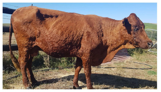
Figure 1. Bonsmara cow with lumpy skin disease—photograph provided by P. B. Kloppers.
2. Host-Range
LSDV is host restricted; its natural hosts are cattle, buffalo and water buffalo [11][12][13]. Antibodies have been detected in black and blue wildebeest, eland, giraffe, greater kudu, African buffalo and other animal species [14]. Giraffe and impala can be experimentally infected and subsequently die from LSD [15]. Recently, LSDV was isolated from nodular lesions of a giraffe which died in a zoo in Vietnam [16]. The authors suggest that it was due to natural infection of the virus, although how this occurred is unknown. The role of wild animals in the epidemiology of LSDV is still not certain.
The host range is slightly broader in cell culture of LSDV. A number of primary cell types derived from lamb and bovine tissue enable the growth of LSDV to high titres and are commonly used for vaccine stock preparations [17][18]. LSDV also grows well in chick chorioallantoic membranes (CAMs) of embryonated eggs, although purification of the virus may need to be extensive which can cause a loss in yields [19]. Various cell lines have been investigated as more convenient alternatives. High titres of LSDV can be obtained when grown in Madin-Darby bovine kidney (MDBK) cells [20]. Unfortunately, this cell line is often contaminated with bovine viral diarrhoea virus (BVDV), and therefore cannot be used for the preparation of vaccine stocks. Removal of BVDV from LSDV cultured in MDBKs can however be achieved after two passages in CAMs [21]. Another is the baby hamster kidney 21 (BHK-21) cell line which was first investigated for LSDV culture in the 1950s–1960s, although it is not frequently used for this purpose [22]. One laboratory very recently demonstrated that BHK-21 cells can be used for the construction of LSDV recombinants [23]. A recently developed embryonic skin of sheep (ESH-L) cell line was also shown to yield high titres of LSDV [18]. LSDV also replicates in Vero (African green monkey kidney) cells and the ovine testis (OA3.Ts) cell line [17][24].
3. Distribution of LSD
Last century, LSD was regarded as a disease endemic to Africa with the first report from Northern Rhodesia (now Zambia) in 1929. In 1989, LSD spread to Israel [25] and then further to other countries in the Middle East [26]. In 2015, LSDV was found in the Balkans and in 2016, in Serbia [27]. In the last decade, LSDV spread to countries in Europe, Bangladesh, China, India and Russia [28][29][30][31][32]. Outbreaks of the disease also occurred more recently in Indonesia and Pakistan last year [33]. Outbreaks in parts of Europe have been controlled [34], but it is likely that LSD will become endemic in most parts of Africa and Asia. The outbreak in the Balkans was controlled by vaccination with a live attenuated LSD vaccine based on the Neethling strain. This vaccine provided protection from infection within 14 days of vaccination [34]. However, there is always the concern of further spread of the disease. Vaccination is the best way to control the spread of LSD as subclinical infections are common and identification and removal of infected animals is not always possible [35].
4. Transmission
The main route of LSDV transmission is via a variety of arthropod vectors: ticks, mosquitoes and biting flies have all been implicated in transmission [36][37][38][39][40][41][42]. Climate change may impact on the spread of the arthropod vectors and as a result, the spread of LSDV [43]. To reduce the risk of insect transmission into areas with no LSDV, disinsectisation of vehicles transporting live animals is important [43].
LSDV is very stable in the environment, persisting for an extended time in necrotic lesions, desiccated crusts and airdried hides [44]. Direct contact between animals was initially deemed as inefficient for LSDV spread [45]. However, more recent work demonstrated the transmission of a virulent recombinant strain of LSDV (Saratov/2017) between infected bulls and in contact cattle in the absence of a vector [46]. There are also gaps in knowledge on how LSDV can persist in the environment, as there is a report in Russia where two almost identical isolates were identified with a two-year gap, with the implication that the virus circulated in cattle in the region or survived in fomites [47]. The presence of LSDV has been reported in semen after virulent challenge experiments [48] and insemination of heifers with infected semen can cause transmission of LSDV [49]. Of note, vaccination prevented LSDV being shed in semen after virulent challenge [48]. In natural outbreaks, LSDV can be transmitted to bovine foetuses and can result in abortion [50].
5. Current LSDV Vaccines Used in the Field
LSD is currently controlled using commercial vaccines which are mainly live attenuated LSDVs [51] which originated from the Neethling field strain of LSDV. This virus was attenuated by serial passage in primary lamb kidney cells and CAMs, carried out at the Onderstepoort Veterinary Research Institute (South Africa) in the 1950s–1960s [22][52]. This is known as the Neethling vaccine strain of LSDV (nLSDV). In South Africa, Neethling-like LSDV vaccine strains are marketed as Lumpyvax (MSD Animal Health), Herbivac LS (Deltamune) and Lumpy Skin Disease Vaccine (Onderstepoort Biological Products—OBP) and share 99.5% DNA sequence homology with each other [53]. nLSDV is also the most commonly used LSDV vaccine strain in Europe [54].
Due to the cross-reactivity between members of the Capripoxvirus genus, live attenuated GTPV and SPPV are also used to vaccinate cattle against LSDV [55]. GTPV-based vaccines have been reported to show the same protection against LSD as LSDV-based vaccines [56][57]. However, this is not the case with SPPV vaccines. In an experiment where groups of sheep, goats and cattle, vaccinated with Romania SPPV vaccine or nLSDV vaccine, were challenged with corresponding virulent strains, goats or sheep receiving the Romania SPPV vaccine were fully protected against challenge with virulent SPPV and GTPV strains, respectively. However, those cattle that were vaccinated with Romania SPPV vaccine showed only partial protection against LSDV challenge compared to full protection in cattle that received the nLSDV vaccine [58]. These results indicate that either LSDV or GTPV based vaccines could be used to protect from LSD in cattle.
6. Efficacy and Safety of Capripoxvirus Vaccines
A trial was conducted to test five LSDV-based live attenuated vaccines, namely: (1) Lumpy Skin Disease Vaccine [59] (Onderstepoort Biological Products OBP; South-Africa; batch 442); (2) Lumpyvax (MSD-Animal Health; South-Africa; batch BNDM07); (3) Kenyavac (Jordan Bioindustries Center Jovac; Jordan; batch 220115-04); (4) Herbivac LS (Deltamune; South-Africa); (5) Vaccin LSD Neethling O vivant (MCI Santé Animale; Morocco, batch 17BLSDN001). Animals were vaccinated and then challenged with virulent LSDV, 21 days after the last vaccination, and were all protected from challenge. They found that vaccination often resulted in a fever, which varied between the groups. Lumpyvax had a greater response whereas MCI- and Herbivac-vaccinated animals had less response. However, despite this, a Neethling response was seen in 43% of the animals vaccinated with Herbivac and 28% of the animals vaccinated with the MCI vaccine [51]. A Neethling response is the presence of superficial and smaller skin lesions which are distinct from those induced by a virulent field strain that disappear within 2–3 weeks without converting into necrotic scabs or ulcers [60]. Although some regard a Neethling response as negligible [60], these mild reactions have resulted in some hesitancy to use the live vaccines.
Adverse reactions in cattle vaccinated with a SPPV and an unverified LSDV vaccine were reported by farmers in Jordan; however, there is suspicion the LSDV vaccine was a smuggled virulent strain [61]. A randomised controlled field study on over 4000 cattle in Israel confirmed the efficacy of nLSDV vaccines compared to SPPV (x10RM65)-based vaccines. The relative risk of getting LSD in x10RM65 vs. nLSDV vaccinated animals was 2.635 (CI 95% = 1.44–4.82) and 11.2 (2.3–54.7) for severe morbidity. The relative risk of disease in x10RM65 vs. nLSDV vaccinated animals for laboratory confirmed cases was even higher at 4.28 (1.59–11.53), leading to the conclusion that nLSDV is significantly more effective at preventing LSD than x10RM65 [62].
References
- Kitching, R.; Smale, C. Comparison of the external dimensions of capripoxvirus isolates. Res. Vet. Sci. 1986, 41, 425–427.
- Tulman, E.R.; Afonso, C.L.; Lu, Z.; Zsak, L.; Sur, J.-H.; Sandybaev, N.T.; Kerembekova, U.Z.; Zaitsev, V.L.; Kutish, G.F.; Rock, D.L. The Genomes of Sheeppox and Goatpox Viruses. J. Virol. 2002, 76, 6054–6061.
- Tulman, E.R.; Afonso, C.L.; Lu, Z.; Zsak, L.; Kutish, G.F.; Rock, D.L. Genome of Lumpy Skin Disease Virus. J. Virol. 2001, 75, 7122–7130.
- McFadden, G. Poxvirus tropism. Nat. Rev. Genet. 2005, 3, 201–213.
- Prozesky, L.; Barnard, B.J. A study of the pathology of lumpy skin disease in cattle. Onderstepoort J. Veter.-Res. 1982, 49, 167–175.
- Aspden, K.; Passmore, J.-A.; Tiedt, F.; Williamson, A.-L. Evaluation of lumpy skin disease virus, a capripoxvirus, as a replication-deficient vaccine vector. J. Gen. Virol. 2003, 84, 1985–1996.
- Abutarbush, S.M.; Ababneh, M.M.; Al Zoubi, I.G.; Al Sheyab, O.M.; Alekish, M.O.; Al Gharabat, R.J. Lumpy Skin Disease in Jordan: Disease Emergence, Clinical Signs, Complications and Preliminary-associated Economic Losses. Transbound. Emerg. Dis. 2013, 62, 549–554.
- Tuppurainen, E.; Dietze, K.; Wolff, J.; Bergmann, H.; Beltran-Alcrudo, D.; Fahrion, A.; Lamien, C.E.; Busch, F.; Sauter-Louis, C.; Conraths, F.J.; et al. Review: Vaccines and Vaccination against Lumpy Skin Disease. Vaccines 2021, 9, 1136.
- Coetzer, J.A.W.; Tuppurainen, E. Lumpy skin disease. Infect. Dis. Livest. 2004, 2, 1268–1276.
- Möller, J.; Moritz, T.; Schlottau, K.; Krstevski, K.; Hoffmann, D.; Beer, M.; Hoffmann, B. Experimental lumpy skin disease virus infection of cattle: Comparison of a field strain and a vaccine strain. Arch. Virol. 2019, 164, 2931–2941.
- Ahmed, E.M.; Eltarabilli, M.M.; Shahein, M.A.; Fawzy, M. Lumpy skin disease outbreaks investigation in Egyptian cattle and buffaloes: Serological evidence and molecular characterization of genome termini. Comp. Immunol. Microbiol. Infect. Dis. 2021, 76, 101639.
- Fagbo, S.; Coetzer, J.A.; Venter, E.H. Seroprevalence of Rift Valley fever and lumpy skin disease in African buffalo (Syncerus caffer) in the Kruger National Park and Hluhluwe-iMfolozi Park, South Africa. J. S. Afr. Veter-Assoc. 2014, 85, 1075.
- De Kock, G. Lumpy skin disease (knopvelsiekte) of cattle in Southern Africa. J. Am. Vet. Med. Assoc. 1948, 112, 57.
- Hedger, R.; Hamblin, C. Neutralising antibodies to lumpy skin disease virus in African wildlife. Comp. Immunol. Microbiol. Infect. Dis. 1983, 6, 209–213.
- Young, E.; Basson, P.A.; Weiss, K.E. Experimental infection of game animals with lumpy skin disease virus (prototype strain Neethling). Onderstepoort J. Vet.-Res. 1970, 37, 79–87.
- Dao, T.D.; Tran, L.H.; Nguyen, H.D.; Hoang, T.T.; Nguyen, G.H.; Tran, K.V.D.; Nguyen, H.X.; Van Dong, H.; Bui, A.N.; Bui, V.N. Characterization of Lumpy skin disease virus isolated from a giraffe in Vietnam. Transbound. Emerg. Dis. 2022, 69, e3268–e3272.
- Rhazi, H.; Safini, N.; Mikou, K.; Alhyane, M.; Lenk, M.; Tadlaoui, K.O.; Elharrak, M. Comparative Sensitivity Study of Primary Cells, Vero, OA3.Tsand ESH-L cell lines to Lumpy Skin Disease, Sheeppox, and Goatpox viruses Detection and Growth. J. Virol. Methods 2021, 293, 114164.
- Binepal, Y.S.; Ongadi, F.A.; Chepkwony, J.C. Alternative cell lines for the propagation of lumpy skin disease virus. Onderstepoort J. Veter.-Res. 2001, 68, 151–153.
- Omar, R. Comparison of the Two Lumpy Skin Disease Virus Vaccines, Neethling and Herbivac, and Construction of a Recombinant Herbivac-Rift Valley Fever Virus Vaccine; University of Cape Town: Sydney, Australia, 2015.
- Fay, P.; Cook, C.; Wijesiriwardana, N.; Tore, G.; Comtet, L.; Carpentier, A.; Shih, B.; Freimanis, G.; Haga, I.R.; Beard, P.M. Madin-Darby bovine kidney (MDBK) cells are a suitable cell line for the propagation and study of the bovine poxvirus lumpy skin disease virus. J. Virol. Methods 2020, 285, 113943.
- Munyanduki, H.; Omar, R.; Douglass, N.; Williamson, A.-L. Removal of bovine viral diarrhea virus (BVDV) from lumpy skin disease virus (LSDV) vaccine stocks by passage on chorioallantoic membranes of fertilized hens’ eggs. J. Virol. Methods 2019, 275, 113752.
- Weiss, K.E. Lumpy Skin Disease Virus. In Cytomegaloviruses. Rinderpest Virus. Lumpy Skin Disease Virus; Springer: Berlin/Heidelberg, Germany, 1968; pp. 111–131.
- van Diepen, M.; Chapman, R.; Douglass, N.; Whittle, L.; Chineka, N.; Galant, S.; Cotchobos, C.; Suzuki, A.; Williamson, A.-L. Advancements in the Growth and Construction of Recombinant Lumpy Skin Disease Virus (LSDV) for Use as a Vaccine Vector. Vaccines 2021, 9, 1131.
- Babiuk, S.; Parkyn, G.; Copps, J.; Larence, J.E.; Sabara, M.I.; Bowden, T.R.; Boyle, D.B.; Kitching, R.P. Evaluation of an Ovine Testis Cell Line (OA3.Ts) for Propagation of Capripoxvirus Isolates and Development of an Immunostaining Technique for Viral Plaque Visualization. J. Vet. Diagn. Investig. 2007, 19, 486–491.
- Yeruham, I.; Nir, O.; Braverman, Y.; Davidson, M.; Grinstein, H.; Haymovitch, M.; Zamir, O. Spread of lumpy skin disease in Israeli dairy herds. Vet. Rec. 1995, 137, 91–93.
- Vandenbussche, F.; Mathijs, E.; Philips, W.; Saduakassova, M.; De Leeuw, I.; Sultanov, A.; Haegeman, A.; De Clercq, K. Recombinant LSDV Strains in Asia: Vaccine Spillover or Natural Emergence? Viruses 2022, 14, 1429.
- Manić, M.; Stojiljković, M.; Petrović, M.; Nišavić, J.; Bacić, D.; Petrović, T.; Vidanović, D.; Obrenović, S. Epizootic features and control measures for lumpy skin disease in south-east Serbia in 2016. Transbound. Emerg. Dis. 2019, 66, 2087–2099.
- Lu, G.; Xie, J.; Luo, J.; Shao, R.; Jia, K.; Li, S. Lumpy skin disease outbreaks in China, since 3 August 2019. Transbound. Emerg. Dis. 2020, 68, 216–219.
- Gupta, T.; Patial, V.; Bali, D.; Angaria, S.; Sharma, M.; Chahota, R. A review: Lumpy skin disease and its emergence in India. Vet. Res. Commun. 2020, 44, 111–118.
- Kayesh, M.E.H.; Hussan, M.T.; Hashem, A.; Eliyas, M.; Anower, A.M. Lumpy Skin Disease Virus Infection: An Emerging Threat to Cattle Health in Bangladesh. Hosts Viruses 2020, 7, 97–108.
- Sprygin, A.; Artyuchova, E.; Babin, Y.; Prutnikov, P.; Kostrova, E.; Byadovskaya, O.; Kononov, A. Epidemiological characterization of lumpy skin disease outbreaks in Russia in 2016. Transbound. Emerg. Dis. 2018, 65, 1514–1521.
- Agianniotaki, E.I.; Mathijs, E.; Vandenbussche, F.; Tasioudi, K.E.; Haegeman, A.; Iliadou, P.; Chaintoutis, S.C.; Dovas, C.I.; Van Borm, S.; Chondrokouki, E.D.; et al. Complete Genome Sequence of the Lumpy Skin Disease Virus Isolated from the First Reported Case in Greece in 2015. Genome Announc. 2017, 5, e00550-17.
- OIE-WAHIS. Events Management. Available online: https://wahis.woah.org/#/event-management (accessed on 1 December 2022).
- Klement, E.; Broglia, A.; Antoniou, S.E.; Tsiamadis, V.; Plevraki, E.; Petrovic, T.; Polacek, V.; Debeljak, Z.; Miteva, A.; Alexandrov, T.; et al. Neethling vaccine proved highly effective in controlling lumpy skin disease epidemics in the Balkans. Prev. Vet. Med. 2020, 181, 104595.
- Kononov, A.; Prutnikov, P.; Shumilova, I.; Kononova, S.; Nesterov, A.; Byadovskaya, O.; Pestova, Y.; Diev, V.; Sprygin, A. Determination of lumpy skin disease virus in bovine meat and offal products following experimental infection. Transbound. Emerg. Dis. 2019, 66, 1332–1340.
- Tuppurainen, E.S.M.; Stoltsz, W.H.; Troskie, M.; Wallace, D.B.; Oura, C.A.L.; Mellor, P.S.; Coetzer, J.A.W.; Venter, E. A Potential Role for Ixodid (Hard) Tick Vectors in the Transmission of Lumpy Skin Disease Virus in Cattle. Transbound. Emerg. Dis. 2011, 58, 93–104.
- Sohier, C.; Haegeman, A.; Mostin, L.; De Leeuw, I.; Van Campe, W.; De Vleeschauwer, A.; Tuppurainen, E.S.M.; van den Berg, T.; De Regge, N.; De Clercq, K. Experimental evidence of mechanical Lumpy Skin Disease virus transmission by Stomoxys calcitrans biting flies and Haematopota spp. horseflies. Sci. Rep. 2019, 9, 20076.
- Paslaru, A.I.; Maurer, L.M.; Vögtlin, A.; Hoffmann, B.; Torgerson, P.R.; Mathis, A.; Veronesi, E. Putative roles of mosquitoes (Culicidae) and biting midges (Culicoides spp.) as mechanical or biological vectors of lumpy skin disease virus. Med. Vet. Entomol. 2022, 36, 381–389.
- Wang, Y.; Zhao, L.; Yang, J.; Shi, M.; Nie, F.; Liu, S.; Wang, Z.; Huang, D.; Wu, H.; Li, D.; et al. Analysis of vaccine-like lumpy skin disease virus from flies near the western border of China. Transbound. Emerg. Dis. 2021, 69, 1813–1823.
- Paslaru, A.I.; Verhulst, N.O.; Maurer, L.M.; Brendle, A.; Pauli, N.; Vögtlin, A.; Renzullo, S.; Ruedin, Y.; Hoffmann, B.; Torgerson, P.R.; et al. Potential mechanical transmission of Lumpy skin disease virus (LSDV) by the stable fly (Stomoxys calcitrans) through regurgitation and defecation. Curr. Res. Insect Sci. 2020, 1, 100007.
- Lubinga, J.C.; Tuppurainen, E.S.M.; Mahlare, R.; Coetzer, J.A.W.; Stoltsz, W.H.; Venter, E.H. Evidence of Transstadial and Mechanical Transmission of Lumpy Skin Disease Virus by Amblyomma hebraeum Ticks. Transbound. Emerg. Dis. 2013, 62, 174–182.
- Kitching, R.P.; Hammond, J.M.; Black, D.N. Studies on the Major Common Precipitating Antigen of Capripoxvirus. J. Gen. Virol. 1986, 67, 139–148.
- Saegerman, C.; Bertagnoli, S.; Meyer, G.; Ganière, J.-P.; Caufour, P.; De Clercq, K.; Jacquiet, P.; Fournié, G.; Hautefeuille, C.; Etore, F.; et al. Risk of introduction of lumpy skin disease in France by the import of vectors in animal trucks. PLoS ONE 2018, 13, e0198506.
- Namazi, F.; Tafti, A.K. Lumpy skin disease, an emerging transboundary viral disease: A review. Vet. Med. Sci. 2021, 7, 888–896.
- Carn, V.M.; Kitching, R.P. An investigation of possible routes of transmission of lumpy skin disease virus (Neethling). Epidemiology Infect. 1995, 114, 219–226.
- Aleksandr, K.; Olga, B.; David, W.B.; Pavel, P.; Yana, P.; Svetlana, K.; Alexander, N.; Vladimir, R.; Dmitriy, L.; Alexander, S. Non-vector-borne transmission of lumpy skin disease virus. Sci. Rep. 2020, 10, 7436.
- Shumilova, I.; Krotova, A.; Nesterov, A.; Byadovskaya, O.; van Schalkwyk, A.; Sprygin, A. Overwintering of recombinant lumpy skin disease virus in northern latitudes, Russia. Transbound. Emerg. Dis. 2022, 69, e3239–e3243.
- Osuagwuh, U.I.; Bagla, V.H.; Venter, E.H.; Annandale, C.H.; Irons, P. Absence of lumpy skin disease virus in semen of vaccinated bulls following vaccination and subsequent experimental infection. Vaccine 2007, 25, 2238–2243.
- Annandale, C.H.; Holm, D.E.; Ebersohn, K.; Venter, E.H. Seminal Transmission of Lumpy Skin Disease Virus in Heifers. Transbound. Emerg. Dis. 2014, 61, 443–448.
- Şevik, M.; Doğan, M. Epidemiological and Molecular Studies on Lumpy Skin Disease Outbreaks in Turkey during 2014-2015. Transbound. Emerg. Dis. 2017, 64, 1268–1279.
- Haegeman, A.; De Leeuw, I.; Mostin, L.; Van Campe, W.; Aerts, L.; Venter, E.; Tuppurainen, E.; Saegerman, C.; De Clercq, K. Comparative Evaluation of Lumpy Skin Disease Virus-Based Live Attenuated Vaccines. Vaccines 2021, 9, 473.
- Van Rooyen, P.J.; Munz, E.K.; Weiss, K.E. The optimal conditions for the multiplication of Neethling-type lumpy skin disease virus in embryonated eggs. Onderstepoort J. Vet. Res. 1969, 36, 165–174.
- Mathijs, E.; Vandenbussche, F.; Haegeman, A.; King, A.; Nthangeni, B.; Potgieter, C.; Maartens, L.; Van Borm, S.; De Clercq, K. Complete Genome Sequences of the Neethling-Like Lumpy Skin Disease Virus Strains Obtained Directly from Three Commercial Live Attenuated Vaccines. Genome Announc. 2016, 4, e01255-16.
- Calistri, P.; De Clercq, K.; Gubbins, S.; Klement, E.; Stegeman, A.; Abrahantes, J.C.; Marojevic, D.; Antoniou, S.; Broglia, A. Lumpy skin disease epidemiological report IV: Data collection and analysis. EFSA J. 2020, 18, e06010.
- Norian, R.; Ahangran, N.A.; Varshovi, H.R.; Azadmehr, A. Comparative efficacy of two heterologous capripox vaccines to control lumpy skin disease in cattle. Bulg. J. Vet. Med. 2019, 22, 171–179.
- Gari, G.; Abie, G.; Gizaw, D.; Wubete, A.; Kidane, M.; Asgedom, H.; Bayissa, B.; Ayelet, G.; Oura, C.A.; Roger, F.; et al. Evaluation of the safety, immunogenicity and efficacy of three capripoxvirus vaccine strains against lumpy skin disease virus. Vaccine 2015, 33, 3256–3261.
- Zhugunissov, K.; Bulatov, Y.; Orynbayev, M.; Kutumbetov, L.; Abduraimov, Y.; Shayakhmetov, Y.; Taranov, D.; Amanova, Z.; Mambetaliyev, M.; Absatova, Z.; et al. Goatpox virus (G20-LKV) vaccine strain elicits a protective response in cattle against lumpy skin disease at challenge with lumpy skin disease virulent field strain in a comparative study. Vet Microbiol. 2020, 245, 108695.
- Hamdi, J.; Bamouh, Z.; Jazouli, M.; Boumart, Z.; Tadlaoui, K.O.; Fihri, O.F.; EL Harrak, M. Experimental evaluation of the cross-protection between Sheeppox and bovine Lumpy skin vaccines. Sci. Rep. 2020, 10, 8888.
- Manual, O. Chapter 3.4.12 Lumpy Skin Disease. Available online: https://www.woah.org/fileadmin/Home/fr/Health_standards/tahm/3.04.12_LSD.pdf (accessed on 10 January 2023).
- Morgenstern, M.; Klement, E. The Effect of Vaccination with Live Attenuated Neethling Lumpy Skin Disease Vaccine on Milk Production and Mortality—An Analysis of 77 Dairy Farms in Israel. Vaccines 2020, 8, 324.
- Abutarbush, S.; Hananeh, W.M.; Ramadan, W.; Al Sheyab, O.M.; Alnajjar, A.R.; Al Zoubi, I.G.; Knowles, N.J.; Tuppurainen, E.S.M.; Bachanek-Bankowska, K. Adverse Reactions to Field Vaccination Against Lumpy Skin Disease in Jordan. Transbound. Emerg. Dis. 2014, 63, e213–e219.
- Ben-Gera, J.; Klement, E.; Khinich, E.; Stram, Y.; Shpigel, N.Y. Comparison of the efficacy of Neethling lumpy skin disease virus and x10RM65 sheep-pox live attenuated vaccines for the prevention of lumpy skin disease—The results of a randomized controlled field study. Vaccine 2015, 33, 4837–4842.
- Tuppurainen, E.; Babiuk, S.; Klement, E. Lumpy Skin Disease; Springer: Berlin/Heidelberg, Germany, 2018.
More
Information
Subjects:
Pathology
Contributors
MDPI registered users' name will be linked to their SciProfiles pages. To register with us, please refer to https://encyclopedia.pub/register
:
View Times:
1.2K
Revisions:
3 times
(View History)
Update Date:
24 Mar 2023
Table of Contents


Notice
You are not a member of the advisory board for this topic. If you want to update advisory board member profile, please contact office@encyclopedia.pub.
OK
Confirm
Only members of the Encyclopedia advisory board for this topic are allowed to note entries. Would you like to become an advisory board member of the Encyclopedia?
Yes
No
${ textCharacter }/${ maxCharacter }
Submit
Cancel
Back
Comments
${ item }
|
${ item.createdUser.fullName }
${ item.createdAt }
${ item.vote }
${ item.reply }
Delete
${ reply.createdUser.fullName }
${ reply.createdAt }
${ reply.vote }
Delete
There is no reply to this comment~
${ item.replyTextCharacter }/${ item.replyMaxCharacter }
Submit
Cancel
More
No more~
There is no comment~
${ textCharacter }/${ maxCharacter }
Submit
Cancel
${ selectedItem.replyTextCharacter }/${ selectedItem.replyMaxCharacter }
Submit
Cancel
Confirm
Are you sure to Delete?
Yes
No