Submitted Successfully!
To reward your contribution, here is a gift for you: A free trial for our video production service.
Thank you for your contribution! You can also upload a video entry or images related to this topic.
Version Summary Created by Modification Content Size Created at Operation
1 -- 1939 2023-02-22 21:15:20 |
2 format correct Meta information modification 1939 2023-02-23 01:57:44 |

Video Upload Options

Do you have a full video?

Confirm

Are you sure to Delete?
Cite
If you have any further questions, please contact Encyclopedia Editorial Office.
Ser, H.; Au Yong, S.; Shafiee, M.N.; Mohd Mokhtar, N.; Raja Ali, R.A. Microbiome and Endometriosis. Encyclopedia. Available online: https://encyclopedia.pub/entry/41560 (accessed on 27 July 2024).
Ser H, Au Yong S, Shafiee MN, Mohd Mokhtar N, Raja Ali RA. Microbiome and Endometriosis. Encyclopedia. Available at: https://encyclopedia.pub/entry/41560. Accessed July 27, 2024.
Ser, Hooi-Leng, Siu-Jung Au Yong, Mohamad Nasir Shafiee, Norfilza Mohd Mokhtar, Raja Affendi Raja Ali. "Microbiome and Endometriosis" Encyclopedia, https://encyclopedia.pub/entry/41560 (accessed July 27, 2024).
Ser, H., Au Yong, S., Shafiee, M.N., Mohd Mokhtar, N., & Raja Ali, R.A. (2023, February 22). Microbiome and Endometriosis. In Encyclopedia. https://encyclopedia.pub/entry/41560
Ser, Hooi-Leng, et al. "Microbiome and Endometriosis." Encyclopedia. Web. 22 February, 2023.
Microbiome and Endometriosis
Edit

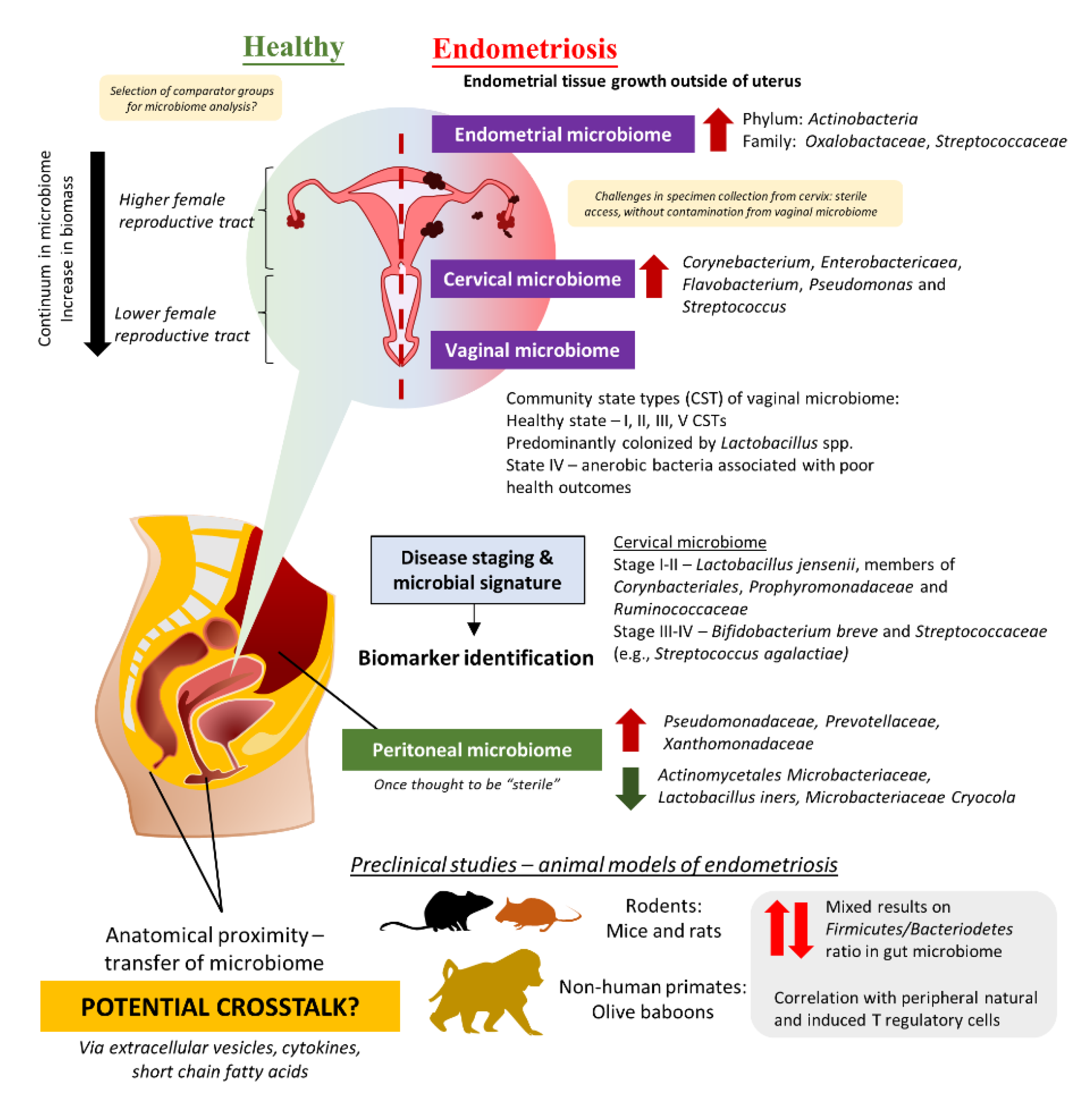
Endometriosis affects approximately 6 to 10% of reproductive-age women globally. Despite much effort invested, the pathogenesis that promotes the development, as well as the progression of this chronic inflammatory disease, is poorly understood. The imbalance in the microbiome or dysbiosis has been implicated in a variety of human diseases, especially the gut microbiome. In the case of endometriosis, emerging evidence suggests that there may be urogenital-gastrointestinal crosstalk that leads to the development of endometriosis. Along with these findings, several studies have reported the potential of probiotics in managing endometriosis, however subsequent investigations on microbial dynamics post administration of probiotics as well as route of administration and formulation of probiotics would be needed to strengthen the rationale of using such microbiome-based intervention in the management of endometriosis.  

endometriosis vaginal microbiome microbiome-based therapeutics gut microbiome

1. The Intricate Relationship between the Female Reproductive Tract Microbiome and Gut Microbiome in the Development and Progression of Endometriosis

The term “microbiome” refers to a collective group of microorganisms that live in a habitat or specific site. Therefore, it is understandable that the microbiome across the human body may demonstrate differences in the microbial population [1]. The classic example would be the gastrointestinal (GI) system (which harbors the largest microbes in the human body)—pH and oxygen availability changes throughout the GI tract which then poses selective pressure on microbes [2][3]. The gastric microbiome of a healthy human adult would consist of a microbial population that could tolerate the low pH in the environment, which include those belonging to the Prevotella, Streptococcus, Veillonella, Rothia, and Haemophilus genus [3][4]. In contrast, the colon microbiome of a healthy human adult is predominantly colonized by members of bacteria belonging to the genera of Lactobacillus, Akkermansia, Enterobacter, Lachnospiraceae, Prevotella, and several more [5]. It is important to note that while microbiome structure may vary between individuals, microbial functions are pretty much conserved which allows researchers to exploit them as disease biomarkers or even therapeutic targets as part of the microbiome-therapeutics strategy. The primary function of the human gastrointestinal system was thought to support the host’s metabolism, including digesting, and absorbing ingested nutrients, and excreting waste products of digestion. However, a growing body of evidence is accentuating the role of the gastrointestinal system as an organ of immunity, particularly in maintaining immune system homeostasis [6]. The gastrointestinal-associated lymphoid tissue acts as the “control center” to manage the immune system in response to massive antigen exposure in the gut and activate adaptive immune responses such as B cell maturation [7].
The role of the gut microbiome in the etiology and pathogenesis of the human disease remains one of the top research areas for the past few decades [8][9]. Nevertheless, emerging evidence highlighted the potential of crosstalk between microbiomes of different sites in several human diseases, including the urogenital and gut microbiome given their anatomical proximity [10][11][12]. Comparable to the continuum observed in the gut microbiome, different parts of the female reproductive tract (FRT) display different distributions of microbes [13][14]. In fact, FRT is categorized into a higher part which consists of the endocervix and uterus proper, and a lower part which comprises the vaginal canal and ectocervix (Figure 1). The lower FRT is dominated by Lactobacillus spp. and these microbes protect the host against pathogens by creating a low pH environment and production of bacteriocins as well as hydrogen peroxide. The vaginal community state type (CST) classification system describes a total of five CSTs, whereby CST I, II, III, and V are dominated by Lactobacillus crispatus, Lactobacillus gasseri, Lactobacillus iners, and Lactobacillus jensenii, respectively [15][16]. These four CSTs are associated with a healthy vaginal microbiome, whereas CST IV, which presents higher proportions of strictly anaerobic bacteria (e.g., Prevotella, Dialister, Atopobium, Gardnerella, Megasphaera, Peptoniphilus, Sneathia, Eggerthella, Aerococcus, Finegoldia, and Mobiluncus), is suggested to be linked with inflammation or dysbiosis in the vagina. Additionally, it is important to note that vaginal CSTs can change throughout women’s lifetimes. Given that some microbes can stimulate the immune system to trigger inflammation while a portion of them helps to maintain homeostasis in the host by the production of antimicrobial compounds or even immunomodulatory compounds, researchers are now exploring the possibility of microbiome involvement in the development and progression of endometriosis.
Figure 1. The potential role of the microbiome in endometriosis.

2. Evidence from Clinical Studies: Are There Any Distinct Microbiome Changes in the Vaginal Microbiome?

Majority of the clinical studies investigating endometriosis and microbiome changes were derived from Asia including Japan, China, Taiwan, Korea, and Turkey [17][18][19][20][21][22][23][24][25][26][27][28]. There were also some studies from the United States, Brazil, Sweden, and Canada [29][30][31][32][33]. Almost all the clinical trials conducted diagnosed endometriosis cases via laparoscopy or histology tests and scored based on the criteria described in the Revised American Society for Reproductive Medicine (r-ASRM) classification of endometriosis. The study by Akiyama et al. in 2019 reflected that even though Lactobacillus spp. dominated the cervical microbiome of endometriosis patients, they still presented a higher abundance of Corynebacterium, Enterobactericaea, Flavobacterium, Pseudomonas, and Streptococcus as compared to control (without endometriosis) [17]. Subsequent quantification of bacteria using real-time PCR confirms the finding from next-generation sequencing, in which Enterobacteriaceae and Streptococcus abundance were statistically different between endometriosis and non-endometriosis control (p > 0.05). Similarly, another team in Taiwan described that not only the cervical microbiome of endometriosis patients was different from healthy women, but there were also some differences between endometriosis patients in Stage I and II as compared to those in Stage III and IV [25]. The team has suggested that potential microbial biomarkers for different stages: (a) Stage I–II: L. jensenii or members in Corynbacteriales, Porphyromonadaceae, and Ruminococcaceae, (b) Stage III–IV: Bifidobacterium breve and Streptococcaceae members (e.g., Streptococcus agalactiae).

Given the challenges in obtaining cervical specimens without cervicovaginal contamination and the nature of biomass in the upper FRT, several teams have attempted to study the differences in the microbiome of the lower FRT. For instance, three studies in Brazil and China studied the vaginal swab or fluid obtained from patients and observed a lower abundance of Lactobacillus in the endometriosis group as compared to the control [24][28][29]. Besides that, the study by Ata et al. discussed the differences in vaginal samples obtained from Stage III or IV endometriosis patients as compared to healthy women [18]. At the genus level, Gemella and Atopobium spp. was absent in the vaginal samples obtained from the endometriosis group. A similar approach was taken by Perrotta et al., but the team took a broader approach to look at the vaginal CST rather than looking at just a specific group of microbes [30]. These data then allowed the team to build a random forest-based classification model with machine-learning methods on microbiota composition to predict r-ASRM stages of endometriosis. Analyzing the changes during follicular and menstrual phases yielded highly predictive taxa which can be used to predict either stage I-II or stage III-IV endometriosis—the genus Anaerococcus (phylum Firmicutes).

3. Connections between Gut Microbiome, Peritoneal Microbiome, and Endometriosis

On the contrary, Chen et al. were unable to identify microbial signatures from FRT microbiome for use as a biomarker but a prediction of metagenome functions using bioinformatic tools indicated a higher proportion of microbes involved in general metabolism, lipid metabolism as well as synthesis and degradation of ketone bodies [19]. Furthermore, Huang et al. conducted a study in China, that recruited patients from June 2019–October 2019, and reported that there are no significant differences in cervical microbiome observed between women without endometriosis and endometriosis patients [22]. In spite of this, the team uncovered differences in fecal microbiome composition between women with endometriosis and those without. Analysis of the fecal microbiome showed depletion of ten taxa including Clostridia Clostridiales, Lachnospiraceae Ruminococcus, Clostridiales Lachnospiraceae, and Ruminococcaceae Ruminococcus, along with an increased abundance of Eggerthella lenta and Eubacterium dolicum in endometriosis patients as compared to women without endometriosis. Likewise, another report by Shan et al. increased Firmicutes/Bacteriodetes ratio in endometriosis group, with enrichment of Actinobacteria, Cynaobacteria, Saccharibacteria, Fusobacteria, and Acidobacteria (p < 0.05) as compared to the control [23]. In essence, these results imply the involvement of the gut microbiome in the development of endometriosis.
Apart from the FRT and gut microbiome, there is another special microbiome that has been investigated to understand the development of the progression of endometriosis—the peritoneal microbiome. Once thought to be “sterile”, the peritoneal microbiome is found to be associated with the etiology of diseases such as end-stage kidney disease and cancer [34][35][36]. As such, it is certainly logical to investigate the changes in the peritoneal microbiome among endometriosis patients, given that the lesion may involve the peritoneum [20][26][37]. In 2022, Yuan et al. reported that there was a total of 276 operational taxonomic units (OTUs) detected in peritoneal fluid collected from endometriosis patients (as compared to 211 OTUs in the control group); out of which, 120 of them were unique to endometriosis group [27]. At the genus level, there was a significantly higher abundance of Acidovorax (p = 0.01), Devosia (p = 0.03), Methylobacterium (p = 0.03), Phascolarctobacterium (p = 0.03), and Streptococcoccus (p = 0.04) in endometriosis group than the control group. In order to investigate potential crosstalk between different microbiomes, a research group in Korea decided to study the extracellular vesicles in the peritoneal fluid of endometriosis patients in comparison to those without endometriosis [21]. The team successfully characterized microbes present in the extracellular vesicles and reported that there was a significant decrease in Actinobacteria at the phylum level among endometriosis patients.

4. Potential Benefits of Probiotics in the Management of Endometriosis

While pharmacotherapy remains critical in the symptomatic management of endometriosis patients, microbiome-based therapeutics may potentially be used in the nearest future to restore the balance in microbiomes and to alleviate chronic inflammation that is commonly observed among endometriosis patients [28][38][39][40]. For instance, L. gasseri OLL2809 was found to be able to inhibit the development of ectopic endometrial cells in the peritoneal cavity via activation of natural killer cells in a rodent model of endometriosis, which most likely occurred via the induction of interleukin-12 production [39]. In the subsequent placebo-controlled study, Itoh et al. also found that taking L. gasseri OLL2809 tablets for three months significantly reduces pain intensity on the visual analog scale (VAS) and dysmenorrhea on the verbal rating scales (VRS) in the active group, recording at −3.28 ± 0.36 and −1.44 ± 0.17, respectively, as compared to the −2.00 ± 0.29 and −1.03 ± 0.16 recorded in the placebo group [41]. Correspondingly, a different study conducted in Japan supported the use of L. gasseri OLL2809 in the treatment of the condition. In this study, endometriosis volume was significantly different between the active group of rats and the control and dienogest-treated groups, with a significant difference in log value of p < 0.05 recorded after four weeks of treatment [42]. Another pilot placebo-controlled randomized clinical trial in Iran evaluated the effects of a multi-strain probiotic capsule known as LactoFem® capsule (containing 109 colony of Lactobacillus acidophilus, Lactobacillus plantarum, Lactobacillus fermentum, and L. gasseri) among Stage III-IV endometriosis patients. The team noted a significant drop in dysmenorrhea scores after 8 weeks of treatment in the probiotic group—from 6.53 ± 2.88 to 3.07 ± 2.49 as compared to 5.60 ± 2.06 to 4.47 ± 2.13 (p = 0.018). Then again, more studies should be conducted to evaluate the actual changes in different microbiomes post-administration of probiotics to monitor the microbial dynamics throughout the period as well as test different administration routes to ensure optimal results from microbiome-based therapeutics. Given that the methods of probiotics preparation differ between administration routes, considerations should also be given to its stability to ensure optimal delivery to the targeted site [43][44]. Along with that, the actions of probiotics on microbiome stability are equally important to counter dysbiosis and subsequently provide beneficial effects in a long-term manner. To date, there is still a lack of guidelines outlining or supporting the standard use of probiotics in the management of endometriosis. Thus, additional investigations into these aspects would enhance the understanding of disease etiology as well as strengthen the rationale for using microbiome-based therapeutics in endometriosis management.

References

  1. Blum, H.E. The human microbiome. Adv. Med. Sci. 2017, 62, 414–420.
  2. Martinez-Guryn, K.; Leone, V.; Chang, E.B. Regional Diversity of the Gastrointestinal Microbiome. Cell Host Microbe 2019, 26, 314–324.
  3. Lopetuso, L.R.; Scaldaferri, F.; Franceschi, F.; Gasbarrini, A. The gastrointestinal microbiome—Functional interference between stomach and intestine. Best Pr. Res. Clin. Gastroenterol. 2014, 28, 995–1002.
  4. Noto, J.M.; Peek, R.M., Jr. The gastric microbiome, its interaction with Helicobacter pylori, and its potential role in the progression to stomach cancer. PLoS Pathog. 2017, 13, e1006573.
  5. Ruan, W.; Engevik, M.A.; Spinler, J.K.; Versalovic, J. Healthy Human Gastrointestinal Microbiome: Composition and Function After a Decade of Exploration. Dig. Dis. Sci. 2020, 65, 695–705.
  6. Mason, K.L.; Huffnagle, G.B.; Noverr, M.C.; Kao, J.Y. Overview of gut immunology. Adv. Exp. Med. Biol. 2008, 635, 1–14.
  7. Morbe, U.M.; Jorgensen, P.B.; Fenton, T.M.; von Burg, N.; Riis, L.B.; Spencer, J.; Agace, W.W. Human gut-associated lymphoid tissues (GALT); diversity, structure, and function. Mucosal Immunol. 2021, 14, 793–802.
  8. Li, S.; Chen, S.; Nie, M.; Wen, L.; Zou, B.; Zhang, L.; Xie, J.; Ser, H.L.; Lee, L.H.; Wang, S.; et al. Salt-Sensitive Ileal Microbiota Plays a Role in Atrial Natriuretic Peptide Deficiency-Induced Cardiac Injury. Nutrients 2022, 14, 3129.
  9. Ser, H.-L.; Wong, J.Y.-J.; Goh, B.-H.; Reginald, K. IDDF2022-ABS-0236 Healing the GUT with probiotics: Can probiotics help relieve allergic rhinitis? Gut 2022, 71, A63–A64.
  10. Joseph, R.J.; Ser, H.L.; Kuai, Y.H.; Tan, L.T.; Arasoo, V.J.T.; Letchumanan, V.; Wang, L.; Pusparajah, P.; Goh, B.H.; Ab Mutalib, N.S.; et al. Finding a Balance in the Vaginal Microbiome: How Do We Treat and Prevent the Occurrence of Bacterial Vaginosis? Antibiotics 2021, 10, 719.
  11. Mestrovic, T.; Matijasic, M.; Peric, M.; Cipcic Paljetak, H.; Baresic, A.; Verbanac, D. The Role of Gut, Vaginal, and Urinary Microbiome in Urinary Tract Infections: From Bench to Bedside. Diagnostics 2020, 11, 7.
  12. Ser, H.-L.; Wong, J.Y.J.; Letchumanan, V.; Law, J.W.-F.; Tan, L.T.-H.; Lee, L.-H. IDDF2021-ABS-0132 Moving beyond the gastrointestinal tract: The involvement of gut microbiome in endometriosis. Gut 2021, 70, A46–A47.
  13. Plesniarski, A.; Siddik, A.B.; Su, R.C. The Microbiome as a Key Regulator of Female Genital Tract Barrier Function. Front. Cell. Infect. Microbiol. 2021, 11, 790627.
  14. Punzon-Jimenez, P.; Labarta, E. The impact of the female genital tract microbiome in women health and reproduction: A review. J. Assist. Reprod. Genet. 2021, 38, 2519–2541.
  15. Gajer, P.; Brotman, R.M.; Bai, G.; Sakamoto, J.; Schutte, U.M.; Zhong, X.; Koenig, S.S.; Fu, L.; Ma, Z.S.; Zhou, X.; et al. Temporal dynamics of the human vaginal microbiota. Sci. Transl. Med. 2012, 4, 132ra152.
  16. Ravel, J.; Gajer, P.; Abdo, Z.; Schneider, G.M.; Koenig, S.S.; McCulle, S.L.; Karlebach, S.; Gorle, R.; Russell, J.; Tacket, C.O.; et al. Vaginal microbiome of reproductive-age women. Proc. Natl. Acad. Sci. USA 2011, 108, 4680–4687.
  17. Akiyama, K.; Nishioka, K.; Khan, K.N.; Tanaka, Y.; Mori, T.; Nakaya, T.; Kitawaki, J. Molecular detection of microbial colonization in cervical mucus of women with and without endometriosis. Am. J. Reprod. Immunol. 2019, 82, e13147.
  18. Ata, B.; Yildiz, S.; Turkgeldi, E.; Brocal, V.P.; Dinleyici, E.C.; Moya, A.; Urman, B. The endobiota study: Comparison of vaginal, cervical and gut microbiota between women with stage 3/4 endometriosis and healthy controls. Sci. Rep. 2019, 9, 1–9.
  19. Chen, S.; Gu, Z.; Zhang, W.; Jia, S.; Wu, Y.; Zheng, P.; Dai, Y.; Leng, J. Microbiome of the lower genital tract in Chinese women with endometriosis by 16s-rRNA sequencing technique: A pilot study. Ann. Transl. Med. 2020, 8, 1440.
  20. Wei, W.; Zhang, X.; Tang, H.; Zeng, L.; Wu, R. Microbiota composition and distribution along the female reproductive tract of women with endometriosis. Ann. Clin. Microbiol. Antimicrob. 2020, 19, 15.
  21. Lee, S.R.; Lee, J.C.; Kim, S.H.; Oh, Y.S.; Chae, H.D.; Seo, H.; Kang, C.S.; Shin, T.S. Altered Composition of Microbiota in Women with Ovarian Endometrioma: Microbiome Analyses of Extracellular Vesicles in the Peritoneal Fluid. Int. J. Mol. Sci. 2021, 22, 4608.
  22. Huang, L.; Liu, B.; Liu, Z.; Feng, W.; Liu, M.; Wang, Y.; Peng, D.; Fu, X.; Zhu, H.; Cui, Z.; et al. Gut Microbiota Exceeds Cervical Microbiota for Early Diagnosis of Endometriosis. Front. Cell. Infect. Microbiol. 2021, 11, 788836.
  23. Shan, J.; Ni, Z.; Cheng, W.; Zhou, L.; Zhai, D.; Sun, S.; Yu, C. Gut microbiota imbalance and its correlations with hormone and inflammatory factors in patients with stage 3/4 endometriosis. Arch. Gynecol. Obs. 2021, 304, 1363–1373.
  24. Chao, X.; Liu, Y.; Fan, Q.; Shi, H.; Wang, S.; Lang, J. The role of the vaginal microbiome in distinguishing female chronic pelvic pain caused by endometriosis/adenomyosis. Ann. Transl. Med. 2021, 9, 771.
  25. Chang, C.Y.; Chiang, A.J.; Lai, M.T.; Yan, M.J.; Tseng, C.C.; Lo, L.C.; Wan, L.; Li, C.J.; Tsui, K.H.; Chen, C.M.; et al. A more diverse cervical microbiome associates with better clinical outcomes in patients with endometriosis: A pilot study. Biomedicines 2022, 10, 174.
  26. Oishi, S.; Mekaru, K.; Tanaka, S.E.; Arai, W.; Ashikawa, K.; Sakuraba, Y.; Nishioka, M.; Nakamura, R.; Miyagi, M.; Akamine, K. Microbiome analysis in women with endometriosis: Does a microbiome exist in peritoneal fluid and ovarian cystic fluid? Reprod. Med. Biol. 2022, 21, e12441.
  27. Yuan, W.; Wu, Y.; Chai, X.; Wu, X. The colonized microbiota composition in the peritoneal fluid in women with endometriosis. Arch. Gynecol. Obs. 2022, 305, 1573–1580.
  28. Lu, F.; Wei, J.; Zhong, Y.; Feng, Y.; Ma, B.; Xiong, Y.; Wei, K.; Tan, B.; Chen, T. Antibiotic Therapy and Vaginal Microbiota Transplantation Reduce Endometriosis Disease Progression in Female Mice via NF-kappaB Signaling Pathway. Front. Med. 2022, 9, 831115.
  29. Hernandes, C.; Silveira, P.; Rodrigues Sereia, A.F.; Christoff, A.P.; Mendes, H.; Valter de Oliveira, L.F.; Podgaec, S. Microbiome Profile of Deep Endometriosis Patients: Comparison of Vaginal Fluid, Endometrium and Lesion. Diagnostics 2020, 10, 163.
  30. Perrotta, A.R.; Borrelli, G.M.; Martins, C.O.; Kallas, E.G.; Sanabani, S.S.; Griffith, L.G.; Alm, E.J.; Abrao, M.S. The vaginal microbiome as a tool to predict rASRM stage of disease in endometriosis: A pilot study. Reprod. Sci. 2020, 27, 1064–1073.
  31. Le, N.; Cregger, M.; Fazleabas, A.; Braundmeier-Fleming, A. Effects of endometriosis on immunity and mucosal microbial community dynamics in female olive baboons. Sci. Rep. 2022, 12, 1590.
  32. Svensson, A.; Brunkwall, L.; Roth, B.; Orho-Melander, M.; Ohlsson, B. Associations between endometriosis and gut microbiota. Reprod. Sci. 2021, 28, 2367–2377.
  33. Wessels, J.M.; Domínguez, M.A.; Leyland, N.A.; Agarwal, S.K.; Foster, W.G. Endometrial microbiota is more diverse in people with endometriosis than symptomatic controls. Sci. Rep. 2021, 11, 1–12.
  34. Simoes-Silva, L.; Araujo, R.; Pestana, M.; Soares-Silva, I.; Sampaio-Maia, B. Peritoneal Microbiome in End-Stage Renal Disease Patients and the Impact of Peritoneal Dialysis Therapy. Microorganisms 2020, 8, 173.
  35. Gilbreath, J.J.; Semino-Mora, C.; Friedline, C.J.; Liu, H.; Bodi, K.L.; McAvoy, T.J.; Francis, J.; Nieroda, C.; Sardi, A.; Dubois, A.; et al. A core microbiome associated with the peritoneal tumors of pseudomyxoma peritonei. Orphanet J. Rare Dis. 2013, 8, 105.
  36. Semino-Mora, C.; Liu, H.; McAvoy, T.; Nieroda, C.; Studeman, K.; Sardi, A.; Dubois, A. Pseudomyxoma peritonei: Is disease progression related to microbial agents? A study of bacteria, MUC2 AND MUC5AC expression in disseminated peritoneal adenomucinosis and peritoneal mucinous carcinomatosis. Ann. Surg. Oncol. 2008, 15, 1414–1423.
  37. Ferrero, S.; Vellone, V.G.; Barra, F. Pathophysiology of pain in patients with peritoneal endometriosis. Ann. Transl. Med. 2019, 7, S8.
  38. Khodaverdi, S.; Mohammadbeigi, R.; Khaledi, M.; Mesdaghinia, L.; Sharifzadeh, F.; Nasiripour, S.; Gorginzadeh, M. Beneficial Effects of Oral Lactobacillus on Pain Severity in Women Suffering from Endometriosis: A Pilot Placebo-Controlled Randomized Clinical Trial. Int. J. Fertil. Steril. 2019, 13, 178–183.
  39. Itoh, H.; Sashihara, T.; Hosono, A.; Kaminogawa, S.; Uchida, M. Lactobacillus gasseri OLL2809 inhibits development of ectopic endometrial cell in peritoneal cavity via activation of NK cells in a murine endometriosis model. Cytotechnology 2011, 63, 205–210.
  40. Ibrahim, A.; Ali, R.A.R.; Manaf, M.R.A.; Ahmad, N.; Tajurruddin, F.W.; Qin, W.Z.; Desa, S.H.M.; Ibrahim, N.M. Multi-strain probiotics (Hexbio) containing MCP BCMC strains improved constipation and gut motility in Parkinson’s disease: A randomised controlled trial. PLoS ONE 2020, 15, e0244680.
  41. Itoh, H.; Uchida, M.; Sashihara, T.; Ji, Z.S.; Li, J.; Tang, Q.; Ni, S.; Song, L.; Kaminogawa, S. Lactobacillus gasseri OLL2809 is effective especially on the menstrual pain and dysmenorrhea in endometriosis patients: Randomized, double-blind, placebo-controlled study. Cytotechnology 2011, 63, 153–161.
  42. Uchida, M.; Kobayashi, O. Effects of Lactobacillus gasseri OLL2809 on the induced endometriosis in rats. Biosci. Biotechnol. Biochem. 2013, 77, 1879–1881.
  43. Del Piano, M.; Carmagnola, S.; Ballare, M.; Sartori, M.; Orsello, M.; Balzarini, M.; Pagliarulo, M.; Tari, R.; Anderloni, A.; Strozzi, G.P.; et al. Is microencapsulation the future of probiotic preparations? The increased efficacy of gastro-protected probiotics. Gut Microbes 2011, 2, 120–123.
  44. Sanders, M.E.; Klaenhammer, T.R.; Ouwehand, A.C.; Pot, B.; Johansen, E.; Heimbach, J.T.; Marco, M.L.; Tennila, J.; Ross, R.P.; Franz, C.; et al. Effects of genetic, processing, or product formulation changes on efficacy and safety of probiotics. Ann. New York Acad. Sci. 2014, 1309, 1–18.
More
Information
Contributors MDPI registered users' name will be linked to their SciProfiles pages. To register with us, please refer to https://encyclopedia.pub/register : , , , ,
View Times: 452
Revisions: 2 times (View History)
Update Date: 23 Feb 2023
1000/1000
Video Production Service