Submitted Successfully!
To reward your contribution, here is a gift for you: A free trial for our video production service.
Thank you for your contribution! You can also upload a video entry or images related to this topic.
Version Summary Created by Modification Content Size Created at Operation
1 -- 2240 2023-02-22 16:48:18 |
2 update references and layout -1 word(s) 2239 2023-02-23 02:30:39 |

Video Upload Options

Do you have a full video?

Confirm

Are you sure to Delete?
Cite
If you have any further questions, please contact Encyclopedia Editorial Office.
Chan, J.W.Y.; Siu, I.C.H.; Chang, A.T.C.; Li, M.S.C.; Lau, R.W.H.; Mok, T.S.K.; Ng, C.S.H. Transbronchial Techniques for Lung Cancer Treatment. Encyclopedia. Available online: https://encyclopedia.pub/entry/41553 (accessed on 07 July 2024).
Chan JWY, Siu ICH, Chang ATC, Li MSC, Lau RWH, Mok TSK, et al. Transbronchial Techniques for Lung Cancer Treatment. Encyclopedia. Available at: https://encyclopedia.pub/entry/41553. Accessed July 07, 2024.
Chan, Joyce W. Y., Ivan C. H. Siu, Aliss T. C. Chang, Molly S. C. Li, Rainbow W. H. Lau, Tony S. K. Mok, Calvin S. H. Ng. "Transbronchial Techniques for Lung Cancer Treatment" Encyclopedia, https://encyclopedia.pub/entry/41553 (accessed July 07, 2024).
Chan, J.W.Y., Siu, I.C.H., Chang, A.T.C., Li, M.S.C., Lau, R.W.H., Mok, T.S.K., & Ng, C.S.H. (2023, February 22). Transbronchial Techniques for Lung Cancer Treatment. In Encyclopedia. https://encyclopedia.pub/entry/41553
Chan, Joyce W. Y., et al. "Transbronchial Techniques for Lung Cancer Treatment." Encyclopedia. Web. 22 February, 2023.
Transbronchial Techniques for Lung Cancer Treatment
Edit

The demand for parenchyma-sparing local therapies for lung cancer is rising owing to an increasing incidence of multifocal lung cancers and patients who are unfit for surgery. With the latest evidence of the efficacy of lung cancer screening, more premalignant or early-stage lung cancers are being discovered and the paradigm has shifted from treatment to prevention. Transbronchial therapy is an important armamentarium in the local treatment of lung cancers, with microwave ablation being the most promising based on early to midterm results. Adjuncts to improve transbronchial ablation efficiency and accuracy include mobile C-arm platforms, software to correct for the CT-to-body divergence, metal-containing nanoparticles, and robotic bronchoscopy.

lung cancer transbronchial ablation techniques robotic bronchoscopy electromagnetic navigation bronchoscopy

1. Introduction

Lobectomy has remained the gold standard treatment for lung cancer for over 20 years, as evidenced by the landmark Lung Cancer Study Group trial in 1995 which reported higher mortality and recurrence rates associated with sublobar resection compared to lobectomy [1]. However, increasing numbers of small lung nodules are being incidentally discovered on computer tomography (CT) scans. Many of these (up to 50%) harbor premalignant or very early-stage lung cancers without regional spread, potentially eradicable with other less invasive forms of local treatment rather than surgery. This phenomenon is fueled both by the increasing availability of CT scans worldwide and the established evidence of lung cancer screening in high-risk populations. The National Lung Screening trial reported 15–20% reduction in lung cancer-specific mortality in 55–74-years-old smokers when low-dose helical CT scans were performed in contrast to chest X-rays [2]. More recently, the NELSON trial demonstrated a decrease in mortality of 26% in high-risk men and up to 61% in high-risk women over a 10-year period by comparing low-dose CT screening to no screening [3]. Of note, the trial reported more early-stage lung cancers discovered in the screened group, which may become the best candidates for less invasive forms of treatment. In addition, it is now not uncommon to encounter patients who have had major lung resection for primary lung cancer in the past and are being followed up for growing and increasing solidity of suspicious lung nodules in the remaining lobes [4]. Very often, in South East Asia, biopsy of these lesions would show different genetic clonality from the previous cancer, indicating that these are patients with a predisposition to multifocal lung cancers due to either genetic or environmental causes [5].

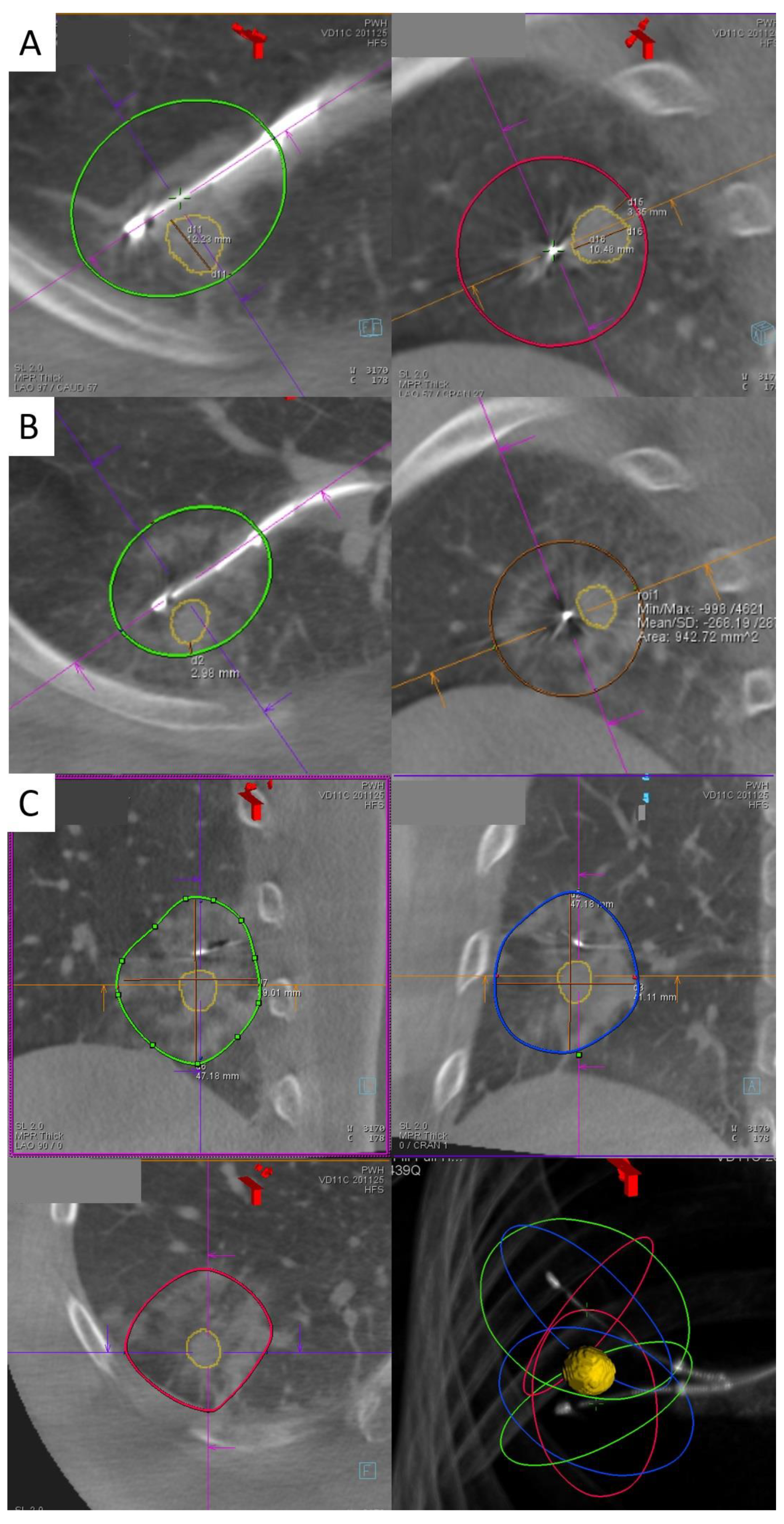
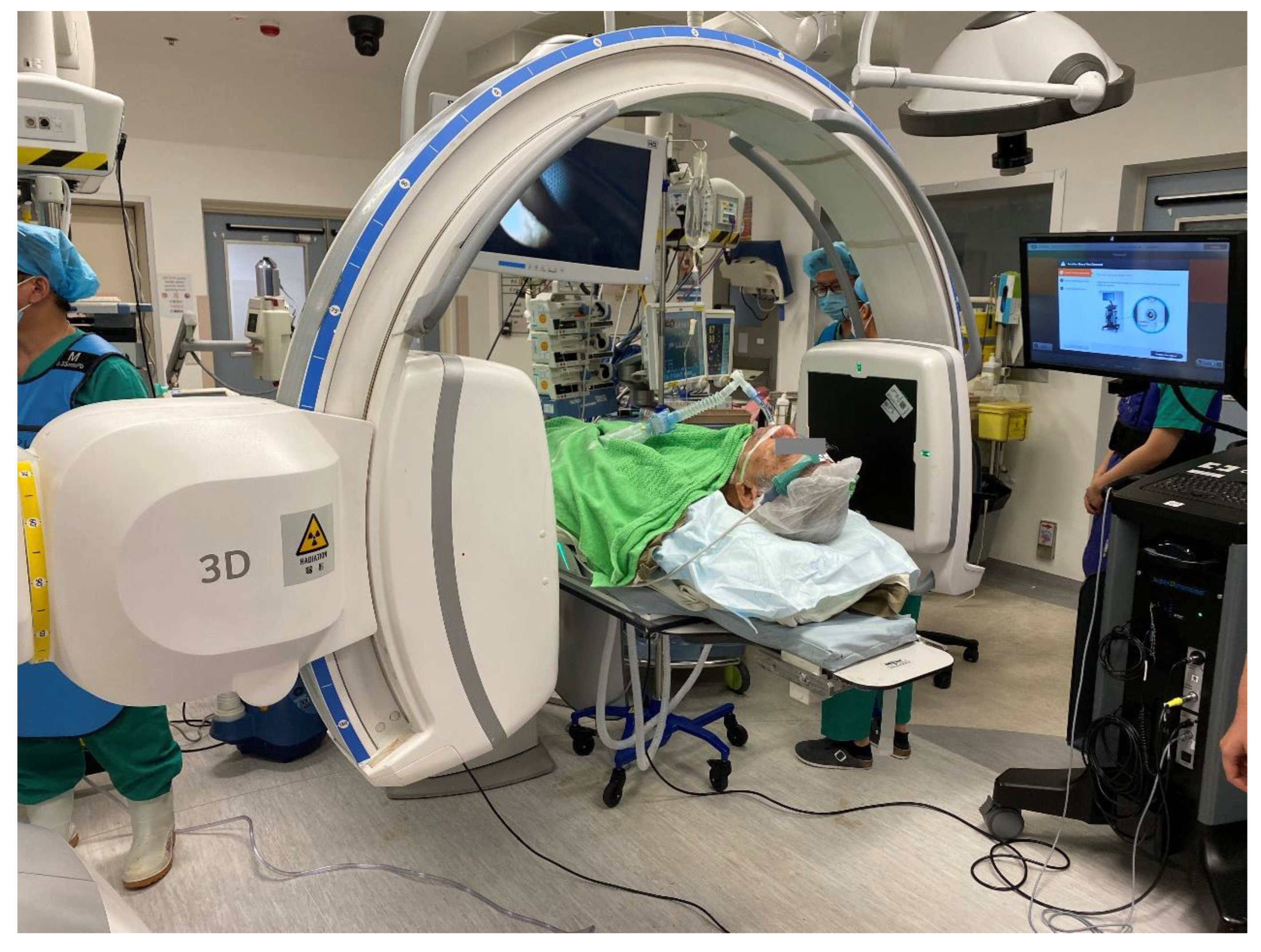
2. Transbronchial Microwave Ablation

There is increasing evidence for local treatment of early lung cancers, especially in patients who decline surgery or those with high surgical risks. Evidence shows that sublobar resection confers similar five-year survival rates, especially in older patients, patients with tumor diameter less than 2 cm and/or pure bronchoalveolar carcinoma [6][7][8]. Meanwhile, SBRT is indicated for patients with stage I or II NSCLC without lymph node involvement and who are surgically inoperable. In multiple retrospective series, SBRT has a decent local control rate of more than 80% [9] and a disease-free survival of 26%. In a multicenter phase II study, an overall survival of 40% at four years has been reported [10]. However, both sublobar resection and SBRT still carry risks such as intra- and postoperative surgical complications and radiation pneumonitis, respectively. Percutaneous ablation of lung tumors has been attempted since the early 2000s [11] following the success of local ablation in hepatocellular carcinomas. The subsequent years have seen the popularization of image-guided local ablation therapies of lung tumors, the first one being radiofrequency ablation (RFA), followed by microwave ablation (MWA) and then cryoablation.
In contrast to the percutaneous route, transbronchial ablation has gained popularity in recent years. A bronchoscopy-guided cooled RFA technique targeted towards lung cancers in human subjects was first pioneered by a Japanese group [12][13]. This was followed by a group in China using electromagnetic navigation bronchoscopy (ENB) guidance [14][15]. The advantage of the transbronchial route over the percutaneous route is the avoidance of pleural puncture, hence fewer pleura-based complications. The data from the Japanese group suggested no pneumothorax, bronchopleural fistula, or pleural effusion in twenty-eight cases of transbronchial RFA [13]. In contrast, the rate of pneumothorax due to percutaneous ablation ranges from 3.5% to 54%. Furthermore, other advantages of transbronchial ablation are the avoidance of needle tract seeding and the ability to reach certain parts of the lung which are difficult or even dangerous with percutaneous access. Examples include areas near the diaphragm, lung apex, mediastinal pleura, or areas shielded by the scapula.
Navigation accuracy has been much improved following the advent of ENB with the support of navigation systems such as SuperDimensionTM (Covidien, Plymouth, MN, USA) and further fine-tuned by position confirmation by fluoroscopy and cone beam CT. A microwave catheter (EmprintTM ablation catheter with the ThermosphereTM technology, Covidien, Plymouth, MN, USA) is inserted within the lung tumor transbronchially and ablated for up to 10 min per burn. For larger tumors, double or triple ablation either in the same position or with catheter position adjustment have been performed to ensure adequate margins.
The Navablate study, in which researchers' institute participated, reported the results of 30 nodules undergoing transbronchial MWA. The median nodule size was 12.5 mm, the procedure day technical success rate was 100%, the mean ablative margin was 9.9 mm, and the technique efficiency (satisfactory ablation as evidenced by a one-month CT scan) was 100% [16]. The technique was safe, with only a 3.3% rate of device-related adverse events (mild hemoptysis), and there were no serious adverse events over 30 days [16]. Since early 2019, the researcher's institute has accumulated 122 cases with 100% technical success rate [17] (Figure 1). The median length of stay was one day only, similar to that reported for the percutaneous approach. Only 3.4% developed pneumothorax requiring chest drainage, and there were two cases of bronchopleural fistula both treated with an endobronchial valve [18]. Post-ablation reaction and fever occurred in 8.9%, minor hemoptysis or hemorrhage—in 4.4%, pleural effusion—in 1.7%, and chest or pleural space infection—in 2.6% [17]. Since the early results were published in 2021 [19], updated midterm data have shown five cases (4.0%) of local ablation site recurrence over a median follow-up of 507 days. On the other hand, a Chinese group who performed transbronchial MWA in 13 patients with different instruments reported a complete ablation rate of 78.6%, a two-year local control rate of 71.4%, and a median progression-free survival of 33 months [20][21].
Figure 1. (A,B) Right lower lobe lung lesion before (A) and after (B) the first transbronchial microwave ablation in two axes on a cone beam CT scan. The lesion is marked by orange tracings while the green and red ovals represent the predicted ablation zone. In (B), ablative changes represented by ground glass opacities are seen surrounding the ablation catheter as predicted; however, the margin was only 2 mm. Therefore, repositioning of the ablation catheter and re-ablation were performed as shown in (C), and the final cone beam computer tomography (CBCT) shows a satisfactory margin of 6.7 mm.
However, the ablation workflow is far from perfect, and further improvements are being developed to smoothen the procedure and reduce the radiation dose. The average number of CBCTs per nodule ablation is 7.1 in the case series [19], yet the minimum number of CBCTs required is only four (pre-procedure, needle position confirmation, ablation catheter position confirmation, post-ablation). The discrepancy is mainly due to incorrect navigation due to the CT-to-body divergence, i.e., the discrepancy between the static preoperative planning CT scan and the dynamic breathing lung, requiring renavigation or redeployment of the needle. The IllumisiteTM platform is the first navigational system on market to correct for the CT-to-body divergence. This digital tomosynthesis-based navigation correction is achieved by utilizing fluoroscopic navigation technology which creates a 3D image enhancing the nodule’s visibility so that the adjusted nodule position can be updated. Throughout the procedure, it provides real-time confirmation that the catheter is aligned to the nodule, and this alignment can be maintained even after the locatable guide (LG) is removed, owing to the presence of position sensor coils embedded in the tip of the extended working channel (EWC) (Figure 2).
Figure 2. Images during ENB navigation using the IllumisiteTM platform. (A) Tip of the locatable guide pointing directly at the lesion (represented by the green ball) after peripheral navigation. In (B), the locatable guide was removed, but there were positional sensors embedded in the extended working channel which is shown pointing slightly off-center at the lesion. In (C), the crosshair is shown, pointing slightly off-center at the lesion which would require further manipulation to ensure target view, while the tip of the catheter is shown to be 2 cm from the target lesion.
The improved navigation and accuracy provided by this platform may also simplify the workflow of transbronchial microwave lung ablation. In previous models without extended working channel (EWC) positional electronics, after the LG is removed, the EWC often returns to its original curvature; thus, subsequent needle deployment is often off-center. With the continuous positional data provided by the tip of an IllumisiteTM EWC, a CrossCountryTM needle can be advanced directly at the green ball in the most bullseye view. Moreover, contrary to previous platforms such as the SuperDimensionTM navigation system, apart from the direction, the exact distance between the center of the nodule and the tip of an EWC is available, too, allowing precise placement of the tip of the ablation catheter (Figure 2B,C).
To further expand the scope of transbronchial ablation, mobile C-arm machines are available to provide high-quality intraoperative reconstructed CT images at institutes without built-in hybrid theatres. Examples include Cios Spin® by Siemens Healthineers [22][23] (Figure 3) and the O-armTM O2 imaging system by Medtronic [24]. In a case series comprising 10 cases utilizing the former system, the navigation success was 80%, while the diagnostic yield for biopsy was 80% [25]. Mobile C-arms had a comparable ease-of-use to floor-mounted CBCT machines and were able to identify most lung lesions, except for two sub-centimeter pure ground glass nodules. However, in order to expand its use to lung nodule ablation, further marking tools, image overlay, and segmentation capabilities are required to ablate nodules that are difficult to visualize on fluoroscopy [25]. Recently, IonTM by Intuitive has integrated Cios Spin® within its own robotic bronchoscopic platform, enabling automatic image transfer from the mobile C-arm machine to an IonTM bronchoscope during procedures [26]. With the increasing popularity of these systems, it is expected that transbronchial treatment can be expanded to less affluent institutions without hybrid operating rooms so as to become more readily available to patients.
Figure 3. Picture of the operating room setup during Cios Spin® mobile C-arm image acquisition.
Ablation zone size is the major limiting factor for the types of early lung tumors that can be treated with transbronchial ablation. The typical CT appearance of early post-ablative morphology of concentric ground glass opacities (GGO) actually contains an outer rim of denser GGO containing congested lung tissue which remains viable [27]. The size observed on CBCT tends to overestimate the area of true coagulation necrosis by 4.1 mm [28]. Thus, a minimal margin of at least 5 mm on a CT scan is recommended to assure adequate tumor kill [28][29][30]. For the EmprintTM ablation catheter, the maximum single ablation produces a predicted oval ablation zone of 4.2 cm in length and 3.5 cm in diameter; thus, the maximum size of a lung nodule taking into account a 5 mm margin on both sides is 2.5 cm.

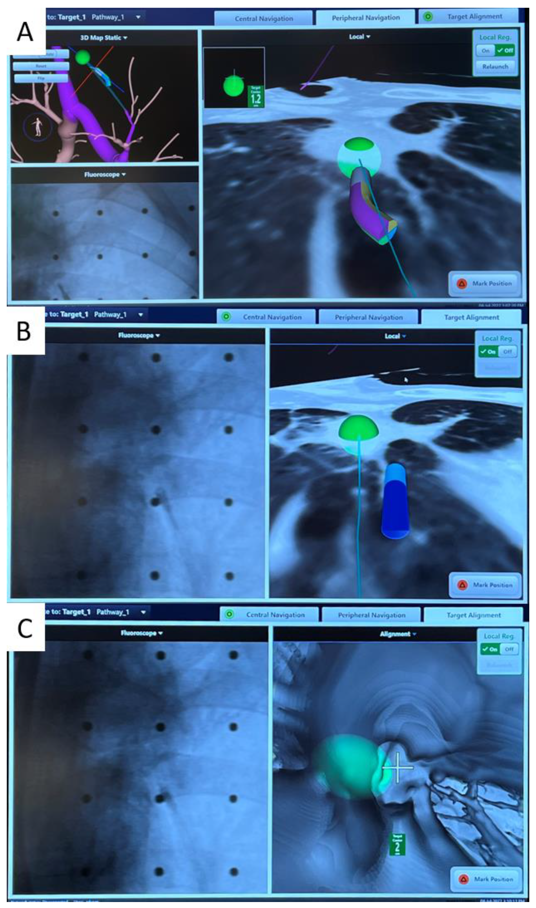
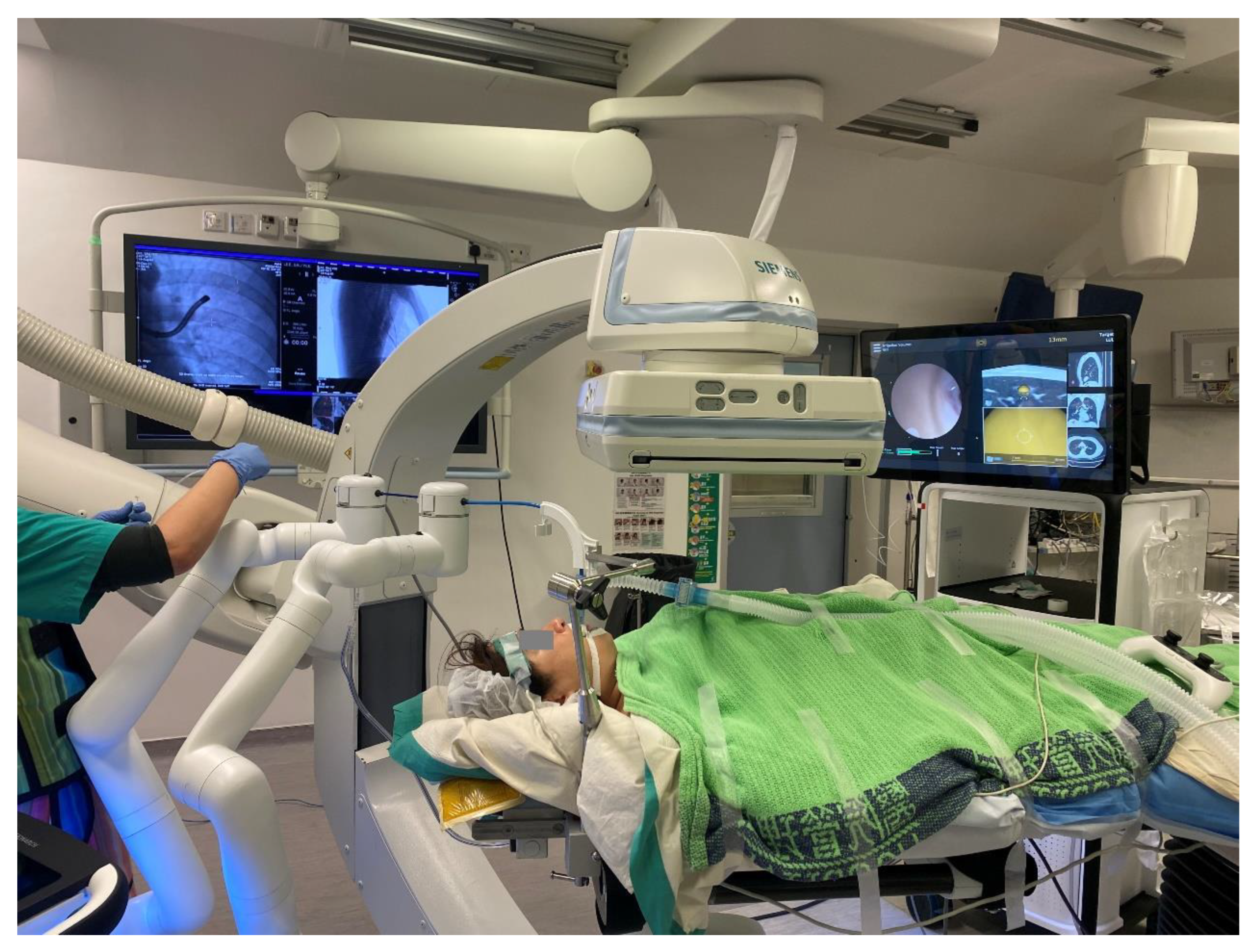
4. Robotic Bronchoscopy to Potentially Improve Transbronchial Ablation Accuracy and Efficacy

Robotic-assisted bronchoscopy (RAB) was originally developed to improve the yield and accuracy of lung nodule biopsy. In comparison to conventional bronchoscopy or other forms of guided bronchoscopy, including virtual bronchoscopy (VB), radial endobronchial ultrasound (r-EBUS), and electromagnetic navigation bronchoscopy (ENB), robotic-assisted bronchoscopy allows a deeper navigation depth up to the ninth airway generation in addition to direct visualization of airways [31]. Currently, there are two major players on the market, the Auris Health MonarchTM platform by Ethicon [32] and the Ion platform by Intuitive [33]. The MonarchTM RAB consists of a telescopic mother–daughter configuration with a 6 mm outer sheath and a 4.2 mm inner scope, utilizing electromagnetic navigation and peripheral vision for guiding navigation (Figure 4). The inner bronchoscope has superior maneuverability to conventional bronchoscopes due to a four-way steering control. While the inner scope is being advanced, structural support is provided by the RAB system, hence reducing the likelihood of scope prolapse into proximal airways. The Ion RAB comprises an articulating catheter with a 3.5 mm outer diameter and a thin 1.8 mm removal visual probe, employing fiberoptic shape sensing for peripheral navigation.
Figure 4. An example of robotic-assisted bronchoscopy by Auris MonarchTM with cone beam CT in a hybrid operating room. This improves the accuracy of navigation, biopsy, or local treatment.
In cadaveric and early human trials of RAB, up to 88–97% navigational success and 77–97% diagnostic yield [34][35][36][37][38][39][40] were reported, particularly when combined with radial EBUS. In lesions without the bronchus sign, the diagnostic yield was higher than that reported using previous nonrobotic systems (54% vs. 31–44%, respectively), such as the SuperDimensionTM navigation system [41]. The latest interim results from a multicenter observational real-world robotic-assisted bronchoscopy biopsy study (TARGET study) reported a 97.5% navigational success and a 91% nodule localization by radial EBUS, while the diagnostic yield data have yet to be published [42]. RAB provides the ability to control the distal end of the scope through multiple active articulation points of the scope, thus allowing enhanced instrument maneuverability (Figure 5). The addition of RAB to transbronchial ablation likely streamlines the navigation process, and in combination with cone beam CT in a hybrid operating room, it further improves the accuracy of ablation catheter placement (Figure 4).
Figure 5. During robotic-assisted bronchoscopy by Auris MonarchTM, multiple panels can be visualized simultaneously: the left-hand panel shows the real-time bronchoscopic view, the middle ones show the bull’s eye view with the centered crosshair and the lesion (in yellow); the right-hand panels show the preoperative CT scan images in three perpendicular axes.

References

  1. Ginsberg, R.J.; Rubinstein, L.V. Randomized Trial of Lobectomy Versus Limited Resection for T1 N0 Non-Small Cell Lung Cancer. Ann. Thorac. Surg. 1995, 60, 615–623. Available online: https://pubmed.ncbi.nlm.nih.gov/7677489/ (accessed on 8 November 2022).
  2. Aberle, D.R.; Adams, A.M.; Berg, C.D.; Black, W.C.; Clapp, J.D.; Fagerstrom, R.M.; The National Lung Screening Trial Research Team. Reduced Lung-Cancer Mortality with Low-Dose Computed Tomographic Screening. N. Engl. J. Med. 2011, 365, 395–409. Available online: http://www.nejm.org/doi/10.1056/NEJMoa1102873 (accessed on 10 April 2020).
  3. De Koning, H.J.; van der Aalst, C.M.; de Jong, P.A.; Scholten, E.T.; Nackaerts, K.; Heuvelmans, M.A.; Lammers, J.W.J.; Weenink, C.; Yousaf-Khan, U.; Horeweg, N.; et al. Reduced Lung-Cancer Mortality with Volume CT Screening in a Randomized Trial. N. Engl. J. Med. 2020, 382, 503–513. Available online: https://pubmed.ncbi.nlm.nih.gov/31995683/ (accessed on 10 May 2022).
  4. Leventakos, K.; Peikert, T.; Midthun, D.E.; Molina, J.R.; Blackmon, S.; Nichols, F.C.; Garces, Y.I.; Hallemeier, C.L.; Murphy, S.J.; Vasmatzis, G.; et al. Management of Multifocal Lung Cancer: Results of a Survey. J. Thorac. Oncol. 2017, 12, 1398–1402. Available online: https://pubmed.ncbi.nlm.nih.gov/28583587/ (accessed on 8 November 2022).
  5. Warth, A.; Macher-Goeppinger, S.; Muley, T.; Thomas, M.; Hoffmann, H.; Schnabel, P.A.; Penzel, R.; Schirmacher, P.; Aulmann, S. Clonality of Multifocal Nonsmall Cell Lung Cancer: Implications for Staging and Therapy. Eur. Respir. J. 2012, 39, 1437–1442. Available online: https://pubmed.ncbi.nlm.nih.gov/22075483/ (accessed on 8 November 2022).
  6. Rami-Porta, R.; Tsuboi, M. Sublobar Resection for Lung Cancer. Eur. Respir. J. 2009, 33, 426–435. Available online: https://pubmed.ncbi.nlm.nih.gov/19181916/ (accessed on 20 September 2020).
  7. El-Sherif, A.; Gooding, W.E.; Santos, R.; Pettiford, B.; Ferson, P.F.; Fernando, H.C.; Urda, S.J.; Luketich, J.D.; Landreneau, R.J. Outcomes of Sublobar Resection Versus Lobectomy for Stage I Non-Small Cell Lung Cancer: A 13-Year Analysis. Ann. Thorac. Surg. 2006, 82, 408–416. Available online: https://pubmed.ncbi.nlm.nih.gov/16863738/ (accessed on 8 August 2020).
  8. Berfield, K.S.; Wood, D.E. Sublobar resection for stage IA non-small cell lung cancer. J. Thorac. Dis. AME Publ. Co. 2017, 9, 208–210.
  9. Abreu, C.E.C.V.; Ferreira, P.P.R.; Moraes, F.Y.D.; Neves, W.F.P.; Gadia, R.; Carvalho, H.D.A. Radioterapia Estereotáxica Extracraniana em Câncer de Pulmão: Atualização. J. Bras. Pneumol. Soc. Bras. Pneumol. E Tisiol. 2015, 41, 376–387. Available online: https://www.jornaldepneumologia.com.br/details/2431/pt-BR/radioterapia-estereotaxica-extracraniana-em-cancer-de-pulmao--atualizacao (accessed on 20 September 2020).
  10. Long-Term Results of Stereotactic Body Radiation Therapy in Medically Inoperable Stage I Non-Small Cell Lung Cancer—PubMed. Available online: https://pubmed.ncbi.nlm.nih.gov/29852036/ (accessed on 20 September 2020).
  11. Dupuy, D.E.; Zagoria, R.J.; Akerley, W.; Mayo-Smith, W.W.; Kavanagh, P.V.; Safran, H. Technical Innovation: Percutaneous Radiofrequency Ablation of Malignancies in the Lung. Am. J. Roentgenol. 2000, 174, 57–59. Available online: https://pubmed.ncbi.nlm.nih.gov/10628454/ (accessed on 20 September 2020).
  12. Tanabe, T.; Koizumi, T.; Tsushima, K.; Ito, M.; Kanda, S.; Kobayashi, T.; Yasuo, M.; Yamazaki, Y.; Kubo, K.; Honda, T.; et al. Comparative Study of Three Different Catheters for ct Imaging-Bronchoscopy- Guided Radiofrequency Ablation as a Potential and Novel Interventional Therapy for Lung Cancer. Chest 2010, 137, 890–897. Available online: https://pubmed.ncbi.nlm.nih.gov/19858232/ (accessed on 8 August 2020).
  13. Koizumi, T.; Tsushima, K.; Tanabe, T.; Agatsuma, T.; Yokoyama, T.; Ito, M.; Kanda, S.; Kobayashi, T.; Yasuo, M. Bronchoscopy-Guided Cooled Radiofrequency Ablation as a Novel Intervention Therapy for Peripheral Lung Cancer. Respiration 2015, 90, 47–55. Available online: https://pubmed.ncbi.nlm.nih.gov/26044954/ (accessed on 8 August 2020).
  14. Xie, F.; Zheng, X.; Xiao, B.; Han, B.; Herth, F.J.F.; Sun, J. Navigation Bronchoscopy-Guided Radiofrequency Ablation for Nonsurgical Peripheral Pulmonary Tumors. Respiration 2017, 94, 293–298. Available online: https://pubmed.ncbi.nlm.nih.gov/28683443/ (accessed on 8 August 2020).
  15. Yuan, H.B.; Wang, X.Y.; Sun, J.Y.; Xie, F.F.; Zheng, X.X.; Tao, G.Y.; Pan, L.; Hogarth, D.K. Flexible Bronchoscopy-Guided Microwave Ablation in Peripheral Porcine Lung: A New Minimally-Invasive Ablation. Transl. Lung Cancer Res. 2019, 8, 787–796. Available online: https://www.ncbi.nlm.nih.gov/pmc/articles/PMC6976383/ (accessed on 8 August 2020).
  16. Medtronic Announces NAVABLATE Study Results Released in Late-Breaking Podium Presentation at European Respiratory Society International Congress 2021—7 September 2021. Available online: https://news.medtronic.com/2021-09-07-Medtronic-Announces-NAVABLATE-Study-Results-Released-in-Late-Breaking-Podium-Presentation-at-European-Respiratory-Society-International-Congress-2021 (accessed on 12 November 2022).
  17. Chan, J.; Yu, P.; Lau, R.; Ng, C. P02.02 Transbronchial Microwave Ablation of Lung Nodules in the Hybrid Operating Room—Mid-Term Follow Up of a Novel Technique. J. Thorac. Oncol. 2021, 16, S977.
  18. Mak, K.L.; Chan, J.W.Y.; Lau, R.W.H.; Ng, C.S.H. Management of Bronchopleural Fistula with Endobronchial Valve in Hybrid Operating Room Following Transbronchial Microwave Ablation. Interact. Cardiovasc. Thorac. Surg. 2021, 33, 992–994. Available online: https://academic.oup.com/icvts/article/33/6/992/6318715 (accessed on 8 November 2022).
  19. Chan, J.W.; Lau, R.W.; Ngai, J.C.; Tsoi, C.; Chu, C.M.; Mok, T.S.; Ng, C.S. Transbronchial Microwave Ablation of Lung Nodules with Electromagnetic Navigation Bronchoscopy Guidance-A Novel Technique and Initial Experience with 30 Cases. Transl. Lung Cancer Res. 2021, 10, 1608–1622. Available online: https://tlcr.amegroups.com/article/view/51418/html (accessed on 8 May 2021).
  20. Xie, F.; Chen, J.; Jiang, Y.; Sun, J.; Hogarth, D.K.; Herth, F.J.F. Microwave Ablation Via a Flexible Catheter for the Treatment of Nonsurgical Peripheral Lung Cancer: A Pilot Study. Thorac. Cancer 2022, 13, 1014–1020. Available online: https://pubmed.ncbi.nlm.nih.gov/35166043/ (accessed on 8 November 2022).
  21. Bao, F.; Yu, F.; Wang, R.; Chen, C.; Zhang, Y.; Lin, B.; Wang, Y.; Hao, X.; Gu, Z.; Fang, W. Electromagnetic Bronchoscopy Guided Microwave Ablation for Early Stage Lung Cancer Presenting as Ground Glass Nodule. Transl. Lung Cancer Res. 2021, 10, 3759–3770. Available online: https://pubmed.ncbi.nlm.nih.gov/34733626/ (accessed on 8 November 2022).
  22. Cios Spin® by Siemens Healthineers. Available online: https://www.siemens-healthineers.com/surgical-c-arms-and-navigation/mobile-c-arms/cios-spin (accessed on 19 November 2022).
  23. Chen, J.; Xie, F.; Zheng, X.; Li, Y.; Liu, S.; Ma, K.C.; Goto, T.; Müller, T.; Chan, E.D.; Sun, J. Mobile 3-Dimensional (3D) C-Arm System-Assisted Transbronchial Biopsy and Ablation for Ground-Glass Opacity Pulmonary Nodules: A case Report. Transl Lung Cancer Res. 2021, 10, 3312–3319. Available online: https://tlcr.amegroups.com/article/view/54398/html (accessed on 7 August 2021).
  24. Cho, R.J.; Senitko, M.; Wong, J.; Dincer, E.H.; Khosravi, H.; Abraham, G.E., III. Feasibility of Using the O-Arm Imaging System During ENB-rEBUS-Guided Peripheral Lung Biopsy: A Dual-Center Experience. J. Bronchol. Interv. Pulmonol. 2020. Available online: https://pubmed.ncbi.nlm.nih.gov/34085805/ (accessed on 11 July 2021).
  25. Chan, J.W.Y.; Lau, R.W.H.; Chu, C.M.; Ng, C.S.H. Expanding the Scope of Electromagnetic Navigation Bronchoscopy-Guided Transbronchial Biopsy and Ablation with Mobile 3D C-Arm Machine Cios Spin ®-Feasibility and Challenges. Transl. lung Cancer Res. 2021, 10, 4043–4046. Available online: https://pubmed.ncbi.nlm.nih.gov/34858793/ (accessed on 10 May 2022).
  26. “Ion Cios Spin”|Search|LinkedIn. Available online: https://www.linkedin.com/search/results/all/?keywords=ionciosspin&origin=GLOBAL_SEARCH_HEADER&sid=9cG (accessed on 19 November 2022).
  27. Chheang, S.; Abtin, F.; Guteirrez, A.; Genshaft, S.; Suh, R. Imaging Features Following Thermal Ablation of Lung Malignancies. Semin. Intervent. Radiol. 2013, 30, 157–168. Available online: https://www.ncbi.nlm.nih.gov/pmc/articles/PMC3709948/ (accessed on 8 August 2020).
  28. Yamamoto, A.; Nakamura, K.; Matsuoka, T.; Toyoshima, M.; Okuma, T.; Oyama, Y.; Ikura, Y.; Ueda, M.; Inoue, Y. Radiofrequency Ablation in a Porcine Lung Model: Correlation Between CT and Histopathologic Findings. Am. J. Roentgenol. 2005, 185, 1299–1306. Available online: http://www.ajronline.org/doi/10.2214/AJR.04.0968 (accessed on 8 August 2020).
  29. Anderson, E.M.; Lees, W.R.; Gillams, A.R. Early Indicators of Treatment Success after Percutaneous Radiofrequency of Pulmonary Tumors. Cardiovasc. Intervent. Radiol. 2009, 32, 478–483. Available online: https://pubmed.ncbi.nlm.nih.gov/19127381/ (accessed on 8 August 2020).
  30. Wolf, F.J.; Grand, D.J.; Machan, J.T.; DiPetrillo, T.A.; Mayo-Smith, W.W.; Dupuy, D.E. Microwave Ablation of Lung Malignancies: Effectiveness, CT Findings, and Safety in 50 Patients. Radiology 2008, 247, 871–879. Available online: https://pubmed.ncbi.nlm.nih.gov/18372457/ (accessed on 8 August 2020).
  31. Chen, A.C.; Gillespie, C.T. Robotic Endoscopic Airway Challenge: REACH Assessment. Ann. Thorac. Surg. 2018, 106, 293–297. Available online: https://pubmed.ncbi.nlm.nih.gov/29486178/ (accessed on 10 May 2022).
  32. Monarch Platform by Auris Health. Available online: https://www.aurishealth.com/monarch-platform (accessed on 19 November 2022).
  33. Intuitive|Robotic-Assisted Bronchoscopy|Ion Platform. Available online: https://www.intuitive.com/en-us/products-and-services/ion (accessed on 12 November 2022).
  34. Rojas-Solano, J.R.; Ugalde-Gamboa, L.; MacHuzak, M. Robotic Bronchoscopy for Diagnosis of Suspected Lung Cancer: A Feasibility Study. J. Bronchol. Interv. Pulmonol. 2018, 25, 168–175. Available online: https://pubmed.ncbi.nlm.nih.gov/29762461/ (accessed on 10 May 2022).
  35. Chen, A.C.; Pastis, N.J.; Machuzak, M.S.; Gildea, T.R.; Simoff, M.J.; Gillespie, C.T.; Mahajan, A.K.; Oh, S.S.; Silvestri, G.A. Accuracy of a Robotic Endoscopic System in Cadaver Models with Simulated Tumor Targets: ACCESS Study. Respiration 2020, 99, 56–61. Available online: https://pubmed.ncbi.nlm.nih.gov/31805570/ (accessed on 10 May 2022).
  36. Chaddha, U.; Kovacs, S.P.; Manley, C.; Hogarth, D.K.; Cumbo-Nacheli, G.; Bhavani, S.V.; Kumar, R.; Shende, M.; Egan, J.P.; Murgu, S. Robot-Assisted Bronchoscopy for Pulmonary Lesion Diagnosis: Results from the Initial Multicenter Experience. BMC Pulm. Med. 2019, 19, 1–7. Available online: https://pubmed.ncbi.nlm.nih.gov/31829148/ (accessed on 10 May 2022).
  37. Agrawal, A.; Hogarth, D.K.; Murgu, S. Robotic Bronchoscopy for Pulmonary Lesions: A Review of Existing Technologies and Clinical Data. J. Thorac. Dis. 2020, 12, 3279–3286. Available online: https://pubmed.ncbi.nlm.nih.gov/32642251/ (accessed on 10 May 2022).
  38. Kalchiem-Dekel, O.; Connolly, J.G.; Lin, I.H.; Husta, B.C.; Adusumilli, P.S.; Beattie, J.A.; Buonocore, D.J.; Dycoco, J.; Fuentes, P.; Jones, D.R.; et al. Shape-Sensing Robotic-Assisted Bronchoscopy in the Diagnosis of Pulmonary Parenchymal Lesions. Chest 2022, 161, 572–582. Available online: https://pubmed.ncbi.nlm.nih.gov/34384789/ (accessed on 8 November 2022).
  39. Benn, B.S.; Romero, A.O.; Lum, M.; Krishna, G. Robotic-Assisted Navigation Bronchoscopy as a Paradigm Shift in Peripheral Lung Access. Lung 2021, 199, 177–186. Available online: https://pubmed.ncbi.nlm.nih.gov/33547938/ (accessed on 8 November 2022).
  40. Fielding, D.I.; Bashirzadeh, F.; Son, J.H.; Todman, M.; Chin, A.; Tan, L.; Steinke, K.; Windsor, M.N.; Sung, A.W. First Human Use of a New Robotic-Assisted Fiber Optic Sensing Navigation System for Small Peripheral Pulmonary Nodules. Respiration 2019, 98, 142–150. Available online: https://pubmed.ncbi.nlm.nih.gov/31352444/ (accessed on 8 November 2022).
  41. Seijo, L.M.; de Torres, J.P.; Lozano, M.D.; Bastarrika, G.; Alcaide, A.B.; Lacunza, M.M.; Zulueta, J.J. Diagnostic Yield of Electromagnetic Navigation Bronchoscopy Is Highly Dependent on the Presence of a Bronchus Sign on CT Imaging: Results from A Prospective Study. Chest 2010, 138, 1316–1321. Available online: https://pubmed.ncbi.nlm.nih.gov/20435658/ (accessed on 11 July 2021).
  42. Transbronchial Biopsy Assisted by Robot Guidance in the Evaluation of Tumors of the Lung—Full Text View—ClinicalTrials.gov. Available online: https://clinicaltrials.gov/ct2/show/NCT04182815 (accessed on 12 November 2022).
More
Information
Subjects: Respiratory System
Contributors MDPI registered users' name will be linked to their SciProfiles pages. To register with us, please refer to https://encyclopedia.pub/register : , , , , , ,
View Times: 237
Revisions: 2 times (View History)
Update Date: 23 Feb 2023
1000/1000
Video Production Service