Submitted Successfully!
To reward your contribution, here is a gift for you: A free trial for our video production service.
Thank you for your contribution! You can also upload a video entry or images related to this topic.
Version Summary Created by Modification Content Size Created at Operation
1 -- 4175 2023-01-11 02:07:36 |
2 update references and layout -2 word(s) 4173 2023-01-11 02:30:59 |

Video Upload Options

Do you have a full video?

Confirm

Are you sure to Delete?
Cite
If you have any further questions, please contact Encyclopedia Editorial Office.
Cai, M.;  Wan, J.;  Cai, K.;  Song, H.;  Wang, Y.;  Sun, W.;  Hu, J. Contribution of Lactate Metabolism in Cancer Progress. Encyclopedia. Available online: https://encyclopedia.pub/entry/39999 (accessed on 03 July 2024).
Cai M,  Wan J,  Cai K,  Song H,  Wang Y,  Sun W, et al. Contribution of Lactate Metabolism in Cancer Progress. Encyclopedia. Available at: https://encyclopedia.pub/entry/39999. Accessed July 03, 2024.
Cai, Ming, Jian Wan, Keren Cai, Haihan Song, Yujiao Wang, Wanju Sun, Jingyun Hu. "Contribution of Lactate Metabolism in Cancer Progress" Encyclopedia, https://encyclopedia.pub/entry/39999 (accessed July 03, 2024).
Cai, M.,  Wan, J.,  Cai, K.,  Song, H.,  Wang, Y.,  Sun, W., & Hu, J. (2023, January 11). Contribution of Lactate Metabolism in Cancer Progress. In Encyclopedia. https://encyclopedia.pub/entry/39999
Cai, Ming, et al. "Contribution of Lactate Metabolism in Cancer Progress." Encyclopedia. Web. 11 January, 2023.
Contribution of Lactate Metabolism in Cancer Progress
Edit

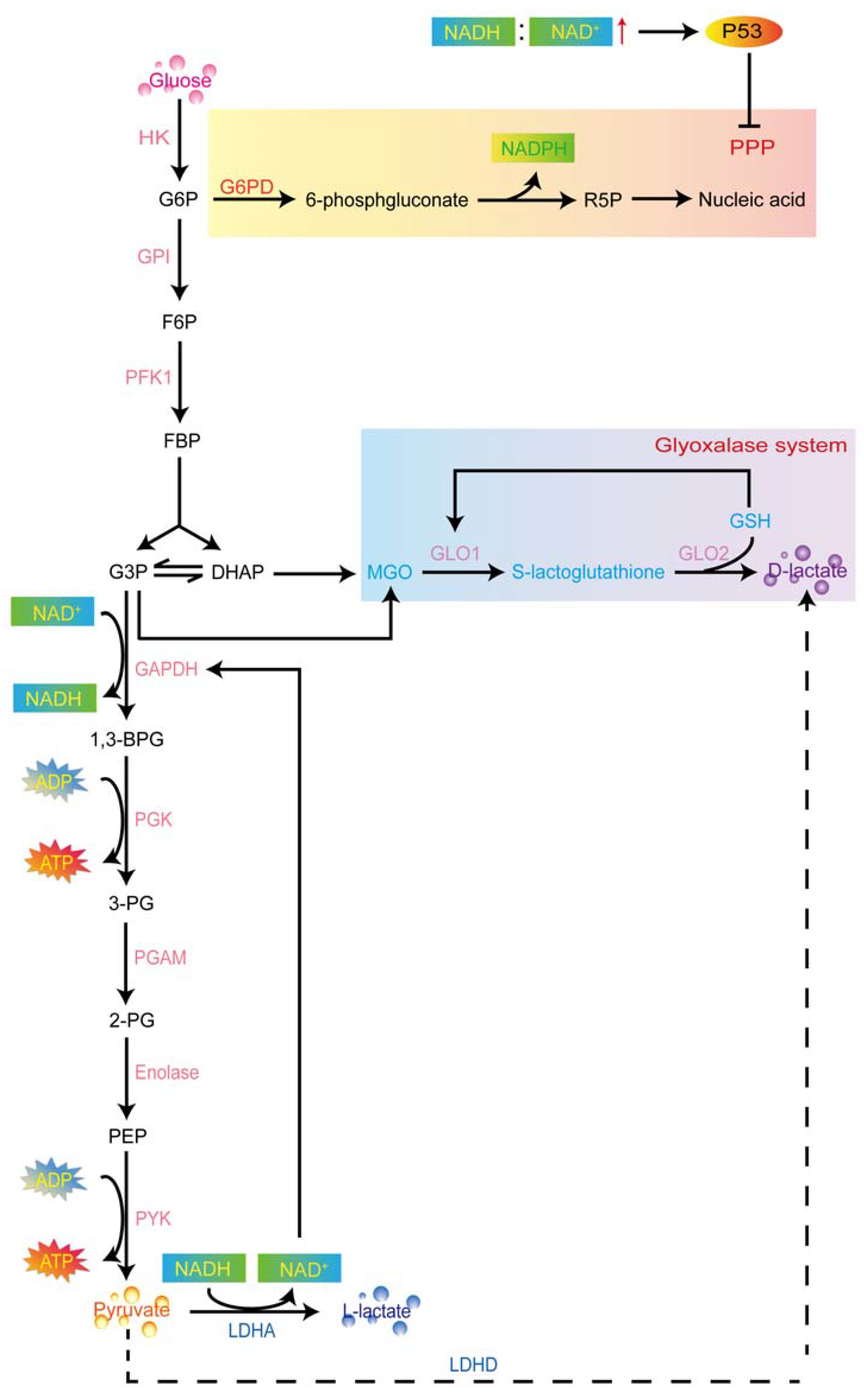
The Warburg effect describes a unique phenomenon that cancers incline to shift the mode of oxidative phosphorylation (OXPHOS) to glycolysis in spite of abundant oxygen. Lactate is the main production of glycolysis, which contains two isomers, L-lactate and D-lactate. The accumulation of high lactate in solid tumors and its extracellular environment is considered as the key and early evidence of malignant development, which is associated with a poor prognosis. Lactate reprograms the tumor microenvironment (TME) to have profound effects on cancer cell phenotype and is conducive to the progress of cancer that involves the eight biological capabilities acquired of cancer: sustaining cell proliferation, promoting growth, resisting cell death, enabling replicative immortality, inducing angiogenesis, activating invasion and metastasis, reprogramming energy metabolism, and evading immune destruction. Lactate’s contribution to cancer is not only the respiratory fuel but also the regulator of intracellular and extracellular molecular signaling in the TME.

lactate metabolism L-lactate D-lactate cancer progression

1. Lactate Metabolism in Carcinoma Cells

1.1. Warburg Effect

The Warburg effect is an extremely common event in many carcinoma cells [1]. This amazing theory was firstly proposed by Otto Warburg and colleagues in the 1920s [2], which has been documented for over 100 years [3]. It describes the unusual metabolic transforming phenomenon in carcinoma cells that, unlike most normal tissues, carcinoma cells tend to metabolize most glucose into lactate for adenosine triphosphate (ATP) production even in the presence of sufficient oxygen, which is termed “aerobic glycolysis” [4][5]. It is not the defective ability of mitochondrial oxidative phosphorylation (OXPHOS) in carcinoma cells leading to no alternative choice. On the contrary, the mitochondrial function is intact [6][7][8], even perhaps with higher-efficiency of OXPHOS in carcinoma cell types [9][10]. In fact, there are several potential advantages of glycolysis in carcinoma cells. For example, glycolysis can provide energy supply more rapidly than the aerobic oxidation for the proliferation of carcinoma cells in spite of less efficient ATP production in this way [3]. Glycolysis reduces the reliance on oxygen for ATP production and thereby, the potentially destructive reactive oxygen species (ROS) produced by the mitochondrial electron transport chain. It also facilitates the generation of NADPH to reducing equivalents for ROS-protective pathways [11]. Except for plentiful ATP synthesis, Pentose phosphate pathway (PPP) is enhanced in the aerobic glycolysis. This pathway provides precursors for lipid and nucleic acid synthesis, which favors cell division [12]. Herein, the metabolic reprogramming can benefit both bioenergetics and biosynthesis, inhibit cellular apoptosis, and generate signal metabolites in favor of carcinoma cell growth.
Since the rate of aerobic glycolysis in carcinoma cells is so high that the speed of lactate production from glucose is approximately 10–100 times faster than the speed of complete oxidation of glucose in the mitochondria [3], it not surprising to observe that the concentration of lactate in the tumor tissues is 100 times as much as the blood [2]. It is estimated that the lactate concentrations range from 5 to 20 mM in the tumor microenvironment [13] and range from 10 to 40 mM in tumors [14]. Here, some questions arise: Is the excess generation of lactate a superfluous metabolic waste in carcinoma cells? If not, what is the pathophysiological function in carcinoma cells? As known, in mammals, lactate possesses two isomers: L- and D-lactate. Of what significance are they in carcinoma cells? In the following section, the content will involve L- and D-lactate production and metabolism in aerobic glycolysis, the research of lactate on cancer progress, hallmarks of cancer associated with the lactate, lactate related molecular signaling to better understand the role of lactate in cancer.

1.2. Metabolism of Lactate Isomers and Aerobic Glycolysis

Most tumor cells can reprogram metabolic procedures associated with increased levels of glycolytic enzymes and intermediates to enhance the glycolysis pathway [15][16]. Lactate is one of the well-known end-products of glycolysis. It is the simplest hydroxyl carboxylic acid and exists as 2 stereoisomers due to the chiral center at C2 [17]. Knowledge of the L- and D-lactate production in the Warburg effect will help us further understand the representative hallmarks in cancer progress and seek for the accurate anticancer targets.

1.2.1. L-Lactate Production in Aerobic Glycolysis

Hexokinase (HK) is the first enzyme involved in glycolysis, catalyzing glucose into glucose 6-phosphate (G6P) [18]. G6P dehydrogenase (G6PD) irreversibly converts partially G6P to 6-phosphgluconate which is also known as the PPP [19]. In tumorigenesis, the utilization of PPP is frequently elevated [7]. In this step, G6P becomes oxidized to generate NADPH and ribose-5-phosphate (R5P)—a structural component of nucleotides. These transketolase reactions in the PPP convert glucose to ribose for nucleic acid synthesis, as well as generates NADPH, a reducing agent needed for synthesis reactions in tumor cells [3]. Yet, the P53 protein is reported to involve the “glycolytic stress response” by sensing an increased NADH: NAD+ ratio in highly glycolytic cells [11] and inhibit PPP by binding to G6PD [7][20]. In parallel to this process, G6P isomerase (GPI) catalyzes G6P to fructose-6-phosphate (F6P) in glycolysis [21]. Then, phosphofructokinase-1 (PFK1) catalyzes the rate-limiting phosphorylation of F6P to fructose-1,6-bisphosphate (FBP) [22]. FBP is cleaved into glyceraldehyde 3-phosphate (G3P) and dihydroxyacetone phosphate (DHAP) catalyzed by aldolase B [23]. G3P-dehydrogenase (GAPDH) can remove hydrogen from G3P to an NAD+ molecule for producing NADH or add a phosphate group to the G3P for producing 1,3-bisphosphoglycerate (1,3-BPG). Then, phosphoglycerate kinase (PGK) catalyzes 1,3-BPG and ADP to produce 3-phosphoglycerate (3-PG) and two ATP molecules. Phosphoglycerate mutase 1 (PGAM1), following, catalyzes the conversion of 3-PG to 2-phosphoglycerate (2-PG) [24]. After that, enolase catalyzes the dehydration of 2-PG into phosphoenolpyruvate (PEP) [25][26]. Finally, as one of the main PEP-consuming reactions, pyruvate synthesis is catalyzed by pyruvate kinase (PYK) [27]. In carcinoma cells, lactate dehydrogenase isoform A (LDHA) preferentially converts synthetic pyruvate to L-lactate by removing hydrogen from the NADH molecule in the final step of the glycolytic pathway [28], thereby regenerating NAD+ to maintain glycolysis [29][30], which serves as a substrate for GAPDH [31]. This is why the decreased GAPDH inhibits glycolysis [32][33], and the accumulation of L-lactate in carcinoma cells implies an increased intracellular NADH: NAD+ ratio [30] (Figure 1).
Figure 1. Lactate production in aerobic glycolysis. HK firstly catalyzes the glucose into GP6. G6PD and GPI convert G6P to 6-phosphgluconate and F6P, respectively. The 6-phosphgluconate finally produces the R5P for nucleotides synthesis which is known as the PPP. PFK1 catalyzes the F6P to FBP for pyruvate synthesis. L-lactate can be produced through the LDHA. FBP can also convert into DHAP and produce the intermediary product—MGO. Glyoxalases are involved in the detoxification of reactive MGO into D-lactate in a two-step reaction using GSH as a cofactor. HK, hexokinase; G6P, glucose 6-phosphate; G6PD, G6P dehydrogenase; GPI, G6P isomerase; F6P, fructose-6-phosphate; NADPH, nicotinamide adenine dinucleotide phosphate; R5P, ribose-5-phosphate; PPP, pentose phosphate pathway; NADH, reduced nicotinamide adenine dinucleotide; NAD+, nicotinamide adenine dinucleotide; PFK1, phosphofructokinase-1; FBP, fructose-1,6-bisphosphate; G3P, glyceraldehyde 3-phosphate; DHAP, dihydroxyacetone phosphate; GAPDH, G3P-dehydrogenase; 1,3-bisphosphoglycerate, 1,3-BPG; PGK, phosphoglycerate kinase; 3-PG, 3-phosphoglycerate; ATP, adenosine triphosphate; PGAM1, phosphoglycerate mutase 1; 2-PG, 2-phosphoglycerate; PEP, phosphoenolpyruvate; PYK, pyruvate kinase; LDHA, lactate dehydrogenase isoform A; MGO, methylglyoxal; GLO1, glyoxalase 1; GLO2, glyoxalase 2; GSH, glutathione.

1.2.2. D-Lactate Production in Aerobic Glycolysis

D-lactate, as an isomer of L-lactate, shares the same mass but has much lower amounts compared with L-lactate in mammals [34]. It is considered the “physiological inertia” in the body [35] due to the absence of metabolizing enzymes [36][37]. Previously, D-lactate is proved to be an important component of the cell wall of a lactic acid bacterium. Besides, bulk D-lactate can be detected in humans and ruminants in the rare metabolic condition of D-lactic acidosis [17]. For the past few years, D-lactate has also reported generation during aerobic glycolysis through the glyoxalase system [38], which is comprised of two enzymes, glyoxalase 1 (GLO1) and glyoxalase 2 (GLO2), and a catalytic amount of reduced glutathione (GSH) as a cofactor [39]. This system converts the metabolic intermediary product—methylglyoxal (MGO) [40] into D-lactate or GSH [38]. In the glycolytic pathway, MGO is a highly reactive three-carbon glycating metabolite [41] that mainly originates from triosephosphates (DHAP and G3P) para-metabolically and para-enzymatically when glucose is degraded [42][43][44]. Glyoxalases are involved in the detoxification of reactive MGO into D-lactate in a two-step reaction using GSH as a cofactor [42][45]. GLO1 (also named S-D-lactoylglutathione lyase) exists in humans, mice, yeast, and elegans [45]. It condensates MGO and reduces GSH to form S-lactoylglutathione [46]. Then, GLO2 hydrolyzes the S-lactoylglutathione and thereby, releasing D-lactate and regenerating GSH [42][46]. In breast carcinoma cells, astrocytoma, and prostate carcinoma cells, the levels of D-lactate are observed as elevated [42][47]. Furthermore, a recent study has demonstrated that produced D-lactate by lung carcinoma cells can shuttle into normal cells to lead to cancer-associated metabolic behavior, implying the role of elevated D-lactate concentration as a hallmark of cancer malignant metabolism [34] (Figure 1).

2. Current Advances of Lactate in Cancer

2.1. Breast Cancer

Breast cancer is the most frequently diagnosed cancer in women and ranks second among causes of cancer-related mortality in females worldwide [48]. The 5-year survival rate is 89% in females with primary breast cancer and less than 5% in patients with metastatic breast cancer [49]. The clinical hallmarks of breast cancer are stromal invasion and metastasis to regional lymph nodes or distant organs [50]. Bone, lung, liver, and brain are generally accepted as the primary target sites of breast cancer metastasis [51]. A previous clinical study has claimed that the lactate concentration is observed depending on the degree of progression of breast tumor tissue. For instance, the lactate concentration is 5.5 ± 2.4 mM in grade II and 7.7 ± 2.9 mM in grade III [52]. Similar to this result, the concentration of L-lactate in malignant breast tumor tissue is higher than in the benign counterparts [53], and tumor lactate in patients with triple negative breast cancer (TNBC) far exceeds that found in circulating blood [54]. The low perfusion or monocarboxylate transporters (MCTs) activity—MCT1 and MCT4 [54] in TNBC, may be the major cause of lactate accumulation in breast tumors and thereby, creates a local tumor microenvironment enriched in lactate produced by aerobic glycolysis [54]. Furthermore, Becker et al. found that L-lactate, produced by cancer-associated fibroblasts (CAFs), was delivered into breast carcinoma cells as fuel for growth and is dependent on the transport of MCT1 [55]. Distinguishment from the common breast cancer, TNBC lacks expression of an estrogen receptor (ER), progesterone receptor (PR), and human epidermal growth factor receptor 2 (HER2) [56]. It is interesting to investigate whether the expression of MCTs is affected by these receptors to influence the lactate shuttle between carcinoma and stroma cells in the tumor microenvironment and thereby, determining the cancer subtypes.
In breast carcinoma cells, the accumulation of lactate can promote the adhesion, migration, and invasion of carcinoma cells by serving as the signal modulator [57]. Lactate receptor—G-protein-coupled receptor 81 (GPR81), expression is observed as a high expression [58][59][60]. A further study demonstrates that GPR81 expression is conducive to multiple malignant phenotypes of carcinoma cells [58], implying the lactate-receptor signal is a potential therapeutic target for breast cancer. In parallel to GPR81, G protein-coupled receptor 132 (GPR132) can also serve as the macrophage sensor of the rising lactate in the acidic breast tumor milieu to promote the alternatively activated macrophage M2-like phenotype, which, in turn, facilitates cancer cell adhesion, migration, and invasion [61]. The M2-like phenotype also can be driven by lactate via the extracellular signaling-regulated kinase (ERK)/STAT3 signaling pathway [61]. Apart from the above molecular signals, 5 mM L-lactate is sufficient to induce the hypoxia induced factor-1 alpha (HIF-1α) expression to promote tumor-associated macrophages (TAMs) via overexpressing the HIF-1α-stabilizing long noncoding RNA [62]. The TAMs further enhance aerobic glycolysis [63] and inhibit apoptosis of breast carcinoma cells [62]. The inter-linked and mutually-reinforcing interaction of L-lactate and macrophages aggravate breast tumor progression. With regard to the role of D-lactate in breast cancer, to our knowledge, few related studies have been investigated. Considering that lactate comprises two isomers—L-lactate and D-lactate, the future research on breast cancer remains to distinguish the biological effect of two types of lactates, especially D-lactate production in glycolysis. Revealing the breast tumor-associated L- and D-lactate production, and their relation with respect to the phenotype of cancer, will provide a better understanding of the whole tumor progression.

2.2. Cervical Cancer

Cervical cancer is the fourth most common malignancy and the disease results in over 300,000 deaths annually worldwide [64]. Recent research has disclosed that, compared to healthy people, the plasmatic lactate concentration is significantly higher in patients with low- and high-grade cervical lesions and cervical cancer [65]. In cervical carcinoma cell lines, the secreted lactate concentration ranges from 1.5 to 3.8 mM after a 24 h period of incubation [65]. Inhibition of lactate synthesis or transport tends to decrease M2 markers of macrophage in the co-cultivated with human papillomavirus (HPV) positive cervical carcinoma cells and macrophages; as a result, the increase the T lymphocyte activation potential in the carcinoma cell lines [65] suggests that lactate inhibition may be a useful tool in anticancer therapies associated with immunomodulatory effects.
Human vaginal secretions have been reported to contain approximately 10–50 mM lactate through bacteria ferment and epithelial cells, of which D-lactate accounts for half of the total lactate [66]. There is no doubt that lactate isomers may play a potential role in the pathological mechanism of cervical cancer. Wagner et al. found that both L- and D-lactate can protect cervical carcinoma cell survival from chemotherapeutic treatment by inhibiting the activity of histone deacetylases (HDACs). The inhibited HDAC activity is beneficial to a more relaxed, transcriptionally permissive chromatin conformation and reduces the DNA damage response (DDR) by modulating the activity of key proteins such as an increased DNA-dependent protein kinase catalytic subunit (DNA-PKcs) [67]. In addition to epigenetic modification, lactate can also activate the GPR81 receptor signal pathway to achieve the survival of carcinoma cells by DNA repair, which is coordinated by MCTs transport [68][69][70]. The notable phenomenon observed by Wagner and his colleagues was that L-lactate primarily inhibited the cAMP accumulation while D-lactate strongly stimulated ERK phosphorylation, which was mainly induced by PKC [67], implying the disparate intrinsic activity of lactate isomers towards the GPR81 receptor signal transduction pathways. Based on the previous studies, Wagner et al. also considered the relationship between drug resistance depending on PKC activity and carcinoma cell survival. Their results suggested that the activated GPR81, stimulated by L- and D-lactate, up-regulated the protein and mRNA expressions of the ATP-binding cassette subfamily B member 1 (ABCB1) to enhance the doxorubicin resistance in the cervical cancer cell [68]. On the contrary, results of L-lactate favoring the progression of cervical cancer, Da et al. declared that the physiological concentration of L-lactate (10 mM and 20 mM) enhanced the phosphorylation of P38 to promote apoptosis in HeLa cells [71]. Wagner et al. declared that both L- and D-lactate (10 mM and 20 mM) may enhance the nuclear localization of DNA-PKcs to suppress retroviral transduction in cervical carcinoma cells [69]. Several factors may attribute to the paradoxical results: Different strains of cells react differently to lactate; for instance, DNA-PKcs-proficient cells among cervical cancer cells are less susceptible to lactate modulation. HeLa and CaSki cells respond to both lactate isomers, while C33A cells respond only to L-lactate [69]; lactate as the signal modulation regulates downstream multiple signal transduction related to cancers; lactate effects may be related to its volume in cancers in a link to the above research. Last but not least, the existence of pH caused acidification in the carcinoma cells and/or tumor microenvironment may affect the modulation of lactate-related signalings [65][71].

2.3. Lung Cancer

Lung cancer is the second most commonly diagnosed cancer after prostate cancer in men and breast cancer in women [72][73]. In North America and other developed countries, it is the leading cause of cancer-related deaths because of the difficulty for diagnosis in the early stage [74]. Higher lactate/3-PG labeling ratios have been noticed in patients with stage I and II lung cancers when they are observed at the time of the original clinical observation. In some cases, years before recurrence or metastases, the primary tumor is even observed with higher lactate/3-PG labeling ratios [14], implying that high lactate is more likely for the progress of lung cancer. In lung cancer model mice, the circulatory turnover flux of lactate exceeds that of glucose by approximately twofold and contributes to the tumor TCA cycle [75], suggesting that lactate can serve as the energy substrate for lung carcinoma cell growth.
Nonsmall cell lung cancer (NSCLC) is the main histologic subtype (85%) of lung cancer [76]. Surgical resections from patients with NSCLC show glucose metabolism-contrasting homeostasis after infusion of 13C-glucose, leading to considerably high levels of lactate [77]. Similarly, in the NSCLC mouse model, the contribution of lactate to the TCA cycle exceeds that of glucose [14]. In lung adenocarcinoma cell lines, upregulated gene expression of TMPRSS11B can enhance the lactate export to promote tumorigenesis [78]. The increased acidic environment along with lactate production promotes the formation of a snail/transcriptional coactivator with PDZ-binding motif (TAZ)/AP-1 complex and contributes to adaptive resistance in NSCLC in the end with the poor prognosis in advanced lung cancer [79]. Recent evidence has identified that lactate, as a characteristic of many NSCLCs, is exploitable for therapeutic targeting and manipulation to reprogram the TME and promote an oncolytic immune response [80]. For example, lactate can bind to its receptor GPR81 to induce the activation of PD-L1 which leads to the reduction of interferon-γ in lung tumor cells and apoptosis of co-cultured Jurkat T-cell leukemia cells for the evading host immunity [81]. Furthermore, 83% of tumor-bearing mice developed lung cancer and showed shorter survival when they were inoculated with the dendritic cells (DCs) treated with lactate. The results suggested that lactate caused the loss of DCs function to weaken the immune surveillance with reduced effector CD8+ T cells [82]. Besides, L-lactate is reported to subtly affect the transcriptome of the pro-inflammatory major histocompatibility complex (MHC)-IIlo TAMs to favor the typical M2 genes expression such as Cd163, Stab1, Lyve1, Tmem26, Folr2, Mmp9, Clec10a, Il4Ra, and Itgb3, that leads to the enhanced T cell suppressive capacity of these TAMs [83]. Of interest, the incubation of MHC-IIlo TAMs with L-lactate showed slightly elevated oxidative phosphorylation (OXPHOS) and enhanced glycolytic capacity, and glycolytic reserve. While in MHC-IIhi TAMs, L-lactate further reduces the ability of OXPHOS [83]. Hence, L-lactate may have different effects on mitochondrial metabolic regulation on the distinct macrophage phenotype in the carcinoma cells.
There are several problems to be solved here: What is the relationship between mitochondria and cancer immune escape? What is the effect of D-lactate on the mitochondria and immunosuppression of lung carcinoma cells? As for the research on D-lactate in lung cancer, Li et al. found that the D-lactate secreted by carcinoma cells can deteriorate the metabolic phenotype of cancer through the co-culture of the carcinoma and normal cells [34]. However, little research has focused on and revealed the molecular mechanisms of D-lactate in regulating lung cancer so far. Except for the immune response, lactate also participates in the mitochondria-related signals in NSCLC [84]. Dynamin-related protein (DRP1), as the regulator of mitochondrial fission, is reported to boost lactate utilization by reducing the production of reactive oxygen species (ROS) and protecting the carcinoma cells from oxidative damage [84]. However, in previous studies, L-lactate treatment can promote modest ROS production to activate PGC-1α mitochondrial biogenesis and NF-E2-related factor 2 (NRF2)—mediated antioxidant and excitotoxic signal transduction in SH-SY5Y [85] and L6 cells [86]. The contrary results may be due to the lactate isomers or the types of cell lines. If a certain proportion of L-and D-lactate treatment indeed has an effect on the tendency of the oxidative stress situation, the ratio of L-lactate/D-lactate may lead to the opposite fate of carcinoma cells. Herein, underlining the subtle metabolic changes of lactate in cancer cells and their TME may be a new direction for cancer treatment.

2.4. Pancreatic Cancer

Pancreatic cancer is the fourth leading cause of cancer death in the USA [87]. The incidence of this type of cancer continually rises with the lowest 5-year survival rate of 9% [73][88], and 95% of pancreatic cancer is classified as pancreatic ductal adenocarcinoma (PDAC). In the mouse model of pancreatic cancer, the activities of glycolytic metabolic-related enzymes (HK, PGK, pyruvate dehydrogenase kinase (PDK1), and LDHA) and the lactate transporter of MCT4 are far higher in the pancreatic tumor than the normal tissue [89], implying the potential role of lactate in tumor pathology. Under the hypoxic condition, in addition to the up-regulated enzymes and transporter, the pancreatic carcinoma cells can consume and release twofold more lactate than the normoxic cells after 48 and 72 h, implying that the pancreatic carcinoma cells possess a high glycolytic rate to produce and extrude lactate into extracellular space for the survival of carcinoma cells, guaranteeing their excellent aggressiveness [89]. For example, the lactate secreted by the PDAC cells can be uptaken by the mesenchymal stem cells as the energy substrate source of the pyruvate, which facilitates the de novo differentiation of mesenchymal stem cells into CAFs for tumor invasion and metastases [90]. Restraining the lactate metabolism by inhibiting the glycolysis or shuttle is reported to prevent tumor growth [91], as well as interfere with the expression of the lactate receptor GPR81 [92]. However, to our knowledge, little research has focused on the vital role of L- and D-lactate in the development of pancreatic cancer.

2.5. Prostate Cancer

Prostate cancer is a leading cause of cancer death among males following lung cancer worldwide [93][94]. Ippolito and his colleagues demonstrated that CAF-derived lactate can reprogram the lipid metabolism in prostate carcinoma cells for growth and metastasis [95]. Recent evidence has demonstrated that the lactate shuttle appeared to be linked to biochemical recurrence after surgery in prostate cancer patients, suggesting that lactate and its metabolism were potentially useful poor prognostic markers [96][97][98]. Fiaschi et al. have found that the prostate cancer cells underwent metabolic reprogramming to support the growth of carcinoma cells that gradually tended to depend on lactate-derived anabolic metabolism by increasing the expression of MCT1 and MCT4 [97]. Ippolito et al. have demonstrated that CAF-derived lactate can promote prostate carcinoma invasion which was dependent on the regulation of MCT1 and LDHB. The intracellular lactate herein induces the HIF-1α stabilization and SIRT1-PGC-1α signaling pathway to enhance the mitochondrial metabolism by altering the NAD+/NADH ratio [99]. Except for involvement in the mitochondrial metabolism via signal mediation, lactate can also work as the direct fuel for mitochondria in the prostate. Bari’s team has revealed the role of L-and D-lactate in mitochondrial metabolism. They claimed that L-lactate can be uptaken by both prostate normal and carcinoma cells, and metabolized by their mitochondria. With a higher mLDH (mitochondrial L-lactate dehydrogenase) activity in carcinoma cells, it can be presumed that a higher volume of pyruvate and NADH production supports the energy demand for the pathological development of prostate cancer [100]. A subsequent study reported that D-lactate can also shuttle into the mitochondria as an energy substrate for malate production in the prostate normal and carcinoma cells. Interestingly, this malate efflux rate caused by D-lactate metabolism is twofold in the prostate carcinoma cells than the normal cells. The process of D-lactate can facilitate the elimination of MGO for ROS reduction, the production of NADPH, and the synthesis of fatty acids which is vital for the viability and proliferation of carcinoma cells [47]. Up to date, the lactate oxidative metabolism in the prostate mitochondria is based on the putative LDH located at the mitochondrial inner (an mLDH for L-lactate metabolism [86][100] and D-lactate dehydrogenase (LDHD) for D-lactate metabolism [47][101][102]); whether the phenomena occur in other cancers remains to be verified. As mentioned in the above context, lactate can influence receptor signaling, immune escape, and DNA repair in cancers. Getting the whole picture of how lactate metabolism shapes the development of prostate cancer may provide a comprehensive knowledge hierarchy and precise treatment strategy.

2.6. Liver Cancer

Liver cancer is an extraordinarily heterogeneous malignant disease among tumors [103], which is the fifth most frequent fatal malignancy worldwide and most patients survive less than a year [104]. Hepatocellular carcinoma accounts for 70–85% of total liver cancer and arises most frequently within the background of chronic liver disease [103]. Recent evidence has revealed that the increased lactate abundance in both plasma and liver tissues was highly associated with the occurrence of hepatocellular carcinoma [105]. The elevated lactate uptake can promote ATP production to supply energy for the growth of hepatocellular carcinoma cells [106]. In addition, the lactate can also be absorbed by Treg cells to promote the nuclear factor of activated T cells 1 (NFAT1) translocation into the nucleus for enhancing the expression of PD-1 in liver tumors and thereby, leading to immune escape [107]. Further supportive evidence for lactate facilitating the development of liver cancer is the application of a genetic tool for interfering the glycolysis. For example, inhibition of lactate production by knockdown of aldolase A (ALDOA) [108] or the HK [109] expression in the process of glycolysis can hamper cell proliferation, migration, and tumorigenesis in the hepatocellular carcinoma cells.
Recent studies have found that L-lactate treatment inhibited the phosphorylation of AMP-activated protein kinase (AMPK) to activate the sterol regulatory element-binding protein 1 (SREBP1) and its downstream stearoyl-coenzyme A (CoA) desaturase-1 (SCD1) in order to drive the ferroptosis resistance and protect the cell from death following the intracellular decreased ratio of AMP: ATP [106]. In addition, exogenous L-lactate treatment can also induce the N-myc downstream-regulated gene family member 3 (NDRG3)/Raf/ERK hypoxia signaling axis to stimulate the angiogenesis and tumor growth of hepatocellular carcinoma cells [110]. From what has been discussed above, interfering with key enzymes or genes of the glycolysis process or reducing L-lactate levels in the tumor microenvironment may exploit an efficient therapy against liver cancer.

References

  1. De Berardinis, R.J.; Lum, J.J.; Hatzivassiliou, G.; Thompson, C.B. The biology of cancer: Metabolic reprogramming fuels cell growth and proliferation. Cell Metab. 2008, 7, 11–20.
  2. Warbug, O. The metabolism of carcinoma cells. J. Cancer Res. 1925, 9, 148–163.
  3. Liberti, M.V.; Locasale, J.W. The Warburg Effect: How Does it Benefit Cancer Cells? Trends Biochem. Sci. 2016, 41, 211–218.
  4. Vander Heiden, M.G.; Cantley, L.C.; Thompson, C.B. Understanding the Warburg effect: The metabolic requirements of cell proliferation. Science 2009, 324, 1029–1033.
  5. Koppenol, W.H.; Bounds, P.L.; Dang, C.V. Otto Warburg’s contributions to current concepts of cancer metabolism. Nat. Rev. Cancer 2011, 11, 325–337.
  6. Cardenas, C.; Lovy, A.; Silva-Pavez, E.; Urra, F.; Mizzoni, C.; Ahumada-Castro, U.; Bustos, G.; Jaňa, F.; Cruz, P.; Foskett, J.K.; et al. Cancer cells with defective oxidative phosphorylation require endoplasmic reticulum-to-mitochondria Ca(2+) transfer for survival. Sci. Signal. 2020, 13, eaay1212.
  7. Pavlova, N.N.; Thompson, C.B. The Emerging Hallmarks of Cancer Metabolism. Cell Metab. 2016, 23, 27–47.
  8. Weinberg, F.; Hamanaka, R.; Wheaton, W.W.; Weinberg, S.; Joseph, J.; Lopez, M.; Kalyanaraman, B.; Mutlu, G.M.; Budinger, G.R.S.; Chandel, N.S. Mitochondrial metabolism and ROS generation are essential for Kras-mediated tumorigenicity. Proc. Natl. Acad. Sci. USA 2010, 107, 8788–8793.
  9. Bonnay, F.; Veloso, A.; Steinmann, V.; Kocher, T.; Abdusselamoglu, M.D.; Bajaj, S.; Rivelles, E.; Landskron, L.; Esterbauer, H.; Zinzen, R.P.; et al. Oxidative Metabolism Drives Immortalization of Neural Stem Cells during Tumorigenesis. Cell 2020, 182, 1490–1507.e19.
  10. Li, T.; Han, J.; Jia, L.; Hu, X.; Chen, L.; Wang, Y. PKM2 coordinates glycolysis with mitochondrial fusion and oxidative phosphorylation. Protein Cell 2019, 10, 583–594.
  11. Birts, C.N.; Banerjee, A.; Darley, M.; Dunlop, C.R.; Nelson, S.; Nijjar, S.K.; Blaydes, J.P. p53 is regulated by aerobic glycolysis in cancer cells by the CtBP family of NADH-dependent transcriptional regulators. Sci. Signal. 2020, 13, eaau9529.
  12. Patra, K.C.; Hay, N. The pentose phosphate pathway and cancer. Trends Biochem. Sci. 2014, 39, 347–354.
  13. Yamagata, M.; Hasuda, K.; Stamato, T.; Tannock, I.F. The contribution of lactic acid to acidification of tumours: Studies of variant cells lacking lactate dehydrogenase. Br. J. Cancer 1998, 77, 1726–1731.
  14. Faubert, B.; Li, K.Y.; Cai, L.; Hensley, C.T.; Kim, J.; Zacharias, L.G.; Yang, C.; Do, Q.N.; Doucette, S.; Burguete, D.; et al. Lactate Metabolism in Human Lung Tumors. Cell 2017, 171, 358–371.e9.
  15. Wang, L.; Bi, R.; Yin, H.; Liu, H.; Li, L. ENO1 silencing impaires hypoxia-induced gemcitabine chemoresistance associated with redox modulation in pancreatic cancer cells. Am. J. Transl. Res. 2019, 11, 4470–4480.
  16. Zhang, M.; Liang, L.; He, J.; He, Z.; Yue, C.; Jin, X.; Gao, M.; Xiao, S.; Zhou, Y. Fra-1 Inhibits Cell Growth and the Warburg Effect in Cervical Cancer Cells via STAT1 Regulation of the p53 Signaling Pathway. Front. Cell Dev. Biol. 2020, 8, 579629.
  17. Zhou, S.; Zheng, Q.; Huang, X.; Wang, Y.; Luo, S.; Jiang, R.; Wang, L.; Ye, W.; Tian, H. Isolation and identification of l/d-lactate-conjugated bufadienolides from toad eggs revealing lactate racemization in amphibians. Org. Biomol. Chem. 2017, 15, 5609–5615.
  18. Mustiere, R.; Vanelle, P.; Primas, N. Plasmodial Kinase Inhibitors Targeting Malaria: Recent Developments. Molecules 2020, 25, 5949.
  19. Karsten, V.; Murray, S.R.; Pike, J.; Troy, K.; Ittensohn, M.; Kondradzhyan, M.; Low, K.B.; Bermudes, D. msbB deletion confers acute sensitivity to CO2 in Salmonella enterica serovar Typhimurium that can be suppressed by a loss-of-function mutation in zwf. BMC Microbiol. 2009, 9, 170.
  20. Jiang, P.; Du, W.; Wang, X.; Mancuso, A.; Gao, X.; Wu, M.; Yang, X. p53 regulates biosynthesis through direct inactivation of glucose-6-phosphate dehydrogenase. Nat. Cell Biol. 2011, 13, 310–316.
  21. Peng, M.; Li, S.; He, Q.; Zhao, J.; Li, L.; Ma, H. Proteomics reveals changes in hepatic proteins during chicken embryonic development: An alternative model to study human obesity. BMC Genom. 2018, 19, 29.
  22. Tiwari, S.; Mishra, M.; Salemi, M.R.; Phinney, B.S.; Newens, J.L.; Gomes, A.V. Gender-specific changes in energy metabolism and protein degradation as major pathways affected in livers of mice treated with ibuprofen. Sci. Rep. 2020, 10, 3386.
  23. Chen, S.M.; Lin, C.E.; Chen, H.H.; Cheng, Y.F.; Cheng, H.W.; Imai, K. Effect of prednisolone on glyoxalase 1 in an inbred mouse model of aristolochic acid nephropathy using a proteomics method with fluorogenic derivatization-liquid chromatography-tandem mass spectrometry. PLoS ONE 2020, 15, e0227838.
  24. Pichitpunpong, C.; Thongkorn, S.; Kanlayaprasit, S.; Yuwattana, W.; Plaingam, W.; Sangsuthum, S.; Aizat, W.M.; Baharum, S.N.; Tencomnao, T.; Hu, V.W.; et al. Phenotypic subgrouping and multi-omics analyses reveal reduced diazepam-binding inhibitor (DBI) protein levels in autism spectrum disorder with severe language impairment. PLoS ONE 2019, 14, e0214198.
  25. Gueugneau, M.; Coudy-Gandilhon, C.; Chambon, C.; Verney, J.; Taillandier, D.; Combaret, L.; Polge, C.; Walrand, S.; Roche, F.; Barthélémy, J.-C.; et al. Muscle Proteomic and Transcriptomic Profiling of Healthy Aging and Metabolic Syndrome in Men. Int. J. Mol. Sci. 2021, 22, 4205.
  26. Yukimoto, R.; Nishida, N.; Hata, T.; Fujino, S.; Ogino, T.; Miyoshi, N.; Takahashi, H.; Uemura, M.; Satoh, T.; Hirofumi, Y.; et al. Specific activation of glycolytic enzyme enolase 2 in BRAF V600E-mutated colorectal cancer. Cancer Sci. 2021, 112, 2884–2894.
  27. Liu, K.; Hu, H.; Wang, W.; Zhang, X. Genetic engineering of Pseudomonas chlororaphis GP72 for the enhanced production of 2-Hydroxyphenazine. Microb. Cell Fact. 2016, 15, 131.
  28. Kim, Y.E.; Jeon, H.J.; Kim, D.; Lee, S.Y.; Kim, K.Y.; Hong, J.; Maeng, P.J.; Kim, K.-R.; Kang, D. Quantitative Proteomic Analysis of 2D and 3D Cultured Colorectal Cancer Cells: Profiling of Tankyrase Inhibitor XAV939-Induced Proteome. Sci. Rep. 2018, 8, 13255.
  29. Zheng, X.; Boyer, L.; Jin, M.; Mertens, J.; Kim, Y.; Ma, L.; Hamm, M.; Gage, F.H.; Hunter, T. Metabolic reprogramming during neuronal differentiation from aerobic glycolysis to neuronal oxidative phosphorylation. Elife 2016, 5, e13374.
  30. Chiarugi, A.; Dolle, C.; Felici, R.; Ziegler, M. The NAD metabolome--a key determinant of cancer cell biology. Nat. Rev. Cancer 2012, 12, 741–752.
  31. Velez, J.; Velasquez, Z.; Silva, L.M.R.; Gartner, U.; Failing, K.; Daugschies, A.; Mazurek, S.; Hermosilla, C.; Taubert, A. Metabolic Signatures of Cryptosporidium parvum-Infected HCT-8 Cells and Impact of Selected Metabolic Inhibitors on C. parvum Infection under Physioxia and Hyperoxia. Biology 2021, 10, 60.
  32. Deng, Y.; Song, P.; Chen, X.; Huang, Y.; Hong, L.; Jin, Q.; Ji, J. 3-Bromopyruvate-Conjugated Nanoplatform-Induced Pro-Death Autophagy for Enhanced Photodynamic Therapy against Hypoxic Tumor. ACS Nano 2020, 14, 9711–9727.
  33. Patgiri, A.; Skinner, O.S.; Miyazaki, Y.; Schleifer, G.; Marutani, E.; Shah, H.; Sharma, R.; Goodman, R.P.; To, T.L.; Bao, X.R.; et al. An engineered enzyme that targets circulating lactate to alleviate intracellular NADH:NAD(+) imbalance. Nat. Biotechnol. 2020, 38, 309–313.
  34. Li, Y.L.; Zhou, B.W.; Cao, Y.Q.; Zhang, J.; Zhang, L.; Guo, Y.L. Chiral Analysis of Lactate during Direct Contact Coculture by Single-Cell On-Probe Enzymatic Dehydrogenation Derivatization: Unraveling Metabolic Changes Caused by d-Lactate. Anal. Chem. 2021, 93, 4576–4583.
  35. Tekkök, S.B.; Brown, A.M.; Westenbroek, R.; Pellerin, L.; Ransom, B.R. Transfer of glycogen-derived lactate from astrocytes to axons via specific monocarboxylate transporters supports mouse optic nerve activity. J. Neurosci. Res. 2005, 81, 644–652.
  36. Ling, B.; Peng, F.; Alcorn, J.; Lohmann, K.; Bandy, B.; Zello, G.A. D-Lactate altered mitochondrial energy production in rat brain and heart but not liver. Nutr. Metab. 2012, 9, 6.
  37. Connor, H.; Woods, H.F.; Ledingham, J.G.G. Comparison of the kinetics and utilisation of D(-)-and L(+)-sodium lactate in normal man. Ann. Nutr. Metab. 1983, 27, 481–487.
  38. Finsterwald, C.; Magistretti, P.J.; Lengacher, S. Astrocytes: New Targets for the Treatment of Neurodegenerative Diseases. Curr. Pharm. Des. 2015, 21, 3570–3581.
  39. Thornalley, P.J. Glutathione-dependent detoxification of alpha-oxoaldehydes by the glyoxalase system: Involvement in disease mechanisms and antiproliferative activity of glyoxalase I inhibitors. Chem. Biol. Interact. 1998, 111–112, 137–151.
  40. Adeva-Andany, M.; López-Ojén, M.; Funcasta-Calderón, R.; Ameneiros-Rodríguez, E.; Donapetry-García, C.; Vila-Altesor, M.; Rodríguez-Seijas, J. Comprehensive review on lactate metabolism in human health. Mitochondrion 2014, 17, 76–100.
  41. Cooper, R.A.; Anderson, A. The formation and catabolism of methylglyoxal during glycolysis in Escherichia coli. FEBS Lett. 1970, 11, 273–276.
  42. Santel, T.; Pflug, G.; Hemdan, N.Y.; Schafer, A.; Hollenbach, M.; Buchold, M.; Hintersdorf, A.; Lindner, I.; Otto, A.; Bigl, M.; et al. Curcumin inhibits glyoxalase 1: A possible link to its anti-inflammatory and anti-tumor activity. PLoS ONE 2008, 3, e3508.
  43. Bellier, J.; Nokin, M.J.; Larde, E.; Karoyan, P.; Peulen, O.; Castronovo, V.; Bellahcène, A. Methylglyoxal, a potent inducer of AGEs, connects between diabetes and cancer. Diabetes Res. Clin. Pract. 2019, 148, 200–211.
  44. Pun, P.B.; Murphy, M.P. Pathological significance of mitochondrial glycation. Int. J. Cell Biol. 2012, 2012, 843505.
  45. Morcos, M.; Du, X.; Pfisterer, F.; Hutter, H.; Sayed, A.A.; Thornalley, P.; Ahmed, N.; Baynes, J.; Thorpe, S.; Kukudov, G.; et al. Glyoxalase-1 prevents mitochondrial protein modification and enhances lifespan in Caenorhabditis elegans. Aging Cell 2008, 7, 260–269.
  46. Birkenmeier, G.; Stegemann, C.; Hoffmann, R.; Gunther, R.; Huse, K.; Birkemeyer, C. Posttranslational modification of human glyoxalase 1 indicates redox-dependent regulation. PLoS ONE 2010, 5, e10399.
  47. de Bari, L.; Moro, L.; Passarella, S. Prostate cancer cells metabolize d-lactate inside mitochondria via a D-lactate dehydrogenase which is more active and highly expressed than in normal cells. FEBS Lett. 2013, 587, 467–473.
  48. Fahad Ullah, M. Breast Cancer: Current Perspectives on the Disease Status. Adv. Exp. Med. Biol. 2019, 1152, 51–64.
  49. Pagani, O.; Senkus, E.; Wood, W.; Colleoni, M.; Cufer, T.; Kyriakides, S.; Costa, A.; Winer, E.P. International guidelines for management of metastatic breast cancer: Can metastatic breast cancer be cured? J. Natl. Cancer Inst. 2010, 102, 456–463.
  50. Veronesi, U.; Boyle, P.; Goldhirsch, A.; Orecchia, R.; Viale, G. Breast cancer. Lancet 2005, 365, 1727–1741.
  51. Liang, Y.; Zhang, H.; Song, X.; Yang, Q. Metastatic heterogeneity of breast cancer: Molecular mechanism and potential therapeutic targets. Semin. Cancer Biol. 2020, 60, 14–27.
  52. Cheung, S.M.; Husain, E.; Masannat, Y.; Miller, I.D.; Wahle, K.; Heys, S.D.; He, J. Lactate concentration in breast cancer using advanced magnetic resonance spectroscopy. Br. J. Cancer 2020, 123, 261–267.
  53. Kalezic, A.; Udicki, M.; Srdic Galic, B.; Aleksic, M.; Korac, A.; Jankovic, A.; Korac, B. Lactate Metabolism in Breast Cancer Microenvironment: Contribution Focused on Associated Adipose Tissue and Obesity. Int. J. Mol. Sci. 2020, 21, 9676.
  54. Ghergurovich, J.M.; Lang, J.D.; Levin, M.K.; Briones, N.; Facista, S.J.; Mueller, C.; Cowan, A.J.; McBride, M.J.; Rodriguez, E.S.R.; Killian, A.; et al. Local production of lactate, ribose phosphate, and amino acids within human triple-negative breast cancer. Med 2021, 2, 736–754.
  55. Becker, L.M.; O’Connell, J.T.; Vo, A.P.; Cain, M.P.; Tampe, D.; Bizarro, L.; Sugimoto, H.; McGow, A.K.; Asara, J.M.; Lovisa, S.; et al. Epigenetic Reprogramming of Cancer-Associated Fibroblasts Deregulates Glucose Metabolism and Facilitates Progression of Breast Cancer. Cell Rep. 2020, 31, 107701.
  56. Waks, A.G.; Winer, E.P. Breast Cancer Treatment: A Review. Jama 2019, 321, 288–300.
  57. Guedes, M.; Araujo, J.R.; Correia-Branco, A.; Gregorio, I.; Martel, F.; Keating, E. Modulation of the uptake of critical nutrients by breast cancer cells by lactate: Impact on cell survival, proliferation and migration. Exp. Cell Res. 2016, 341, 111–122.
  58. Lee, Y.J.; Shin, K.J.; Park, S.A.; Park, K.S.; Park, S.; Heo, K.; Seo, Y.K.; Noh, D.Y.; Ryu, S.O.; Suh, P.G. G-protein-coupled receptor 81 promotes a malignant phenotype in breast cancer through angiogenic factor secretion. Oncotarget 2016, 7, 70898–70911.
  59. Stäubert, C.; Broom, O.J.; Nordström, A. Hydroxycarboxylic acid receptors are essential for breast cancer cells to control their lipid/fatty acid metabolism. Oncotarget 2015, 6, 19706–19720.
  60. Ishihara, S.; Hata, K.; Hirose, K.; Okui, T.; Toyosawa, S.; Uzawa, N.; Nishimura, R.; Yoneda, T. The lactate sensor GPR81 regulates glycolysis and tumor growth of breast cancer. Sci. Rep. 2022, 12, 6261.
  61. Chen, P.; Zuo, H.; Xiong, H.; Kolar, M.J.; Chu, Q.; Saghatelian, A.; Siegwart, D.J.; Wan, Y. Gpr132 sensing of lactate mediates tumor-macrophage interplay to promote breast cancer metastasis. Proc. Natl. Acad. Sci. USA 2017, 114, 580–585.
  62. Chen, F.; Chen, J.; Yang, L.; Liu, J.; Zhang, X.; Zhang, Y.; Tu, Q.; Yin, D.; Lin, D.; Wong, P.P.; et al. Extracellular vesicle-packaged HIF-1alpha-stabilizing lncRNA from tumour-associated macrophages regulates aerobic glycolysis of breast cancer cells. Nat. Cell Biol. 2019, 21, 498–510.
  63. Jeong, H.; Kim, S.; Hong, B.J.; Lee, C.J.; Kim, Y.E.; Bok, S.; Oh, J.M.; Gwak, S.H.; Yoo, M.Y.; Lee, M.S.; et al. Tumor-Associated Macrophages Enhance Tumor Hypoxia and Aerobic Glycolysis. Cancer Res. 2019, 79, 795–806.
  64. Cohen, P.A.; Jhingran, A.; Oaknin, A.; Denny, L. Cervical cancer. Lancet 2019, 393, 169–182.
  65. Stone, S.C.; Rossetti, R.A.M.; Alvarez, K.L.F.; Carvalho, J.P.; Margarido, P.F.R.; Baracat, E.C.; Tacla, M.; Boccardo, E.; Yokochi, K.; Lorenzi, N.P.; et al. Lactate secreted by cervical cancer cells modulates macrophage phenotype. J. Leukoc. Biol. 2019, 105, 1041–1054.
  66. Boskey, E.R.; Cone, R.A.; Whaley, K.J.; Moench, T.R. Origins of vaginal acidity: High D/L lactate ratio is consistent with bacteria being the primary source. Hum. Reprod. 2001, 16, 1809–1813.
  67. Wagner, W.; Ciszewski, W.M.; Kania, K.D. L- and D-lactate enhance DNA repair and modulate the resistance of cervical carcinoma cells to anticancer drugs via histone deacetylase inhibition and hydroxycarboxylic acid receptor 1 activation. Cell Commun. Signal. CCS 2015, 13, 36.
  68. Wagner, W.; Kania, K.D.; Blauz, A.; Ciszewski, W.M. The lactate receptor (hcar1/gpr81) contributes to doxorubicin chemoresistance via abcb1 transporter up-regulation in human cervical cancer hela cells. J. Physiol. Pharmacol. 2017, 68, 555–564.
  69. Wagner, W.; Sobierajska, K.; Kania, K.D.; Paradowska, E.; Ciszewski, W.M. Lactate Suppresses Retroviral Transduction in Cervical Epithelial Cells through DNA-PKcs Modulation. Int. J. Mol. Sci. 2021, 22, 13194.
  70. Wagner, W.; Kania, K.D.; Ciszewski, W.M. Stimulation of lactate receptor (HCAR1) affects cellular DNA repair capacity. DNA Repair 2017, 52, 49–58.
  71. Da, Q.; Yan, Z.; Li, Z.; Han, Z.; Ren, M.; Huang, L.; Zhang, X.; Liu, J.; Wang, T. TAK1 is involved in sodium L-lactate-stimulated p38 signaling and promotes apoptosis. Mol. Cell. Biochem. 2021, 476, 873–882.
  72. Torre, L.A.; Bray, F.; Siegel, R.L.; Ferlay, J.; Lortet-Tieulent, J.; Jemal, A. Global cancer statistics, 2012. CA Cancer J. Clin. 2015, 65, 87–108.
  73. Siegel, R.L.; Miller, K.D.; Jemal, A. Cancer statistics, 2020. CA Cancer J. Clin. 2020, 70, 7–30.
  74. Nooreldeen, R.; Bach, H. Current and Future Development in Lung Cancer Diagnosis. Int. J. Mol. Sci. 2021, 22, 8661.
  75. Hui, S.; Ghergurovich, J.M.; Morscher, R.J.; Jang, C.; Teng, X.; Lu, W.; Esparza, L.A.; Reya, T.; Zhan, L.; White, E.; et al. Glucose feeds the TCA cycle via circulating lactate. Nature 2017, 551, 115–118.
  76. Sung, H.; Ferlay, J.; Siegel, R.L.; Laversanne, M.; Soerjomataram, I.; Jemal, A.; Bray, F. Global Cancer Statistics 2020: GLOBOCAN Estimates of Incidence and Mortality Worldwide for 36 Cancers in 185 Countries. CA Cancer J. Clin. 2021, 71, 209–249.
  77. Fan, T.W.; Lane, A.N.; Higashi, R.M.; Farag, M.A.; Gao, H.; Bousamra, M.; Miller, D.M. Altered regulation of metabolic pathways in human lung cancer discerned by (13)C stable isotope-resolved metabolomics (SIRM). Mol. Cancer 2009, 8, 41.
  78. Updegraff, B.L.; Zhou, X.; Guo, Y.; Padanad, M.S.; Chen, P.H.; Yang, C.; Sudderth, J.; Rodriguez-Tirado, C.; Girard, L.; Minna, J.D.; et al. Transmembrane Protease TMPRSS11B Promotes Lung Cancer Growth by Enhancing Lactate Export and Glycolytic Metabolism. Cell reports 2018, 25, 2223–2233.e6.
  79. Dong, Q.; Zhou, C.; Ren, H.; Zhang, Z.; Cheng, F.; Xiong, Z.; Wu, Z. Lactate-induced MRP1 expression contributes to metabolism-based etoposide resistance in non-small cell lung cancer cells. Cell Commun. Signal. 2020, 18, 167.
  80. Liao, Z.X.; Kempson, I.M.; Hsieh, C.C.; Tseng, S.J.; Yang, P.C. Potential therapeutics using tumor-secreted lactate in nonsmall cell lung cancer. Drug Discov. Today 2021, 26, 2508–2514.
  81. Feng, J.; Yang, H.; Zhang, Y.; Wei, H.; Zhu, Z.; Zhu, B.; Yang, M.; Cao, W.; Wang, L.; Wu, Z. Tumor cell-derived lactate induces TAZ-dependent upregulation of PD-L1 through GPR81 in human lung cancer cells. Oncogene 2017, 36, 5829–5839.
  82. Caronni, N.; Simoncello, F.; Stafetta, F.; Guarnaccia, C.; Ruiz-Moreno, J.S.; Opitz, B.; Galli, T.; Proux-Gillardeaux, V.; Benvenuti, F. Downregulation of Membrane Trafficking Proteins and Lactate Conditioning Determine Loss of Dendritic Cell Function in Lung Cancer. Cancer Res. 2018, 78, 1685–1699.
  83. Geeraerts, X.; Fernandez-Garcia, J.; Hartmann, F.J.; de Goede, K.E.; Martens, L.; Elkrim, Y.; Debraekeleer, A.; Stijlemans, B.; Vandekeere, A.; Rinaldi, G.; et al. Macrophages are metabolically heterogeneous within the tumor microenvironment. Cell Rep. 2021, 37, 110171.
  84. Hu, M.; Zhao, Y.; Cao, Y.; Tang, Q.; Feng, Z.; Ni, J.; Zhou, X. DRP1 promotes lactate utilization in KRAS-mutant non-small-cell lung cancer cells. Cancer Sci. 2020, 111, 3588–3599.
  85. Tauffenberger, A.; Fiumelli, H.; Almustafa, S.; Magistretti, P.J. Lactate and pyruvate promote oxidative stress resistance through hormetic ROS signaling. Cell Death Dis. 2019, 10, 653.
  86. Hashimoto, T.; Hussien, R.; Oommen, S.; Gohil, K.; Brooks, G.A. Lactate sensitive transcription factor network in L6 cells: Activation of MCT1 and mitochondrial biogenesis. FASEB J. Off. Publ. Fed. Am. Soc. Exp. Biol. 2007, 21, 2602–2612.
  87. Tempero, M.A. NCCN Guidelines Updates: Pancreatic Cancer. J. Natl. Compr. Cancer Netw. 2019, 17, 603–605.
  88. Vincent, A.; Herman, J.; Schulick, R.; Hruban, R.H.; Goggins, M. Pancreatic cancer. Lancet 2011, 378, 607–620.
  89. Guillaumond, F.; Leca, J.; Olivares, O.; Lavaut, M.-N.; Vidal, N.; Berthezène, P.; Dusetti, N.J.; Loncle, C.; Calvo, E.; Turrini, O.; et al. Strengthened glycolysis under hypoxia supports tumor symbiosis and hexosamine biosynthesis in pancreatic adenocarcinoma. Proc. Natl. Acad. Sci. USA 2013, 110, 3919–3924.
  90. Bhagat, T.D.; Von Ahrens, D.; Dawlaty, M.; Zou, Y.; Baddour, J.; Achreja, A.; Verma, A. Lactate-mediated epigenetic reprogramming regulates formation of human pancreatic cancer-associated fibroblasts. eLife 2019, 8, e50663.
  91. Kumstel, S.; Schreiber, T.; Goldstein, L.; Stenzel, J.; Lindner, T.; Joksch, M.; Zhang, X.; Wendt, E.H.U.; Schönrogge, M.; Krause, B.; et al. Targeting pancreatic cancer with combinatorial treatment of CPI-613 and inhibitors of lactate metabolism. PLoS ONE 2022, 17, e0266601.
  92. Roland, C.L.; Arumugam, T.; Deng, D.; Liu, S.H.; Philip, B.; Gomez, S.; Burns, W.R.; Ramachandran, V.; Wang, H.; Cruz-Monserrate, Z.; et al. Cell surface lactate receptor GPR81 is crucial for cancer cell survival. Cancer Res. 2014, 74, 5301–5310.
  93. Yamada, Y.; Beltran, H. The treatment landscape of metastatic prostate cancer. Cancer Lett. 2021, 519, 20–29.
  94. Bray, F.; Ferlay, J.; Soerjomataram, I.; Siegel, R.L.; Torre, L.A.; Jemal, A. Global cancer statistics 2018: GLOBOCAN estimates of incidence and mortality worldwide for 36 cancers in 185 countries. CA Cancer J. Clin. 2018, 68, 394–424.
  95. Ippolito, L.; Comito, G.; Parri, M.; Iozzo, M.; Duatti, A.; Virgilio, F.; Lorito, N.; Bacci, M.; Pardella, E.; Sandrini, G.; et al. Lactate Rewires Lipid Metabolism and Sustains a Metabolic-Epigenetic Axis in Prostate Cancer. Cancer Res. 2022, 82, 1267–1282.
  96. Pertega-Gomes, N.; Baltazar, F. Lactate transporters in the context of prostate cancer metabolism: What do we know? Int. J. Mol. Sci. 2014, 15, 18333–18348.
  97. Fiaschi, T.; Marini, A.; Giannoni, E.; Taddei, M.L.; Gandellini, P.; De Donatis, A.; Lanciotti, M.; Serni, S.; Cirri, P.; Chiarugi, P. Reciprocal metabolic reprogramming through lactate shuttle coordinately influences tumor-stroma interplay. Cancer Res. 2012, 72, 5130–5140.
  98. Zacharias, N.; Lee, J.; Ramachandran, S.; Shanmugavelandy, S.; McHenry, J.; Dutta, P.; Millward, S.; Gammon, S.; Efstathiou, E.; Troncoso, P.; et al. Androgen Receptor Signaling in Castration-Resistant Prostate Cancer Alters Hyperpolarized Pyruvate to Lactate Conversion and Lactate Levels In Vivo. Mol. Imag. Biol. 2019, 21, 86–94.
  99. Brauer, H.A.; Makowski, L.; Hoadley, K.A.; Casbas-Hernandez, P.; Lang, L.J.; Roman-Perez, E.; D’Arcy, M.; Freemerman, A.J.; Perou, C.M.; Troester, M.A. Impact of tumor microenvironment and epithelial phenotypes on metabolism in breast cancer. Clin. Cancer Res. Off. J. Am. Assoc. Cancer Res. 2013, 19, 571–585.
  100. De Bari, L.; Chieppa, G.; Marra, E.; Passarella, S. L-lactate metabolism can occur in normal and cancer prostate cells via the novel mitochondrial L-lactate dehydrogenase. Int. J. Oncol. 2010, 37, 1607–1620.
  101. Flick, M.J.; Konieczny, S.F. Identification of putative mammalian D-lactate dehydrogenase enzymes. Biochem. Biophys. Res. Commun. 2002, 295, 910–916.
  102. de Bari, L.; Atlante, A.; Guaragnella, N.; Principato, G.; Passarella, S. D-Lactate transport and metabolism in rat liver mitochondria. Biochem. J. 2002, 365, 391–403.
  103. Li, L.; Wang, H. Heterogeneity of liver cancer and personalized therapy. Cancer Lett. 2016, 379, 191–197.
  104. Marengo, A.; Rosso, C.; Bugianesi, E. Liver Cancer: Connections with Obesity, Fatty Liver, and Cirrhosis. Annu. Rev. Med. 2016, 67, 103–117.
  105. Broadfield, L.A.; Duarte, J.A.G.; Schmieder, R.; Broekaert, D.; Veys, K.; Planque, M.; Vriens, K.; Karasawa, Y.; Napolitano, F.; Fujita, S.; et al. Fat Induces Glucose Metabolism in Nontransformed Liver Cells and Promotes Liver Tumorigenesis. Cancer Res. 2021, 81, 1988–2001.
  106. Zhao, Y.; Li, M.; Yao, X.; Fei, Y.; Lin, Z.; Li, Z.; Cai, K.; Zhao, Y.; Luo, Z. HCAR1/MCT1 Regulates Tumor Ferroptosis through the Lactate-Mediated AMPK-SCD1 Activity and Its Therapeutic Implications. Cell Rep. 2020, 33, 108487.
  107. Kumagai, S.; Koyama, S.; Itahashi, K.; Tanegashima, T.; Lin, Y.T.; Togashi, Y.; Kamada, T.; Irie, T.; Okumura, G.; Kono, H.; et al. Lactic acid promotes PD-1 expression in regulatory T cells in highly glycolytic tumor microenvironments. Cancer Cell 2022, 40, 201–218.e9.
  108. Niu, Y.; Lin, Z.; Wan, A.; Sun, L.; Yan, S.; Liang, H.; Zhan, S.; Chen, D.; Bu, X.; Liu, P.; et al. Loss-of-Function Genetic Screening Identifies Aldolase A as an Essential Driver for Liver Cancer Cell Growth Under Hypoxia. Hepatology 2021, 74, 1461–1479.
  109. Guo, W.; Qiu, Z.; Wang, Z.; Wang, Q.; Tan, N.; Chen, T.; Chen, Z.; Huang, S.; Gu, J.; Li, J.; et al. MiR-199a-5p is negatively associated with malignancies and regulates glycolysis and lactate production by targeting hexokinase 2 in liver cancer. Hepatology 2015, 62, 1132–1144.
  110. Lee, D.C.; Sohn, H.A.; Park, Z.-Y.; Oh, S.; Kang, Y.K.; Lee, K.-M.; Kang, M.; Jang, Y.J.; Yang, S.-J.; Hong, Y.K.; et al. A lactate-induced response to hypoxia. Cell 2015, 161, 595–609.
More
Information
Subjects: Cell Biology
Contributors MDPI registered users' name will be linked to their SciProfiles pages. To register with us, please refer to https://encyclopedia.pub/register : , , , , , ,
View Times: 497
Revisions: 2 times (View History)
Update Date: 11 Jan 2023
1000/1000
Video Production Service