Submitted Successfully!
To reward your contribution, here is a gift for you: A free trial for our video production service.
Thank you for your contribution! You can also upload a video entry or images related to this topic.
Version Summary Created by Modification Content Size Created at Operation
1 -- 4110 2022-11-30 12:26:45 |
2 Reference format revised. + 7 word(s) 4117 2022-12-01 08:18:05 |

Video Upload Options

Do you have a full video?

Confirm

Are you sure to Delete?
Cite
If you have any further questions, please contact Encyclopedia Editorial Office.
Kashyap, D.;  Panda, M.;  Baral, B.;  Varshney, N.;  R, S.;  Bhandari, V.;  Parmar, H.S.;  Prasad, A.;  Jha, H.C. Outer Membrane Vesicles as an Emerging Vaccine Platform. Encyclopedia. Available online: https://encyclopedia.pub/entry/37334 (accessed on 27 July 2024).
Kashyap D,  Panda M,  Baral B,  Varshney N,  R S,  Bhandari V, et al. Outer Membrane Vesicles as an Emerging Vaccine Platform. Encyclopedia. Available at: https://encyclopedia.pub/entry/37334. Accessed July 27, 2024.
Kashyap, Dharmendra, Mrutyunjaya Panda, Budhadev Baral, Nidhi Varshney, Sajitha R, Vasundhra Bhandari, Hamendra Singh Parmar, Amit Prasad, Hem Chandra Jha. "Outer Membrane Vesicles as an Emerging Vaccine Platform" Encyclopedia, https://encyclopedia.pub/entry/37334 (accessed July 27, 2024).
Kashyap, D.,  Panda, M.,  Baral, B.,  Varshney, N.,  R, S.,  Bhandari, V.,  Parmar, H.S.,  Prasad, A., & Jha, H.C. (2022, November 30). Outer Membrane Vesicles as an Emerging Vaccine Platform. In Encyclopedia. https://encyclopedia.pub/entry/37334
Kashyap, Dharmendra, et al. "Outer Membrane Vesicles as an Emerging Vaccine Platform." Encyclopedia. Web. 30 November, 2022.
Outer Membrane Vesicles as an Emerging Vaccine Platform
Edit

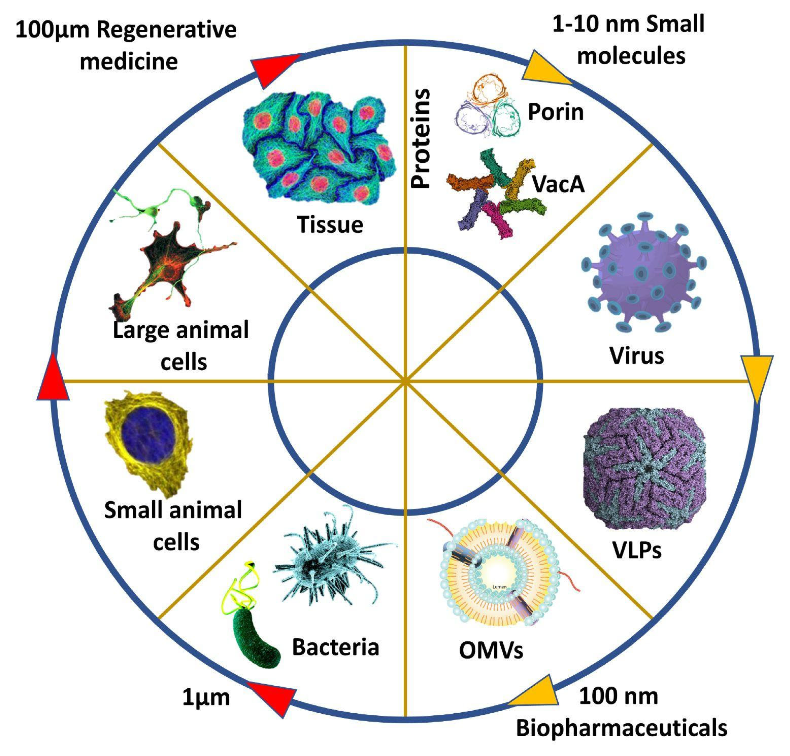
Vaccine adjuvants are substances that improve the immune capacity of a recombinant vaccine to a great extent and have been in use since the early 1900s; they are primarily short-lived and initiate antigen activity, mainly an inflammatory response. With the developing technologies and innovation, early options such as alum were modified, yet the inorganic nature of major vaccine adjuvants caused several side effects. Outer membrane vesicles, which respond to the stressed environment, are small nano-sized particles secreted by gram-negative bacteria. The secretory nature of outer membrane vesicles (OMV) gives us many benefits in terms of infection bioengineering. 

outer membrane vesicles (OMV) vaccines adjuvants

1. Introduction

The outer membrane vesicles (OMVs) are outer membrane vesicles of gram-negative bacteria, having stellar intrinsic immunostimulatory properties; they are nano-sized particles having a mean size between 20 to 200 nm; they are spherical buds of the outer membrane filled with periplasm and are usually bilayered. The bilayered nature of OMVs protects the lumen from immediate degradation by extracellular enzymes. OMVs can fuse with other cells allowing for the transfer of lumen contents and, therefore, providing a host-pathogen interaction. OMVs have several biological functions, including the delivery of proteins and toxins to target cells, transport of various effectors between bacterial cells, protection of nucleic acid during intercellular transport, and bacterial defense [1]. Several bacterial species produce OMVs, such as Escherichia coli, Pseudomonas aeruginosa, Shigella sp., Salmonella sp., Helicobacter pylori (H. pylori), Campylobacter jejuni, Borrelia burgdorferi, Vibrio sp., Neisseria sp. [2]. For the first time in 1959, it was observed that the cell-free filtrate of V. cholera induces an immune response in rabbits. Then in 1966, secretion of lipopolysaccharide in the form of spherical-shaped pouches was observed in E. coli culture grown on a lysine-limiting medium [3]. Indian Scientists Smriti Narayan Chatterjee and J. Das 1966–67 used Transmission-electron microscopy and discovered OMVs in V. Cholerae as membrane-bound vesicles produced by blebbing out and pinching off of outer membranes [4]; these OMVs have been alternatively termed “microvesicles”/“outer membrane fragments”, “blebs”, and “extracellular vesicles” [5].
Analysis of OMVs shows the presence of various outer membranes (OmpA, OmpC, and OmpF) and periplasmic proteins (AcrA and alkaline phosphatase). Several virulence factors are also present, which aid in the adhesion and invasion of host tissue. Periplasmic proteins present in the inner leaflet of the outer membrane show increased incorporation within OMVs compared to the tightly bound proteins of the inner membrane [2]. It was demonstrated that the lipid composition, SDS page protein profile, and specific activities of several membrane enzymes were similar in both the membrane vesicles and the outer membrane. Moreover, proteins including lipoproteins were less abundant in OMVs than in the outer membrane, indicating the likely origin of OMVs from specific outer membrane regions [6]. Multiple proteomic analyses reveal that inner membrane proteins and protoplasmic proteins are also abundant in OMVs. A recent study showed that OMVs are likely formed by cell lysis (The cell wall is degraded by endolysin, triggering explosive cell lysis, allowing for the fragmented membranes to round up forming OMVs), explaining the presence of inner membrane and cytoplasmic contents in OMVs [7]. Preferential packaging of proteins to OMVs is regulated by the total protein content of OMVs. Enrichment of proteins to OMVs takes place when protein contents of OMV are significantly higher with respect to the cellular concentration [8].
Nonetheless, lipids are integral structural components of OMVs. Just as in proteins, there are occurrences of lipids in the outer membrane but not in OMVs. Glycerophospholipids, phosphatidylglycerol, phosphatidylethanolamine, and cardiolipin are all important lipids present in OMVs and are associated with their curvature. Notably, a higher proportion of fatty acids results in their rigid structure [2]. OMVs carry luminal and surface-associated DNA, and a clear distinction is observed during DNase treatment; luminal DNA is present even after treatment compared to surface-associated DNA. Besides DNA, RNA, plasmid; and chromosomal DNA;phage DNA is also present in OMVs. Similar to protein incorporation, DNA is thought to be incorporated into OMVs after cell lysis during biogenesis [2] (Figure 1).
Figure 1. A vaccine as therapeutic candidates from well-defined small pharmaceuticals protein to OMV and large undefined regenerative medicines.

2. Formation of OMVs

There are three major models behind the formation of OMVs. The first is based on the loss or relocation of covalent linkages between the peptidoglycan layer and the outer membrane; as lipoprotein is associated with the linkage, it is hypothesized that a mutation in the lipoprotein gene could lead to potential hyper vesiculation. The missing cross-links along with an outer membrane grow faster, allowing the outer membrane to protrude and initiate vesiculation. In the second model, peptidoglycan fragments, misfolded proteins protrude into the periplasmic space, which exerts a turgor pressure on the outer membrane leading to the pinching off of OMVs. Another lipoprotein, vfgl, contributes to an increased peptidoglycan production or downregulation of transglycosylases, which again leads to an increased turgor pressure on the outer membrane, which then pinches off OMVs to decrease the pressure exerted [2]. The enrichment of the curvature-inducing molecules such as B-band lipopolysaccharide and the quinolone PQS of Pseudomonas aeruginosa forms the basis of the third model of OMV production. PQS is thought to increase anionic repulsions between lipopolysaccharide molecules leading to membrane budding due to isolated divalent cations [9]. Hydrophobic molecules such as PQS allow the formation of OMVs by inducing the outer membrane curvature, thus allowing the spread of these hydrophobic molecules in a hydrophilic environment. OMVs allow soluble proteins to be encased in an insoluble protective membrane sheath, often enabling the transport of these proteins in a hostile environment (having high temperatures or degrading enzymes such as proteases). Moreover, encasing such soluble proteins allows them to travel a greater distance while maintaining their concentration [10].
Another mechanism by which PQS causes blebbing is the asymmetric expansion of the outer layer of the outer membrane. However, this model is restricted as it is species-specific [9]. OMV-mediated secretion involves the presence of an insoluble membrane surrounding the soluble material. It can be regulated both temporally and spatially. In pathogenic bacteria, proteins known as adhesins present in the outer membrane, are crucial for colonization of the host tissue as they cause coaggregation. OMVs present multivalent complexes of membrane adhesins [10]. The bacterial interaction with the host triggers the release of OMVs containing adhesin proteins, which promote the adhesion of the bacterial cells to the epithelial linings of the host [2]. In addition to reaching a particular site, OMVs also have the advantage of targeting a particular site via the binding proteins present on the surface of the OMVs. Three different types of OMV delivery have been known. First is the targeted lysis, thus releasing the contents of the vesicles. Second is the attachment and subsequent fusion of the membrane with another cell allowing the lumen contents to be transferred; this mechanism carries importance regarding horizontal gene transfer between the bacterial strains and occasionally among bacterial species. Thus, having an important role to play in evolution and population dynamics. Another delivery mechanism is endocytosis within eukaryotic cells, which results in a display of bacterial epitopes to the immune system [10].

3. Species Producing OMVs

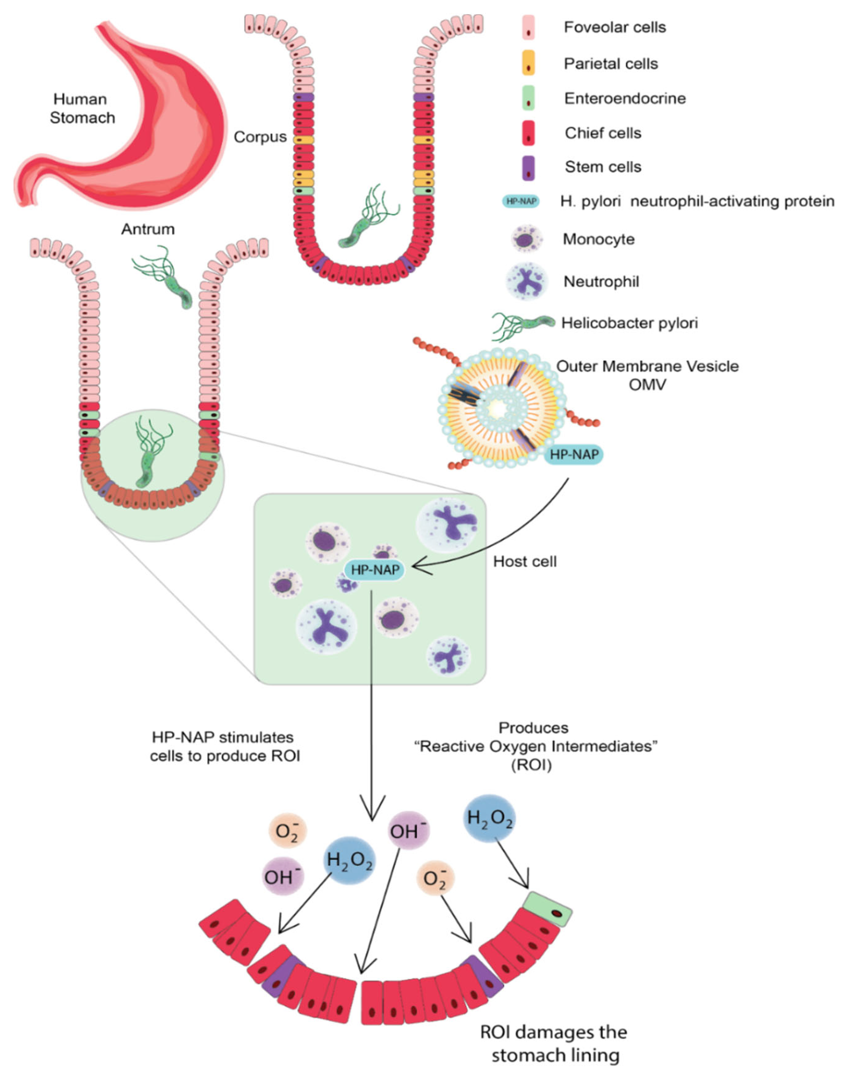
3.1. Helicobacter pylori

Helicobacter pylori (H. pylori), is a helical-shaped, microaerophilic, gram-negative bacteria [11]. It is catalase, urease, and oxidase-positive and contains 3–5 polar flagella for motility. H. pylori can colonize the stomach’s highly acidic environment by converting urea into ammonia with the help of urease; this ammonia neutralizes the acidic environment making it more hospitable for the bacterium to survive. It has evolved ways to interfere with the host’s immune responses, making it ineffective in eliminating the bacterium. Furthermore, their helical shape burrows into the stomach’s mucus lining and develops the infection. Usually, there are no symptoms of H. pylori infection, but it sometimes causes gastritis (inflammation of the stomach lining) or stomach ulcers. The infection for a longer time increases the risk of gastric cancer development [12]. H. pylori are observed to release OMV in both in vivo and in vitro conditions. OMV of H. pylori consists of phosphatidylglycerol (PG), phosphatidylcholine (PC), lysophosphatidylcholine (LPC), phosphatidylethanolamine (PE), lysophosphatidylethanolamine (LPE), cardiolipin, and lipopolysaccharide (LPS) [13]. Proteomic analysis suggests the presence of an allelic form of vacuolating cytotoxin (VacA), several porins and lipoprotein 20 (Lpp20), various adhesions such as sialic acid-binding adhesion (SabA), blood group antigen-binding adhesin (BabA), adherence-associated lipoprotein A (AlpA), OipA, adherence associated lipoprotein (AlpA), H. pylori neutrophil-activating protein (HP-NAP), urease, and some other associated virulence factors [14][15].
The cytotoxin-associated protein (CagA) is present on the surface of H. pylori OMVs; CagA is a virulence factor encoded inside the 40-kb cag pathogenicity island (PAI); this PAI also contains a type-IV secretion system that delivers CagA protein into the host cell. H. pylori neutrophil-activating protein (HP-NAP) is chemotactic for monocytes and neutrophils of humans and stimulates the cell to produce reactive oxygen species (ROS); ROS damages the gastric epithelium by releasing nutrients to the bacterium; therefore, delivering HP-NAP through OMV to gastric mucosa can increase the availability of nutrients to bacteria via ROS-mediated mucosal damage [11]. H. pylori OMV can trigger serum IgG response that contains antibodies having specificity for bacterial Lewis’s epitopes. Continuous release of OMVs in the cellular milieu contributes to the persistence of H. pylori in the stomach through maintaining biofilm. OMV of H. pylori contributes to chronic inflammation by delivering certain virulence factors and toxins to the gastric epithelium; they also deliver PE, LPS, and porins to the gastric epithelium, which respond by secreting IL-8 [16]. H. pylori OMV can induce apoptosis of gastric epithelium cells in a mitochondrial-independent and caspase-dependent pathway. It is observed that OMVs are internalized into the AGS cells in, which OMV-mediated delivery of peptidoglycan to cytosolic NOD-1 leads to the activation of NFkB and IL-8, this cytokine is acting as a chemoattractant to involve more neutrophils in the site of production; these H. pylori OMVs also regulated the proliferation of gastric epithelium cells, resulting in growth arrest and decreased cell viability. Once these OMVs are inside AGS cells, the VacA-mediated alkalization of late endosomal compartments leads to increased cytoplasmic iron levels and a decrease in GHS [11] (Figure 2).
Figure 2. Helicobacter pylori Neutrophil Activating Proteins (HP-NAP) stimulates the release of Reactive Oxygen species (ROS), which damages the gastric epithelium. The delivery of HP-NAP through OMV to gastric mucosa thus increases the nutrient availability to bacteria via ROS-mediated mucosal damage. Gastric Epithelium Lining Figure Inspiration from [17].

3.2. Neisseria meningitidis

Neisseria meningitides (N. meningitidis) is a gram-negative bacterium that causes severe disease in humans called meningococcal meningitis. It is an inflammation of the meninges, which are membranous coverings around the brain and spinal cord. The most common symptoms of meningitis include high fever, stiff neck, headache, vomiting, confusion, and sensitivity to light. The bacteria are transmitted through respiratory droplets from carriers. 12 serogroups have been identified in N. meningitides; among them, six major pathogenic serogroups can cause an epidemic, they are A, B, C, W, X, and Y (WHO). Vaccines developed using capsular polysaccharides of the pathogen for serogroup A and C have been in use since the 1960s and for serogroups A, C, W, X, and Y since the 1980s [18]; these capsular polysaccharide structures have high immunogenic properties and are conjugated with carrier proteins that induce a high immune response. However, this capsular polysaccharide vaccine approach is not suitable for serogroup B of N. meningitidis due to the low immunogenicity and the risk of autoimmune response due to structural homology of this capsular polysaccharide structure with polysialylated form of neural cell adhesion molecule (PSA-NCAM) present in brain tissue of fetus. Therefore, the vaccine approach for N. meningitidis serogroup B has been broadly focused on outer membrane vesicles proteins (OMVp) [19].

3.3. Campylobacter jejuni

Campylobacter jejuni (C. jejuni) is a spiral-shaped, rod-shaped, S-shaped, or curved-shaped gram-negative bacterium that is a significant cause of acute food-borne gastroenteritis found in animals; pigs, cattle, poultry, cats, and dogs. Clinical symptoms of the infection include fever, headache, nausea, abdominal pain, non-inflammatory diarrhea to inflammatory diarrhea with blood. It rarely causes death, mostly confined to infants, elderly persons, or patients with other severe diseases (WHO). The synchronized delivery of virulence factors such as adherence, the ability of invasion, production, and motility of toxins is a general mechanism through, which bacterial pathogens interact and cause damage to host cells and increase their survival rate. The mechanism of secretion of bacterial proteins and non-protein substances directly into the host cytoplasm requires direct contact of enteric bacteria with the host cells. Bacterial toxin such as cytolethal distending toxin (CDT) has been considered an essential factor for the pathogenesis of infection by C. jejuni; these CDTs belong to AB2-type toxins, consisting of three subunits-Cdt A, Cdt B, Cdt C [20]. Cdt A and C are binding proteins that deliver catalytic subunit Cdt B into the host cell; this Cdt B showcases DNase I-like activity and causes double-stranded DNA damage; this leads to cell cycle arrest at the G2/M stage and activation of DNA repair response [21].

4. OMV-Based Vaccine Delivery

Host immune responses are the first line of barrier to stopping any infection, but occasionally immoderate responses could lead to lethargic tissue damage. Thus, there is a need for a system that avoids excessive immune responses. Along with the adjuvant properties, OMVs also provide complete immunity as they carry the antigen of pathogens. Moreover, the non-replicative nature of OMVs makes them advantageous for antigen delivery to the host thus denying any fright of infection associated with whole cell vaccine against disease-causing pathogens. OMVs stimulate the innate immune system of the host via the activation of TLRs and NLRs as they contain various PAMPs such as lipoproteins, LPS, and pathogenic DNA fragments [22].

Host immune responses are the first line of barrier to stopping any infection, but occasionally immoderate responses could lead to lethargic tissue damage. Thus, there is a need for a system that avoids excessive immune responses. Along with the adjuvant properties, OMVs also provide complete immunity as they carry the antigen of pathogens. Moreover, the non-replicative nature of OMVs makes them advantageous for antigen delivery to the host thus denying any fright of infection associated with whole cell vaccine against disease-causing pathogens. OMVs stimulate the innate immune system of the host via the activation of TLRs and NLRs as they contain various PAMPs such as lipoproteins, LPS, and pathogenic DNA fragments [22].

Host immune responses are the first line of barrier to stopping any infection, but occasionally immoderate responses could lead to lethargic tissue damage. Thus, there is a need for a system that avoids excessive immune responses. Along with the adjuvant properties, OMVs also provide complete immunity as they carry the antigen of pathogens. Moreover, the non-replicative nature of OMVs makes them advantageous for antigen delivery to the host thus denying any fright of infection associated with whole cell vaccine against disease-causing pathogens. OMVs stimulate the innate immune system of the host via the activation of TLRs and NLRs as they contain various PAMPs such as lipoproteins, LPS, and pathogenic DNA fragments [22].

5. Uptake of OMVs by Host Cells

OMVs can enter through many ways: macropinocytosis, lipid raft-dependent or independent endocytosis, clathrin, caveolin, and dynamin-dependent entry [23]. LPS is usually delivered through endocytosis; O antigen structural region is crucial to OMV entry; if OMVs lack antigen, they can use clathrin-dependent endocytosis to enter the host cell. PAMPs facilitate TLR signaling to facilitate OMV entry into host cells [24]. OMVs mimic the pathogenicity of bacteria; these invade the epithelial lining of the host cells and present themselves to the body’s immune cells, such as neutrophils, macrophages, and dendritic cells in the submucosa, thus activating the immune response. B and T lymphocytes will also be stimulated, thus, enabling a comprehensive immune response. When treated with E. coli, OMVs can also cause apoptosis of host cells by developing the G2 phase arrest, or they can carry virulence factors that cause cell death of epithelial cells of the gut [25]. Neisseria meningitidis OMVs can stimulate the human neutrophils to produce cytokines and chemokines such as interleukin 1-beta, IL-8, tumor necrosis factor-alpha, macrophage inflammatory proteins 1 alpha, and 1 beta [26]. Gamma interferon-stimulated can maintain or increase the inflammation reaction. E. coli OMVs have cytotoxic necrotizing factors 1; these can reduce the membrane fluidity of polymorphonuclear leukocytes, thereby decreasing the levels of cytokines and chemokines [27]. OMVs make macrophages secrete proinflammatory substances such as chemokines and cytokines; they are phagocytosed by macrophages, activating them, then induce other immune molecules such as interleukin 1-beta, IL-8, tumor necrosis factor-alpha, and macrophage inflammatory proteins 1 alpha and 1 beta [28]. Legionella pneumophila OMVs facilitate the replication of the pathogen in the host macrophages [29].

OMVs can also induce macrophage remodeling leading to dysfunction of immune cells. OMVs can play an important role in secreting anti-inflammatory molecules such as IL 10. Porphyromonas gingivalis, Treponema denticola, and Tannerella forsythia OMVs phagocytosed by macrophages; they also stimulate the macrophages to release immune molecules such as TNFα, IL-8, and IL-1β and activate the NF-κB complex; they also primed and activated the inflammasome complex [30].

OMVs can also induce macrophage remodeling leading to dysfunction of immune cells. OMVs can play an important role in secreting anti-inflammatory molecules such as IL 10. Porphyromonas gingivalis, Treponema denticola, and Tannerella forsythia OMVs phagocytosed by macrophages; they also stimulate the macrophages to release immune molecules such as TNFα, IL-8, and IL-1β and activate the NF-κB complex; they also primed and activated the inflammasome complex [30].

6. OMVs-Based Therapeutics

The production of OMVs is very cost-effective, and it is quite easy to scale up the production of OMVs in inexpensive liquid media. OMV production can be increased by inducing hyper-vesiculation in the bacteria. Notably, hyper-vesiculation-based increased production of OMVs was successfully done in Neisseria meningitidis. OMVs are quite stable under various conditions due to the protective lipid outer membrane. N. meningitidis OMVs are shown to retain their antigenicity and enzymatic activity while stored at 4 °C. However, storage at 37 °C for three months led to a certain loss in antigenicity [31]. Phosphotriesterase, upon packaging into OMVs of E. coli retained enzyme activity 100 times more than free phosphotriesterase [32]. Notably, OMVs maintain the activity of phosphodiesterase even after multiple cycles of freeze and thaw.

7. Native OMV Vaccines

The benefits of OMVs in vaccines can be increased multiple folds by necessary modifications genetically. Meningitis B vaccine study has also proved safe and effective by engineering the adverse effects of lipopolysaccharide. The simultaneous reaction of multiple antigens/important antigens and heterologous antigens promotes their application extensively [33]. Natural OMVs are purified and concentrated, and the detergent-based extraction of OMVs helps to reduce the toxicity of the LPS complex. The term native OMV is used to describe intact OMV generation from cell supernatant and for concentration OMVs from dead cells using detergent-free disruption methods [34]. Currently, there are vaccines comprising capsular polysaccharides coupled to a carrier protein for several serogroups of N. meningitides; however, this is not possible for serogroup B because it mimics the molecular structures in the brain, thus leading to greater risks of autoimmunity [35]. Polysaccharide combined with antigen becomes immunogenic in infants and prime for memory anticapsular antibody response. To combat these meningococcal OMV vaccines have been developed using DOC detergents to release the OMVs, this also had the added benefit of reducing the toxicity of the LPS complex, however, there was the loss of certain important outer membrane lipoproteins and increased contamination with the inner membrane proteins and cytoplasmic contents [33].
The immune response in infants and children is largely targeted against surface accessible loops on a porin protein called Por A. Thus, these OMV vaccines are effective against one particular strain. To combat multiple strains OMV vaccines must be prepared from more than one strain of mutants expressing more than one type of Por A molecule. However, there are more than 20 different Por A molecules in many outbreaks, making it difficult to create an effective OMV vaccine. However, since the adult immune response is wider than that of children, an OMV vaccine can still be effective in adults [36].
In addition to serving as vaccines against meningococcal pathogens, OMVs also act as vaccines for other pathogens such as the Shigella flexneri; this vaccine could protect mice against shigellosis. With enhanced protection when nOMVs were encapsulated in poly nanoparticles. Shigella sonnei mdOMV is in clinical trials and can elicit anti-LPS O antigen-specific antibodies in healthy adults. nOMV vaccines derived from S. typhimurium protected against various serotypes due to antibodies being elicited against the outer membrane proteins. mdOMV vaccines produced high anti-O antigen-specific IgG responses, which were much more diverse when compared to those induced by glycoconjugates [37]. Demand for the Bordetella pertussis vaccine has grown over the past few years with an increased incidence of whooping cough; this has been associated with the change from whole-cell vaccines to acellular pertussis vaccines, which consist of few antigens associated with alum; though there has been a reduction in mortality rates, immunity was not provided against circulating strains of the bacteria [38].

8. Heterologous Vaccines

There are two ways by, which the antigenic content of OMVs can be enhanced. One is by inducing mutations so that the proteins present on the outer membrane are not released. The second is by introducing antigens onto the membrane by using autotransporters [33]. Surface-associated antigens would bind with B cells, and luminal antigens can trigger the cytotoxic T-cell response. Thus, depending upon the desired immune response, the OMV can be designed.
In addition to luminal antigen loading, surface-bound antigens can also elicit a strong immune response. Lipoprotein OspA in Neisseria meningitidis is a surface-exposed antigen. The surface-exposed antigen elicited a more robust OspA-specific antibody immune response against outer membrane proteins and LPS than the luminal antigen [39]. O antigen from Francisella tularensis has resulted in O antigen-specific IgG production in mice, protecting them against the lethal strain. Heterologous proteins can also be attached post OMV production by a spy catcher coupled with a protein attached to the spy tag [40]. OMVs activate pathogen-associated molecular patterns, including TLR2, TLR5, TLR 9, and TLR13, which recognize lipoproteins, flagellin proteins, and unmethylated CpG motifs; these then stimulate the corresponding Pattern associated Receptors on Antigen Presenting Cells. Human kidney cells have been transfected by expressing TLRs from Shigella, Salmonella, and Neisseria meningitides, mdOMVs from these bacteria have been modified to remove acyl chains from the LPS to reduce endotoxicity. Most of these trigger TLR2, TL4, and TLR5 to induce an immune response. The contribution of the other TLRs to stimulate the Pathogen Receptor cells is low [41].

9. OMV-Based Novel Adjuvants

OMV as an adjuvant is based on Pathogen Associated Molecular Pattern (PAMP), induction of dangerous molecules, and geographic concept. The PAMPs activate both the Pattern Recognizing Receptors (PRRs) and Toll Signalling Receptors (TLRs); these then recruit the cells into active immunity and stimulate the Antigen Presenting Cells (APCs). It is thus thought that OMVs could increase antigen uptake, cell surface expression, and immunostimulatory molecules, aiding in T cell production [42]. Danger molecules affect host cells, leading to mature T cells engaged in an enhanced immune response [43]. The geographic concept involves diverting antigens from the injection site to the tissue-draining lymph node by dendritic cells. Thus, OMVs have immunogenic properties, act as carriers, and show an inherent adjuvant effect [44].

Meningitidis MenB OMVs was used as an adjuvant with group A meningococcal capsular polysaccharide. Unlike other adjuvants causing hypersensitivity, these vaccines had low toxicity and elicited a strong T cell response. [45]. OMV derived from E. coli can stimulate the humoral and cell-mediated immunity mediated via the IFN-g and IL-17 T cell-dependent response production [46]. In HIV, in addition to inducing IFN-g and IL-17, T cell-dependent response and performing Th1 oriented response. Other particles such as Virus-Like Particles (VLP) induced a high anti-HIV IgG production by using OMV vaccines [47][48]. OMVs have been explored as an adjuvant by mixing them with other known antigens such as keyhole limpet hemocyanin and ovalbumin in the hepatitis B vaccine, triggering an enhanced immune response in the host cells. Lipoprotein OspA in Neisseria meningitidis is a surface-exposed antigen. The surface-exposed antigen elicited a more robust OspA-specific antibody immune response against outer membrane proteins and LPS than the luminal antigen [49]

10. Conclusions

OMVs are an emerging and promising platform for vaccine development, especially in non-regenerative and acellular vaccines. OMVs-based vaccines are more immunogenic in comparison to non-regenerative and VLP vaccines. OMV-based vaccines are safer than the whole pathogen attenuated vaccine as OMVs do not have the self-replicative capacity, and there is no evolutionary escaping. OMVs vaccines are potential candidates where whole-cell treatment approaches are not applicable. Moreover, the cons of OMV-based vaccines are their low yields and endotoxic effects. Notably, there is continuous improvement in extraction methods of OMVs to increase vesicle formation and genetic modifications to avoid possible endotoxicity. Various factors are responsible for elevated spontaneous vesicle formation in bacteria, such as temperature, stress, and antibiotic treatments. Host-associated antimicrobial peptides have immunomodulatory potential; they act by directing and evoking the proinflammatory (Th1/Th17) response. Importantly, OMV-based vaccines are a potential platform for vaccine development against various bacterial infections. Conclusively, OMV-based vaccines are the future of antibacterial vaccine development.

References

  1. Fulsundar, S.; Harms, K.; Flaten, G.E.; Johnsen, P.J.; Chopade, B.A.; Nielsen, K.M. Gene Transfer Potential of Outer Membrane Vesicles of Acinetobacter Baylyi and Effects of Stress on Vesiculation. Appl. Environ. Microbiol. 2014, 80, 3469–3483.
  2. Jan, A.T. Outer Membrane Vesicles (OMVs) of Gram-Negative Bacteria: A Perspective Update. Front. Microbiol. 2017, 8, 1053.
  3. Kulkarni, H.M.; Jagannadham, M.V.Y. Biogenesis and Multifaceted Roles of Outer Membrane Vesicles from Gram-Negative Bacteria. Microbiology 2014, 160, 2109–2121.
  4. Chatterjee, S.N.; Das, J.Y. Electron Microscopic Observations on the Excretion of Cell-Wall Material by Vibrio Cholerae. Microbiology 1967, 49, 1–11.
  5. Mayrand, D.; Grenier, D. Biological Activities of Outer Membrane Vesicles. Can. J. Microbiol. 1989, 35, 607–613.
  6. Hussain, S.; Bernstein, H.D. The Bam Complex Catalyzes Efficient Insertion of Bacterial Outer Membrane Proteins into Membrane Vesicles of Variable Lipid Composition. J. Biol. Chem. 2018, 293, 2959–2973.
  7. Turner, L.; Bitto, N.J.; Steer, D.L.; Lo, C.; D’Costa, K.; Ramm, G.; Shambrook, M.; Hill, A.F.; Ferrero, R.L.; Kaparakis-Liaskos, M. Helicobacter Pylori Outer Membrane Vesicle Size Determines Their Mechanisms of Host Cell Entry and Protein Content. Front. Immunol. 2018, 9, 1466.
  8. Bonnington, K.E.; Kuehn, M.J. Protein Selection and Export via Outer Membrane Vesicles. Biochim. Et Biophys. Acta (BBA) Mol. Cell Res. 2014, 1843, 1612–1619.
  9. Roier, S.; Zingl, F.G.; Cakar, F.; Durakovic, S.; Kohl, P.; Eichmann, T.O.; Klug, L.; Gadermaier, B.; Weinzerl, K.; Prassl, R.; et al. A Novel Mechanism for the Biogenesis of Outer Membrane Vesicles in Gram-Negative Bacteria. Nat. Commun. 2016, 7, 10515.
  10. Kulp, A.; Kuehn, M.J. Biological Functions and Biogenesis of Secreted Bacterial Outer Membrane Vesicles. Annu. Rev. Microbiol. 2010, 64, 163–184.
  11. Alfarouk, K.O.; Bashir, A.H.H.; Aljarbou, A.N.; Ramadan, A.M.; Muddathir, A.K.; AlHoufie, S.T.S.; Hifny, A.; Elhassan, G.O.; Ibrahim, M.E.; Alqahtani, S.S.; et al. The Possible Role of Helicobacter Pylori in Gastric Cancer and Its Management. Front. Oncol. 2019, 9, 75.
  12. Parker, H.; Keenan, J.I. Composition and Function of Helicobacter Pylori Outer Membrane Vesicles. Microbes Infect. 2012, 14, 9–16.
  13. Chmiela, M.; Walczak, N.; Rudnicka, K. Helicobacter Pylori Outer Membrane Vesicles Involvement in the Infection Development and Helicobacter Pylori-Related Diseases. J. Biomed. Sci. 2018, 25, 78.
  14. Olofsson, A.; Vallström, A.; Petzold, K.; Tegtmeyer, N.; Schleucher, J.; Carlsson, S.; Haas, R.; Backert, S.; Wai, S.N.; Gröbner, G.; et al. Biochemical and Functional Characterization of Helicobacter Pylori Vesicles. Mol. Microbiol. 2010, 77, 1539–1555.
  15. Mullaney, E.; Brown, P.A.; Smith, S.M.; Botting, C.H.; Yamaoka, Y.Y.; Terres, A.M.; Kelleher, D.P.; Windle, H.J. Proteomic and Functional Characterization of the Outer Membrane Vesicles from the Gastric Pathogen Helicobacter Pylori. Proteom. Clin. Appl. 2009, 3, 785–796.
  16. Ismail, S.; Hampton, M.B.; Keenan, J.I. Helicobacter Pylori Outer Membrane Vesicles Modulate Proliferation and Interleukin-8 Production by Gastric Epithelial Cells. Infect. Immun. 2003, 71, 5670–5675.
  17. Hancock, R.E.W.; Haney, E.F.; Gill, E.E. The Immunology of Host Defence Peptides: Beyond Antimicrobial Activity. Nat. Rev. Immunol. 2016, 16, 321–334.
  18. Acevedo, R.; Fernandez, S.; Zayas, C.; Acosta, A.; Sarmiento, M.; Ferro, V.; Rosenqvist, E.; Campa, C.; Cardoso, D.; Garcia, L.; et al. Bacterial Outer Membrane Vesicles and Vaccine Applications. Front. Immunol. 2014, 5, 121.
  19. Holst, J.; Martin, D.; Arnold, R.; Huergo, C.C.; Oster, P.; O’Hallahan, J.; Rosenqvist, E. Properties and Clinical Performance of Vaccines Containing Outer Membrane Vesicles from Neisseria Meningitidis. Vaccine 2009, 27, B3–B12.
  20. Ge, Z.; Schauer, D.B.; Fox, J.G. In Vivo Virulence Properties of Bacterial Cytolethal-Distending Toxin. Cell. Microbiol. 2008, 10, 1599–1607.
  21. Lara-Tejero, M.; Galán, J.E. CdtA, CdtB, and CdtC Form a Tripartite Complex That Is Required for Cytolethal Distending Toxin Activity. Infect. Immun. 2001, 69, 4358–4365.
  22. Kaparakis-Liaskos, M.; Ferrero, R.L. Immune Modulation by Bacterial Outer Membrane Vesicles. Nat. Rev. Immunol. 2015, 15, 375–387.
  23. Alaniz, R.C.; Deatherage, B.L.; Lara, J.C.; Cookson, B.T. Membrane Vesicles Are Immunogenic Facsimiles of Salmonella Typhimurium That Potently Activate Dendritic Cells, Prime B and T Cell Responses, and Stimulate Protective Immunity in Vivo. J. Immunol. 2007, 179, 7692–7701.
  24. van der Pol, L.; Stork, M.; van der Ley, P. Outer Membrane Vesicles as Platform Vaccine Technology. Biotechnol. J. 2015, 10, 1689–1706.
  25. Cheng, K.; Zhao, R.; Li, Y.; Qi, Y.; Wang, Y.; Zhang, Y.; Qin, H.; Qin, Y.; Chen, L.; Li, C.; et al. Bioengineered Bacteria-Derived Outer Membrane Vesicles as a Versatile Antigen Display Platform for Tumor Vaccination via Plug-and-Display Technology. Nat. Commun. 2021, 12, 2041.
  26. Rabets, A.; Bila, G.; Grytsko, R.; Samborskyy, M.; Rebets, Y.; Vari, S.G.; Pagneux, Q.; Barras, A.; Boukherroub, R.; Szunerits, S.; et al. The Potential of Developing Pan-Coronaviral Antibodies to Spike Peptides in Convalescent COVID-19 Patients. Arch. Immunol. Ther. Exp. 2021, 69, 5.
  27. Qing, G.; Gong, N.; Chen, X.; Chen, J.; Zhang, H.; Wang, Y.; Wang, R.; Zhang, S.; Zhang, Z.; Zhao, X.; et al. Natural and Engineered Bacterial Outer Membrane Vesicles. Biophys. Rep. 2019, 5, 184–198.
  28. Klimentová, J.; Stulík, J. Methods of Isolation and Purification of Outer Membrane Vesicles from Gram-Negative Bacteria. Microbiol. Res. 2015, 170, 1–9.
  29. Dragovic, R.A.; Gardiner, C.; Brooks, A.S.; Tannetta, D.S.; Ferguson, D.J.P.; Hole, P.; Carr, B.; Redman, C.W.G.; Harris, A.L.; Dobson, P.J.; et al. Sizing and Phenotyping of Cellular Vesicles Using Nanoparticle Tracking Analysis. Nanomed. Nanotechnol. Biol. Med. 2011, 7, 780–788.
  30. Gerritzen, M.J.H.; Salverda, M.L.M.; Martens, D.E.; Wijffels, R.H.; Stork, M. Spontaneously Released Neisseria Meningitidis Outer Membrane Vesicles as Vaccine Platform: Production and Purification. Vaccine 2019, 37, 6978–6986.
  31. Chandler, C.E.; Ernst, R.K. Bacterial Lipids: Powerful Modifiers of the Innate Immune Response. F1000Research 2017, 6, 1334.
  32. Ko, S.H.; Rho, D.J.; Jeon, J.I.; Kim, Y.-J.; Woo, H.A.; Kim, N.; Kim, J.M. Crude Preparations of Helicobacter Pylori Outer Membrane Vesicles Induce Upregulation of Heme Oxygenase-1 via Activating Akt-Nrf2 and MTOR–IκB Kinase–NF-ΚB Pathways in Dendritic Cells. Infect. Immun. 2016, 84, 2162–2174.
  33. Schetters, S.T.T.; Jong, W.S.P.; Horrevorts, S.K.; Kruijssen, L.J.W.; Engels, S.; Stolk, D.; Daleke-Schermerhorn, M.H.; Garcia-Vallejo, J.; Houben, D.; Unger, W.W.J.; et al. Outer Membrane Vesicles Engineered to Express Membrane-Bound Antigen Program Dendritic Cells for Cross-Presentation to CD8+ T Cells. Acta Biomater. 2019, 91, 248–257.
  34. Durant, L.; Stentz, R.; Noble, A.; Brooks, J.; Gicheva, N.; Reddi, D.; O’Connor, M.J.; Hoyles, L.; McCartney, A.L.; Man, R.; et al. Bacteroides Thetaiotaomicron-Derived Outer Membrane Vesicles Promote Regulatory Dendritic Cell Responses in Health but Not in Inflammatory Bowel Disease. Microbiome 2020, 8, 88.
  35. Laughlin, R.C.; Alaniz, R.C. Outer Membrane Vesicles in Service as Protein Shuttles, Biotic Defenders, and Immunological Doppelgängers. Gut Microbes 2016, 7, 450–454.
  36. Vidakovics, M.L.A.P.; Jendholm, J.; Mörgelin, M.; Månsson, A.; Larsson, C.; Cardell, L.-O.; Riesbeck, K. B Cell Activation by Outer Membrane Vesicles—A Novel Virulence Mechanism. PLoS Pathog. 2010, 6, e1000724.
  37. Bottero, D.; Gaillard, M.E.; Zurita, E.; Moreno, G.; Martinez, D.S.; Bartel, E.; Bravo, S.; Carriquiriborde, F.; Errea, A.; Castuma, C.; et al. Characterization of the Immune Response Induced by Pertussis OMVs-Based Vaccine. Vaccine 2016, 34, 3303–3309.
  38. Finne, J.; Leinonen, M.; Mäkelä, P.H. ANTIGENIC SIMILARITIES BETWEEN BRAIN COMPONENTS AND BACTERIA CAUSING MENINGITIS: Implications for Vaccine Development and Pathogenesis. Lancet 1983, 322, 355–357.
  39. Granoff, D.M. Review of Meningococcal Group B Vaccines. Clin. Infect. Dis. 2010, 50 (Suppl. S2), S54–S65.
  40. Tunheim, G.; Arnemo, M.; Næss, L.M.; Fjeldheim, Å.K.; Nome, L.; Bolstad, K.; Aase, A.; Mandiarote, A.; González, H.; González, D.; et al. Preclinical Immunogenicity and Functional Activity Studies of an A+W Meningococcal Outer Membrane Vesicle (OMV) Vaccine and Comparisons with Existing Meningococcal Conjugate- and Polysaccharide Vaccines. Vaccine 2013, 31, 6097–6106.
  41. Jäger, J.; Marwitz, S.; Tiefenau, J.; Rasch, J.; Shevchuk, O.; Kugler, C.; Goldmann, T.; Steinert, M. Human Lung Tissue Explants Reveal Novel Interactions during Legionella Pneumophila Infections. Infect. Immun. 2014, 82, 275–285.
  42. Martins, P.; Machado, D.; Theizen, T.H.; Guarnieri, J.P.O.; Bernardes, B.G.; Gomide, G.P.; Corat, M.A.F.; Abbehausen, C.; Módena, J.L.P.; Melo, C.F.O.R.; et al. Outer Membrane Vesicles from Neisseria Meningitidis (Proteossome) Used for Nanostructured Zika Virus Vaccine Production. Sci. Rep. 2018, 8, 8290.
  43. Muralinath, M.; Kuehn, M.J.; Roland, K.L.; Curtiss, R. Immunization with Salmonella Enterica Serovar Typhimurium-Derived Outer Membrane Vesicles Delivering the Pneumococcal Protein PspA Confers Protection against Challenge with Streptococcus Pneumoniae. Infect. Immun. 2011, 79, 887–894.
  44. Schild, S.; Nelson, E.J.; Bishop, A.L.; Camilli, A. Characterization of Vibrio Cholerae Outer Membrane Vesicles as a Candidate Vaccine for Cholera. Infect. Immun. 2009, 77, 472–484.
  45. Chen, L.; Valentine, J.L.; Huang, C.-J.; Endicott, C.E.; Moeller, T.D.; Rasmussen, J.A.; Fletcher, J.R.; Boll, J.M.; Rosenthal, J.A.; Dobruchowska, J.; et al. Outer Membrane Vesicles Displaying Engineered Glycotopes Elicit Protective Antibodies. Proc. Natl. Acad. Sci. USA 2016, 113, E3609–E3618.
  46. Chen, D.J.; Osterrieder, N.; Metzger, S.M.; Buckles, E.; Doody, A.M.; DeLisa, M.P.; Putnam, D. Delivery of Foreign Antigens by Engineered Outer Membrane Vesicle Vaccines. Proc. Natl. Acad. Sci. USA 2010, 107, 3099–3104.
  47. Shen, Y.; Torchia, M.L.G.; Lawson, G.W.; Karp, C.L.; Ashwell, J.D.; Mazmanian, S.K. Outer Membrane Vesicles of a Human Commensal Mediate Immune Regulation and Disease Protection. Cell Host Microbe 2012, 12, 509–520.
  48. Siegemund, S.; Schütze, N.; Freudenberg, M.A.; Lutz, M.B.; Straubinger, R.K.; Alber, G. Production of IL-12, IL-23 and IL-27p28 by Bone Marrow-Derived Conventional Dendritic Cells Rather than Macrophages after LPS/TLR4-Dependent Induction by Salmonella Enteritidis. Immunobiol. 2008, 212, 739–750.
  49. Tan, K.; Li, R.; Huang, X.; Liu, Q. Outer Membrane Vesicles: Current Status and Future Direction of These Novel Vaccine Adjuvants. Front. Microbiol. 2018, 9, 783.
More
Information
Contributors MDPI registered users' name will be linked to their SciProfiles pages. To register with us, please refer to https://encyclopedia.pub/register : , , , , , , , ,
View Times: 514
Revisions: 2 times (View History)
Update Date: 01 Dec 2022
1000/1000
Video Production Service