Submitted Successfully!
To reward your contribution, here is a gift for you: A free trial for our video production service.
Thank you for your contribution! You can also upload a video entry or images related to this topic.
Version Summary Created by Modification Content Size Created at Operation
1 -- 5561 2022-11-26 06:02:58 |
2 format change -20 word(s) 5541 2022-11-28 03:23:17 |

Video Upload Options

Do you have a full video?

Confirm

Are you sure to Delete?
Cite
If you have any further questions, please contact Encyclopedia Editorial Office.
Christodoulou, M.C.;  Palacios, J.C.O.;  Hesami, G.;  Jafarzadeh, S.;  Lorenzo, J.M.;  Domínguez, R.;  Moreno, A.;  Hadidi, M. Spectrophotometric Methods for Measurement of Antioxidant Activity. Encyclopedia. Available online: https://encyclopedia.pub/entry/36644 (accessed on 16 May 2024).
Christodoulou MC,  Palacios JCO,  Hesami G,  Jafarzadeh S,  Lorenzo JM,  Domínguez R, et al. Spectrophotometric Methods for Measurement of Antioxidant Activity. Encyclopedia. Available at: https://encyclopedia.pub/entry/36644. Accessed May 16, 2024.
Christodoulou, Marios C., Jose C. Orellana Palacios, Golnaz Hesami, Shima Jafarzadeh, José M. Lorenzo, Rubén Domínguez, Andres Moreno, Milad Hadidi. "Spectrophotometric Methods for Measurement of Antioxidant Activity" Encyclopedia, https://encyclopedia.pub/entry/36644 (accessed May 16, 2024).
Christodoulou, M.C.,  Palacios, J.C.O.,  Hesami, G.,  Jafarzadeh, S.,  Lorenzo, J.M.,  Domínguez, R.,  Moreno, A., & Hadidi, M. (2022, November 26). Spectrophotometric Methods for Measurement of Antioxidant Activity. In Encyclopedia. https://encyclopedia.pub/entry/36644
Christodoulou, Marios C., et al. "Spectrophotometric Methods for Measurement of Antioxidant Activity." Encyclopedia. Web. 26 November, 2022.
Spectrophotometric Methods for Measurement of Antioxidant Activity
Edit

The antioxidant potential can be measured by various assays with specific mechanisms of action, including hydrogen atom transfer, single electron transfer, and targeted scavenging activities. Understanding the chemistry of mechanisms, advantages, and limitations of the methods is critical for the proper selection of techniques for the valid assessment of antioxidant activity in specific samples or conditions. There are various analytical techniques available for determining the antioxidant activity of biological samples, including food and plant extracts. The different methods are categorized into three main groups, such as spectrometry, chromatography, and electrochemistry techniques. Among these assays, spectrophotometric methods are considered the most common analytical technique for the determination of the antioxidant potential due to their sensitivity, rapidness, low cost, and reproducibility.

antioxidative activity determination free radicals plant-based antioxidant phenolic compounds colorimetry

1. Introduction

Many studies have been conducted related to the oxidation origin of free radicals and the general role of antioxidants since the beginning of the 21st century. This interest was generated because free radicals are highly reactive and unstable molecules with a significant impact on the human biological system even though they are neutral. In fact, some important lipid-derived compounds, such as aldehydes, can produce negative effects on human health, although these compounds can naturally form during food processing, mainly linked to thermal treatments [1]. Radicals’ significant activity is an outcome of an atom that carries an unpaired electron. Due to this lack of outer-shell electrons, they are constantly searching to bind with another atom or molecule to stabilize themselves [2]. Despite antioxidant defense mechanisms, human cell damage accelerates aging and can play a critical role in the development of other diseases [3][4]. Further oxidative modification of biological macromolecules (e.g., lipids, proteins, and DNA) can result in tissue injury [5]. In understanding these occurrences and preventing them, a higher quality of life may be gained.
Recently, extensive research has been classified into different types of free radicals. The three main categories are: reactive oxygen species (ROS), reactive nitrogen species (RNS), and reactive sulfur species (RSS), which are formed from oxygen, nitrogen, and sulfur atoms, respectively [6]. Examples of ROS, RNS, and RSS include hydrogen oxide, hydrogen peroxide, singlet oxygen, alko-xyradical, peroxyl-radical, nitrogen monoxide, nitric oxide superoxide anion, hydroxyl anions, alkyl-thiol, etc. [2][6][7]. More specifically, the ROS group includes lipid peroxidation products and protein carbonyl species while the RNS group includes nitric oxide and peroxynitrites. Nitric oxide plays a key role in DNA damage, inflammation, cancer cell growth, and apoptotic malfunction, even though it has a lifespan of only a fraction of a second. In addition, peroxynitrites have the potential to cause lipid peroxidation, DNA damage, and long-term damage to all biomolecules. Similarly, sulfur species (RSS) may act in unison to damage biomolecules and, hence, extensive damage to genes in DNA may result in genes that produce ineffective proteins [4][8]. The origin of radicals is not yet well defined, but our own body often produces free radicals in the process of breaking down nutrients to create the energy that allows our bodies to function. Endogenous sources are multifaction in mitochondria, peroxisomes, endoplasmic reticulum, phagocytic cells, etc. while some exogenous sources may be air pollution, ultraviolet radiation, alcohol, smoking, contact with heavy metals, pesticides, and certain drugs such as halothane and paracetamol [9].
Fortunately, antioxidants can neutralize free radicals and reduce the risk of damage [10]. Antioxidants have become rapidly known for their health-promoting capabilities. By definition, the term “antioxidant” refers to a class of compounds with synthetic or natural orientation, which can act as chain-breaking antioxidant inhibitors, stopping the chain reaction of free radicals by complexing with them [11]. Apart from stopping the formation mechanism, an antioxidant compound must be able to scavenge radicals and form new ones that are stable [12]. Natural compounds such as these can be found in fruits, roots, vegetables, and plants [13][14][15][16]. In addition to this, a study conducted by Yashin et al. [17] suggests that the most biologically active compounds are contained in various spices and herbs.
The main representatives of antioxidants are vitamins A, C, and E; beta-carotene; anthocyanidins; phenols; flavonoids; phenolic acids, etc. [17]. Certainly, natural antioxidants that are consumed daily in our diet can protect our bodies and act as anticarcinogenic agents. Higher antioxidant and anticancer activities are also demonstrated in cases where there is a synergetic effect between different antioxidant natural molecules [18][19][20]. Endogenous defenses in humans have gradually improved over time, resulting in a balance between free radicals and oxidative stress. Enzymatic antioxidants and non-enzymatic oxidants are the two main types of antioxidants found in humans [6]. Antioxidant defense mechanisms attempt to scavenge reactive oxygen species and prevent their formation, although they are not always successful. The antioxidant network is complex, containing substances that are both endogenous and ingested. Enzymes called superoxide dismutase (SOD) convert O2 to H2O2 and eliminate it from the body [21]. However, because of the blood–brain barrier, antioxidants sometimes fail to provide adequate protection [22]. Numerous studies have demonstrated the necessity of antioxidants, but currently, the preferred choice of determination method is a controversial challenge. Antibody, fluorescence, light emission, spectrophotometric, chromatography, and electrophoretic techniques are the most widely used quantification procedures for determining the total antioxidant activity (TAA) [6][23][24].

2. Oxidation Process and Radicals

As mentioned before, free radicals are chemical entities (atoms, molecules, or ions), that have a single unpaired electron in one of their outer orbits, which makes them unstable and reactive [25]. The outcome is the formation of more radicals or unwanted side products as an electron attempts to bind with another. The majority have a half-life that depends on their environment. For example, the half-life of NO• or most oxygen species is a few minutes whereas the half-life of sulfur anion free radical is seconds. The initiation of the generation of radicals is any source of heat, ultraviolet irradiation, and air pollution, or naturally occurs in mitochondria. In humans, small molecules, peptides, proteins, and enzymes mostly contain nitrogen, oxygen, and sulfur, which serve a variety of functions in living creatures [26]. Nitrogen and oxygen are usually bonded in ‘chains’ of two to three atoms (peroxides, ozone, dinitrogen trioxide, etc.) while sulfur chains can be considerably longer (Table 1). These atoms have many oxidation states, and sometimes, during certain events and under certain circumstances, they release free radicals as side products [2][25][26]. During the propagation step, free radicals react with other molecules until their termination, where the free radicals bind together in a way that the chain is no longer propagated.
Table 1. Reactive oxygen (ROS), nitrogen (RNS), and sulfur (RSS) species and their non-free-radical species.
Reactive oxygen (ROS), nitrogen (RNS), and sulfur (RSS) species are formed both endogenously and exogenously [2]. The most prevalent free radicals that cause harm to biological systems are oxygen-free radicals, also known as ROS [27]. They are produced as a by-product of biochemical reactions by neutrophils and macrophages in mitochondria, peroxisomes, and other organelles [28]. Activated forms of ROS are illustrated in Table 1 and are usually separated as small particles that do not contain carbon atoms, such as •OH, in contrast with forms such as ROO•. Reactive sulfur species have also received attention for their role in oxidative stress, a phenomenon caused by an imbalance between the production and accumulation of reactive species in cells and tissues and the ability of a biological system to detoxify these reactive products [29]. Normally, sulfur radicals can be produced by hydrogen donation, enzymatic oxidation, and interaction with reactive oxygen species such as hydrogen peroxide, singlet oxygen, peroxynitrite, and superoxide [26]. When cellular thiols are oxidized, they form species that can oxidize and inhibit the action of proteins and enzymes. Currently, known sulfur-free radicals include disulfides and monosulifes, disulfide-S-oxides, and sulfenic acid agents (Table 1). According to Abedinzadeh et al. [30], the formation of highly reactive sulfur radicals participates in several different reactions, which lead to disulfide radical anion, thiyl peroxyl radical, and others. On the other hand, reactive nitrogen species (RNS) are a class of antimicrobial compounds produced by nitric oxide and superoxide. When they combine, they are converted to a peroxynitrite free radical. Rapid protonation of peroxynitrite anion in vivo gives peroxynitrous acid (ONOOH), which acts as an electrophilic nitrating agent for tyrosine and tryptophan sidechains in proteins. The decomposition of peroxynitrous acid can generate hydroxyl radicals, which can subsequently damage human DNA [31]. Further damage to DNA clones has been reported as the presence of nitric oxide free radical is related to dose-dependent DNA strand breaks and the transformation from cytosine to uracil and 5-methylcytosine to thymine [32]. The endogenous antioxidant defense system can also be overwhelmed by ROS, RNS, and RSS, resulting in cellular damage and dysfunction, which leads to a variety of illnesses. ROS and RNS are key regulatory mediators in signaling pathways at low concentrations, but they are toxic in moderate and high quantities, inactivating critical cellular components [33].
Nitric oxide (NO•) has two purposes in health and sickness and its level influences both. Nitric oxide has the potential to act as an active marker of cancer progression during physiological and pathological processes by encouraging angiogenesis or the production of new blood vessels [34]. Furthermore, by upregulating p53, poly (ADP-ribose) polymerase, and DNA-dependent protein kinase, NO• may affect tumor DNA repair processes (DNA-PK). The use of NO• in cancer research has significant therapeutic implications for disease detection and treatment. At the same time, the ratio of ROS/RNS is engaged in a range of physiological activities, including immunological function (i.e., protection against harmful microorganisms), cellular signaling pathways, mitogenic response, and redox regulation, and has beneficial effects at moderate or low levels. However, at higher ratios of ROS/RNS, oxidative and nitrosative stress can occur, which can destroy biomolecules as the antioxidant and oxidant levels are unbalanced [9][35]. Increasingly more free radicals build up, causing extensive damage to macromolecules, including nucleic acids, proteins, and lipids.
Peroxidation of lipid products and protein carbonyls is only one of the side effects of ROS when nitric oxide and peroxynitrites are produced from nitrogen radicals [4]. Therefore, any anomalies that occur in these crucial structural components have been related to the onset of a variety of neurodegenerative diseases, such as Alzheimer’s, Parkinson’s, etc. [36]. A study by Porter et al. [37] indicated that malondialdehyde (MDA, ROS agent) interacts with low-density lipoproteins and, as a result, lipid peroxidation forms, which indirectly causes atherosclerosis. Additionally, ONOO is another major RNS player that acts as a lipid peroxidation catalyst, which causes membrane and lipoprotein disruption. In the growth of cancer, ONOO and MDA act as cytotoxic and mutagenic agents, promoting DNA damage through mutations, resulting in decreased tumor suppressor gene expression or enhanced oncogene expression [38]. The possible consequences of the effects of lipids and proteins include tissue damage, neurological illnesses, cancer, cardiovascular diseases, cataracts, rheumatoid arthritis, asthma, stroke, myocardial infarction, chronic heart failure, diabetes, and many other neurodegenerative disorders [4][22][31].

3. Classification of Natural Antioxidants

Antioxidants can be separated into two main categories: synthetic and natural, which are derivatives of fruits, herbs, and plants [39][40][41]. Additionally, different plant by-products are also an economic source of natural antioxidants [42]. Nowadays, the employment of synthetic antioxidants in food, such as butylated hydroxytoluene (BHT) or butylated hydroxyanisole (BHA), has raised social concern, as these compounds are effective and relatively cheap to produce, but they can generate allergies and serious problems for human health in the long term [43][44]. This is also the major reason why many companies are trying to replace compounds synthesized in the laboratory with natural antioxidants to prevent oxidation in food products and secure a healthier lifestyle [45]. However, antioxidants must meet some of the following criteria to be utilized in this task. Firstly, they must be efficient, and their utilization must be cost-effective. Secondly, large-scale usage is not practicable if they are too expensive or complex to synthesize or isolate. Finally, they need to be kept at low concentrations, as these substances can be harmful to people at extremely high levels.
It is well known that plants, fruits, vegetables, herbs, seeds, and other natural sources present a large cocktail of antioxidants, such as phenolic compounds, carotenoids, and vitamins [16]. Due to this fact, a diet rich in fruits and vegetables is usually recommended in order to receive all the benefits. By doing so, it is possible to prevent or delay certain diseases, such as cardiovascular diseases or diabetes [43][46]. This variety of phenolic acids and flavonoids, along with a general classification of antioxidants, is shown in Figure 1. Additionally, some by-products of the food and agricultural industries, such as shells or peels, have been and continue to be investigated for the extraction of antioxidants. It is also important to note that, depending on the type of the plant and its morphological parts, the antioxidant capacity can vary significantly, as the antioxidant capacity of the leaves is not the same as that of the stem [47]. Based on this, different processes such as extraction, separation, and characterization of natural antioxidants have been investigated over the years.
Figure 1. Classification of natural antioxidants [43].
According to Figure 1, natural antioxidants can be classified into endogenous and exogenous antioxidants. Endogenous antioxidants are synthesized internally by the metabolism while exogenous antioxidants are obtained mostly in plants. The combination of endogenous and exogenous antioxidants in the human body helps maintain the nucleophilic tone, which translates into a healthy physical state [46]. Endogenous antioxidants can be divided into enzymatic and non-enzymatic. Enzymatic antioxidants act as the first line of defense in the human body while non-enzymatic antioxidants usually act as the second line of defense. The main representatives of enzymatic antioxidants with the highest effectiveness are superoxide dismutase (SOD) and catalase (CAT). SOD is responsible for obtaining O2 and H2O2 from the O2 radical. Then, CAT takes H2O2 and converts it to H2O and O2 [48]. Non-enzymatic proteins such as albumin and transferrin are also endogenous antioxidants. Proteins of this type are capable of trapping metal ions, avoiding the formation of new reactive species [46]. Exogenous antioxidants can also be divided into many different compounds. The most significant ones that are present in the diet of an average person are phenolic compounds such as flavonoids and phenolic acids, vitamins such as ascorbic acid (C) and tocopherol (E), and carotenoids [44].
In the case of vitamins, those that are water-soluble, such as ascorbic acid, are responsible for stopping free radicals present in the aqueous phase. Fat-soluble vitamins, such as tocopherol, are present in cell membranes, preventing their degradation [48]. The largest groups of exogenous natural antioxidants are phenolic structures. Within them, a distinction can be made between phenolic acids and flavonoids. Both are present in plants and this is also the reason why plant-derived foods contain a large amount of these exogenous antioxidants. As can be seen in Figure 1, these acids are divided into two groups depending on whether they are formed from benzoic or cinnamic acid. Some examples are caffeic acid (derived from hydroxycinnamic acid) and vanillic acid (derived from hydroxybenzoic acid). It should be noted that compounds derived from hydroxybenzoic acid have a lower antioxidant capacity than those derived from hydroxycinnamic acid. In general, it is estimated that the average person consumes about 200 mg/day of phenolic acid during the day [43].
On the other hand, flavonoids are present in large amounts in plants, formed from primary metabolites. Plants can transform the amino acids tyrosine and phenylalanine into new compounds. All of them have a general structure consisting of 3 phenyl rings and another heterocyclic ring containing an oxygen atom, forming a 15-carbon skeleton. Many of these compounds with variations in the two phenyl rings are found in nature [49]. Flavonoids are also present in the diets of people, reaching higher levels of daily intake than phenolic acids. Delving a little deeper into this group of compounds, flavonols are more common than flavones. They accumulate in the leaves and skin of plants and can act as complex-forming agents with metal ions due to the presence of carbonyl and hydroxyl groups in their structure [43].

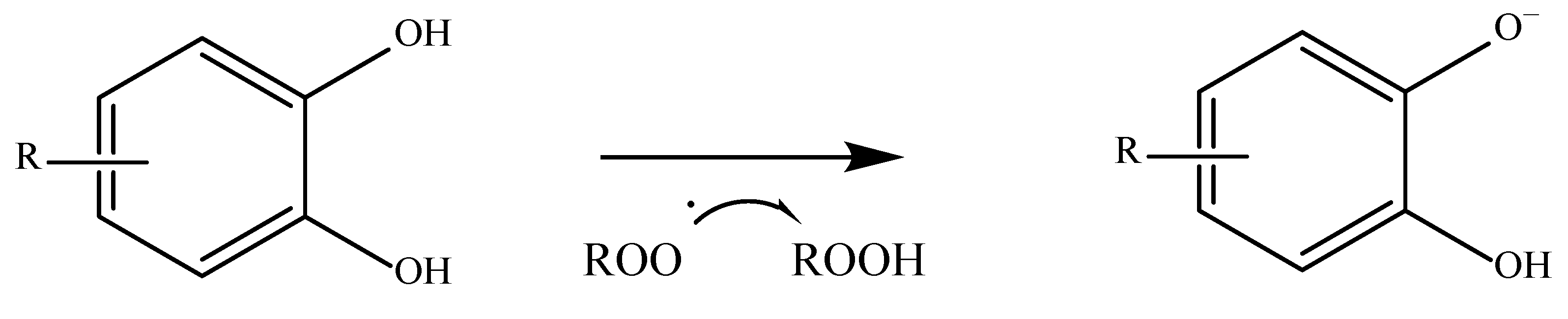
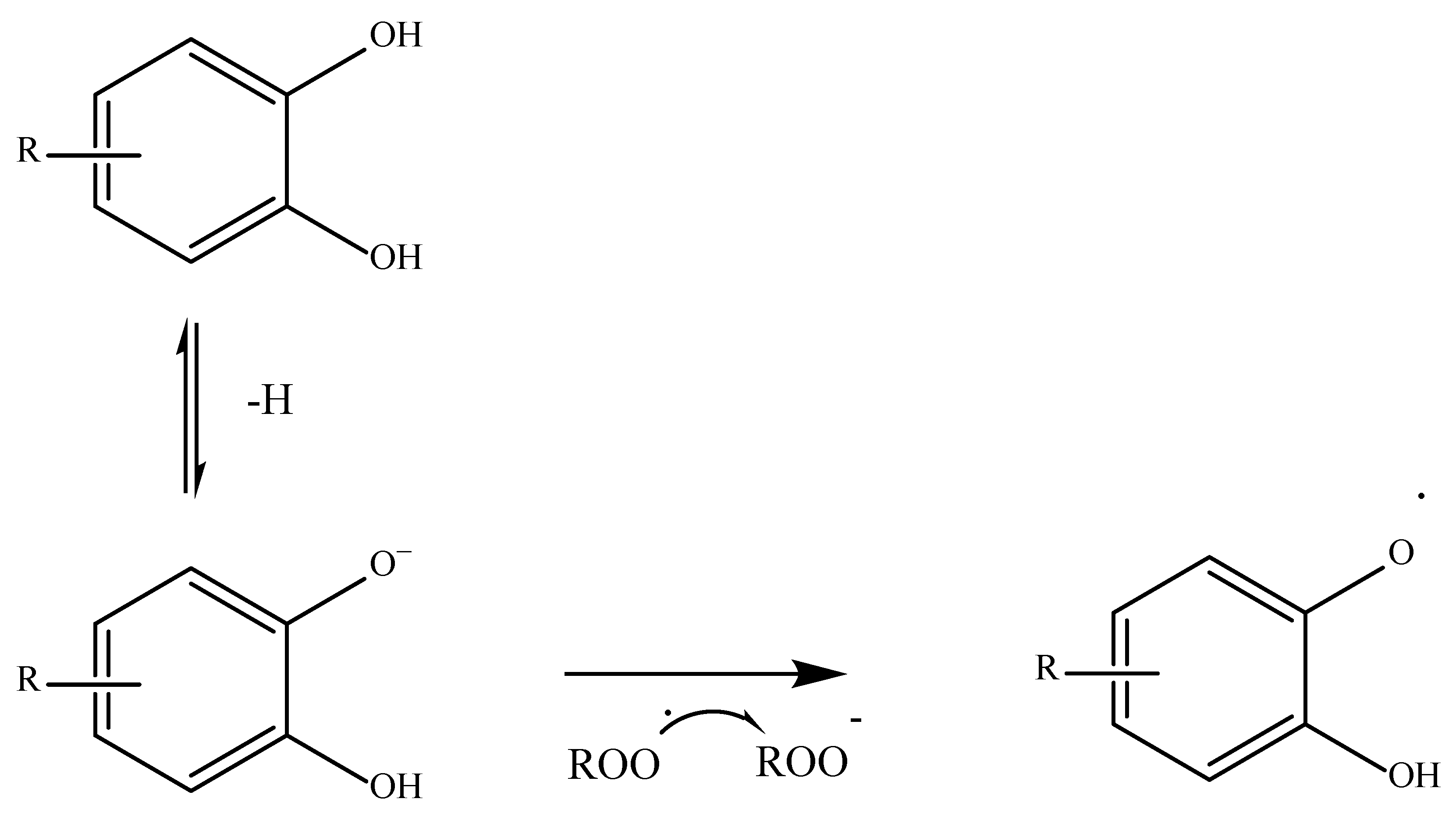
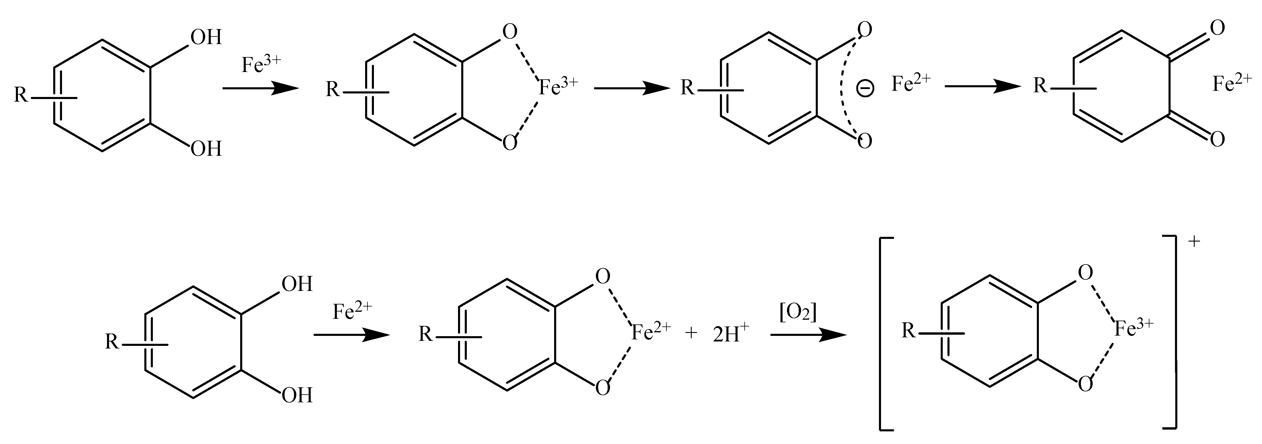
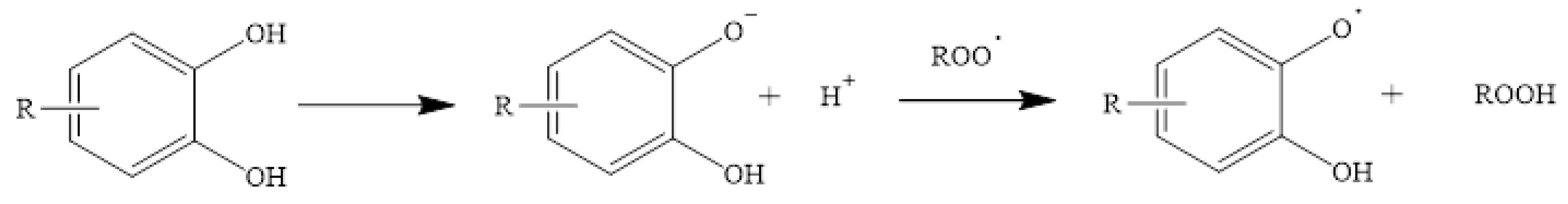
4. Natural Antioxidant Mechanism in Radical Scavenging

Antioxidants have a differing capacity to stop the propagation of free radicals. The important factors influencing this are both the structure of the antioxidant and the structure of the compound to be oxidized, the presence of pro-oxidants, and the concentration of all of them. In addition, the region in the organism where all these substances are present and react together must be considered. There are various ways in which antioxidants carry out their work and there are numerous variables that can affect the antioxidant capacity [48]. This section will show some of the known mechanisms used by natural antioxidants in dealing with the propagation of free radicals. As the number of natural antioxidants is very large, only a selection of the best-known and most important ones will be discussed.
Starting with phenolic acids, which are among the most abundant exogenous antioxidants, their ability to neutralize free radicals depends on several things, including the number of hydroxyl groups present on their aromatic ring, their position on it, and the presence of other substituents. As a general rule, the greater the substitution of the aromatic ring, the greater the difference in the antioxidant activity of these compounds. An aromatic ring that is unsubstituted cannot act as a hydrogen donor and therefore has a lower antioxidant capacity [50][51]. There are many different mechanisms by which an antioxidant compound can perform its function. The first and most common is known as HAT or hydrogen-electron transfer. This mechanism is used not only by phenolic acids but by all antioxidants that have a labile hydrogen atom in their structure. These compounds give up their hydrogen to stabilize the radical. Once this is carried out, the antioxidant compound becomes a radical itself, but it can be stabilized and reach a state where it is harmless [52]. A way of measuring how easily a hydrogen atom of an antioxidant compound can react with a free radical is the bond dissociation energy. The lower the value, the easier a hydrogen atom is transferred [53]. An example of this mechanism is shown in Figure 2.
Figure 2. Phenolic acid derivative neutralizing a free radical via HAT [43].
Another mechanism widely used by phenolic acids is the so-called SET or single electron transfer. In this case, the antioxidant gives up an electron to the free radical to stabilize it in an anionic form as shown in Figure 3 [43][52].
Figure 3. Phenolic acid derivative neutralizing a free radical via SET [43][52].
The third and fourth mechanisms by which the various phenolic acids exert their antioxidant capacity are known as transition metal chelation and sequential proton loss electron transfer (SPLET). The metal chelation mechanism is the ability of certain antioxidants to chelate transition metals, preventing them from catalyzing reactions that produce free radicals inside an organism (Figure 4). Some of these metals are Fe, Cu, and Mn [52][54].
Figure 4. Phenolic acid derivative chelating Fe3+ and Fe2+ [43][52].
On the other hand, SPLET, or sequential proton loss electron transfer, occurs when the antioxidant compound donates a proton to a free radical and transforms into an anion, which subsequently donates an electron to stabilize itself [52]. An example of this is shown in Figure 5.
Figure 5. Phenolic acid derivative neutralizing a free radical via SPLET [52].
In the previous section, it was pointed out that antioxidants derived directly from cinnamic acid have a higher antioxidant capacity than those derived from benzoic acid. This is due to the presence of the double bond of cinnamic acid, which can conjugate with the electron cloud of the aromatic ring, giving it a greater capacity to stabilize reactive species. Moreover, since the carbonyl group of cinnamic acid derivatives is distant from the aromatic ring, their antioxidant activity is higher than that of benzoic acid derivatives [48]. Flavonoids, as phenolic structures, also present these mechanisms. Their ability to neutralize free radicals is influenced both by the type of catechol ring present in their structure and by the presence of hydroxyl groups. Additionally, their double bond plays a critical role, as it can be conjugated and provide greater structural stability [43]. An example of a typical flavonoid structure is shown in Figure 6.
Figure 6. Most common structure of flavonoids. R1-R5 can range from hydrogen atoms to hydroxyl or methoxy groups [43].
Apart from phenolic structures, there are many other compounds in exogenous antioxidants. Among vitamins, tocopherol, or vitamin E, is a fat-soluble vitamin present in cell membranes. Although this compound has a phenolic structure, it is classified within the group of vitamins. There are, in turn, different classes of tocopherols depending on their structure. α-Tocopherol is the most abundant form of vitamin E in nature [43][46]. This compound uses the SPLET mechanism to exert its antioxidant activity (Figure 7).
Figure 7. α-Tocopherol neutralizing a free radical via SPLET [43][46].
In the case of endogenous natural antioxidants, the non-enzymatic ones, such as transferrin and albumin, use the mechanism of metal chelation to trap metal ions as already mentioned. As for those that are enzymatic, such as SOD and CAT, they catalyze a series of reactions essential for the correct functioning of the organism [55]:

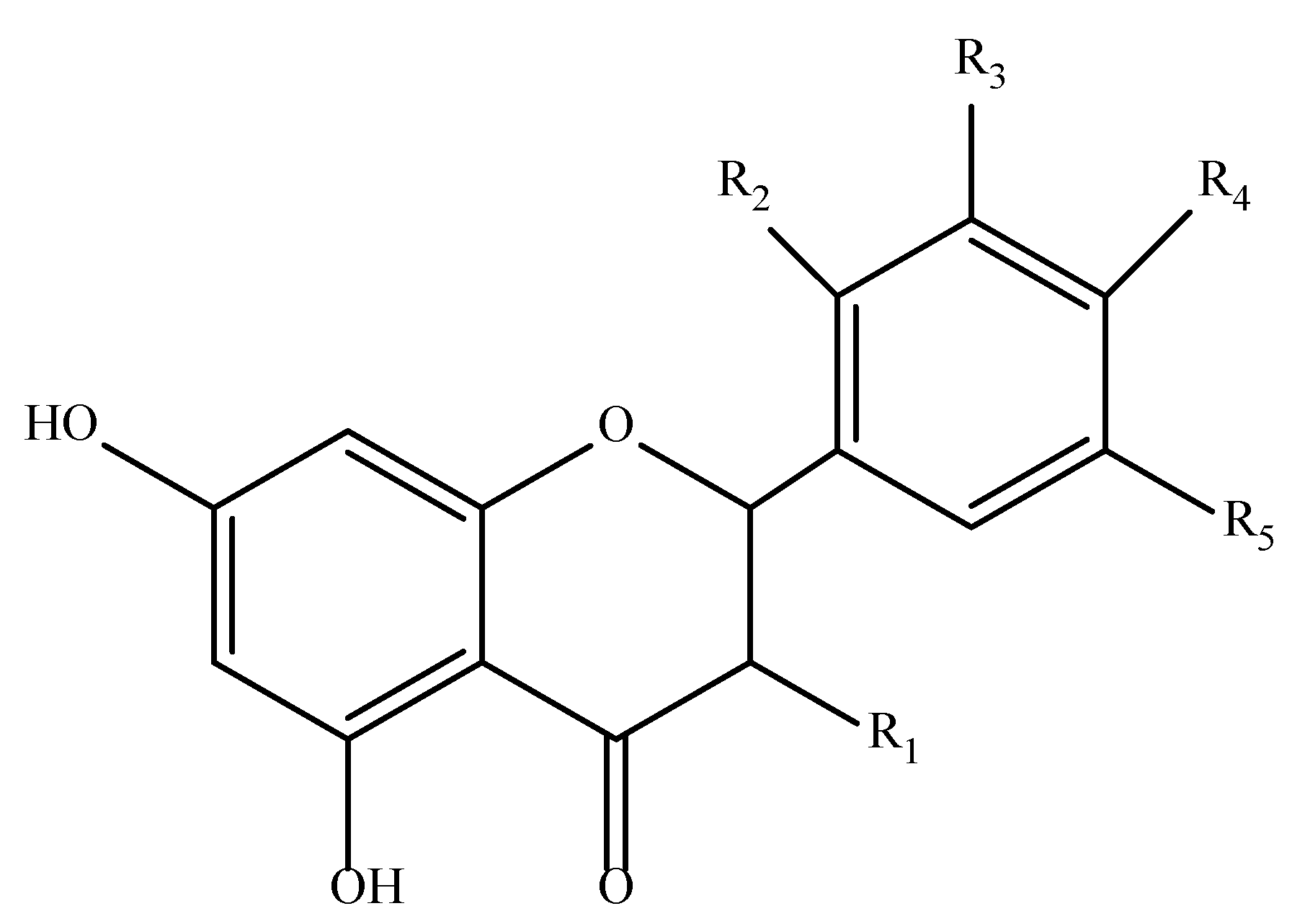
5. Spectrophotometric Methods for Measuring Antioxidant Activity

Based on the previous discussion, it clear that it is critical to investigate the techniques that can be used to determine the total antioxidant activity (TAC) and total phenolic content (TPC). In this context, spectrophotometric (colorimetric and fluorescence) tests have received more attention as they are fast, reproducible, easy, and cheap [6][48][56]. Colorimetric assays, which are the most famous, such as DPPH, FRAP, ABTS, etc., change their color due to an electronic transition in atoms or molecules. A change in the electronic transitions affects how much light is absorbed by the molecules, which in turn alters the color of the molecules. Numerous complexes enter an excitation state after receiving an electron. The production of brightly colored complexes is caused by the fact that the excitation energy needed for an electron to transition from one energy level to another frequently falls in the visible area of the electromagnetic spectrum. Because of the different types of donors and acceptors involved, the absorption wavelength of the transition bands is unique. Any wavelength from 400 to 750 nm is visible as red, orange, yellow, green, blue, and violet [57]. All spectrophotometric methods are quantification techniques as a result of the regression line and regression equation of different concentrations of standards [58]. Depending on each case, the antioxidant structure and properties and the solubility and partition coefficient dictate the prevailing mechanism in a given system and guide the selection of the optimum assay [56][59]. Table 2 illustrates the spectrophotometric assays that will be addressed in more detail, along with their absorption maxima (λmax), fundamental principle, and any observed color shifting.
Table 2. Spectrophotometric parameters of each assay.

Assay

nm

Principle of Method

Determination

Color Shifting

Reference

From

To


DPPH

515–520

Antioxidant reaction with free organic radicals

Colorimetry

Antioxidants 11 02213 i001

[10][58][60]

Folin–Ciocalteu

760–765

The reductive capacity of antioxidants to determine the total phenolic content

Colorimetry

Antioxidants 11 02213 i002

[58][61][62]

CUPRAC

450–490

Measures TAC of the reduction of Cu (II) to Cu (I) by antioxidants

Colorimetry

Antioxidants 11 02213 i003

[48][63][64]

FRAP

593

Measures the antioxidant potential through the reduction of Fe (III) to Fe (II) by antioxidants

Colorimetry

Antioxidants 11 02213 i004

[23][43][48][64]

ABTS

414, 645–650, 734, 815–820

Measures the relative ability of antioxidants to scavenge the ABTS generated in the aqueous phase

Colorimetry

Antioxidants 11 02213 i005

[6][23][63][64][65][66]

ORAC and HORAC

485–525 and 485–535

Antioxidant reaction with peroxyl radicals and quench OH radicals generated by a Co(II)-based Fenton-like system

Loss of fluorescence of fluorescein

Antioxidants 11 02213 i006

[5][24][48][65][67]

TBA-TBARS

532–535

Based on the reactivity of malondialdehyde (MDA) with TBA to produce a red color

Colorimetry

Antioxidants 11 02213 i007

[23][68][69][70]

FOX

550–560

Measure the levels of hydrogen peroxide in biological systems by the oxidation of Fe(II) to Fe(III)

Colorimetry

Antioxidants 11 02213 i008

[23][71][72]

FTC

500

Measure the levels of hydrogen peroxide as the ferric ion is converted by an oxidant from a ferrous ion

Colorimetry

Antioxidants 11 02213 i009

[23][70][73]

β-Carotene Bleaching Assay

440

Measure the levels of peroxyl radicals as β-carotene blenched

Colorimetry

Antioxidants 11 02213 i010

[23][74]

Hydrogen peroxide scavenging

460

Total oxidant scavenging capacity of antioxidants

Fluorescence

Antioxidants 11 02213 i011

[10][75]

Superoxide radical scavenging

560–562

Total oxidant scavenging capacity of antioxidants

Colorimetry

Antioxidants 11 02213 i012

[75][76]

Nitric oxide radical scavenging

540

Total oxidant scavenging capacity of antioxidants

Colorimetry

Antioxidants 11 02213 i013

[75]

Peroxynitrite Scavenging

485, 505, 529–530, 611

Total oxidant scavenging capacity of antioxidants

Fluorescence

Antioxidants 11 02213 i014

[77][78]

6. Advantages and Limitations of Spectrophotometric Assays

Antioxidant scavenging is becoming more interesting to scientists, who are attempting to understand the mechanisms involved in biological systems, the antioxidant capacity of food, and the free radicals of various substrates. Alternative methods for the measurement of antioxidant activity are still required (liquid and gas chromatography, electrophoretic procedures, etc.), although spectrophotometric applications are important. The benefits of employing spectrophotometric TAC assays are numerous and include their ease of use, low cost per sample, quick turnaround times, and the ability to be carried out manually, semi-automatically, or automatically. However, many parameters can affect the results of the measurements, such as the working pH area, the temperature of the current reaction, the applicability of the assay to both hydrophilic and lipophilic compounds, and others [48][66][79][80]. Therefore, there is an imperative need to select the applicable assay in each case, as the ABTS and CUPRAC tests can detect both hydrophilic and lipophilic antioxidants and FRAP and FC exclusively detect hydrophilic antioxidants while others such as DPPH can only be applied to hydrophobic systems. At the same time, interferences that may appear could affect the color of the food matrix and can be absorbed in the same area as the antioxidants [81].
The TAC tests have the benefit of being able to quantitatively assess the antioxidant components of a sample. It takes a lot of time and effort to measure each antioxidant component separately [82]. Several studies on food, plants, and human body fluids have been the subject of several years of investigation. Cao et al. [83] and Prior et al. [59] observed no link between serum ORAC and TEAC or between serum FRAP and serum TEAC. Furthermore, to demonstrate how these approaches differ from one another, a comparative analysis of the antioxidant capacities of 30 plant extracts was performed using the DPPH, ABTS, and FRAP tests [84]. The FRAP and ABTS assays had the highest correlation (0.946) while the ABTS and DPPH assays had the lowest correlation (0.906).
Undoubtedly, one of the most common assays is the DPPH approach. The application of this test facilitates an understanding of a variety of chemical processes and offers several obvious advantages, such as affordability, experiment simplicity, reproducibility, applicability at room temperature, and automation possibilities [85]. However, the overlapping spectra of substances that are absorbed in the same wavelength range as DPPH is a significant drawback. For instance, anthocyanins exhibit significant absorption in the same wavelength range (500–550 nm) as DPPH, which could introduce interference into the data and affect how it is interpreted [56]. On the other hand, CUPRAC reagent is more stable and convenient to use than other chromogenic reagents (e.g., ABTS, DPPH). The CUPRAC assay works best at a pH of 7.0, which is very similar to the physiological pH (7.4) and simulates antioxidant action in natural settings. Furthermore, it is characterized by robustness in contrast to free radical reagents, such as DPPH, as it is not affected by physiological conditions such as air, humidity, and sunshine [48]. Additionally, CUBRAC, like the ABTS assay, is very selective because it has a lower redox potential than the Folin or ferric ion-based approaches. Additionally, the CUPRAC reagent does not cause oxidation of simple sugars or citric acid, which are not considered as real antioxidants, but the majority of phenolic antioxidants are readily oxidized due to their advantageous redox potentials. Moreover, the CUPRAC reagent can easily oxidize several antioxidants that are resistant to the FRAP, FOX, and FTC assays, with perfectly linear absorbance–concentration curves [81]. Although, this assay does not assess antioxidant enzymes or certain thiol antioxidants, such as glutathione [48][63].
Another favorable assay is the Folin–Ciocalteu test. Numerous benefits also exist for the use of FC to quantify TPC, including its ease of use, repeatability, and robustness. In fact, according to a previous report, there is a strong correlation in the Folin-determined concentration between FRAP and ABTS assays (0.946) in contrast with ABTS and DPPH assays (0.906) [17]. It does, however, have significant shortcomings. This test is sensitive to pH, temperature, and the reaction duration. Therefore, careful selection of the reaction state is crucial for achieving consistent and trustworthy findings. Due to the involvement of non-phenolic reducing agents present in the system when reducing the Folin–Ciocalteu reagent, TPC overestimation is a significant concern for the Folin–Ciocalteu test. Reducing sugars and certain amino acids are some of these pollutants [85].
Indirect measures, which are based on determining a sample’s capability to reduce a metal complex, can also be performed with FRAP. One major limitation of the FRAP assay is that an aqueous testing apparatus is required, but it provides quick, repeatable findings. Consequently, a water-soluble antioxidant must be used as the reference [23]. In addition, the propensity of blue to precipitate, form a suspension, and stain the measurement vat is a drawback of this FRAP test. Because of this, the timing of the addition of Fe3+ (FeCl3) is crucial and may lead to inaccuracies in the interpretation of the outcomes [48]. In fact, FRAP results can vary tremendously depending on the timescale of analysis. Moreover, after several hours of reaction time, the absorption of polyphenols such as caffeic acid, tannic acid, ferulic acid, ascorbic acid, and quercetin gradually increases. Some polyphenols have slower reactions and need more time to be detected while others react rapidly with iron complexes, leading to degradation into other compounds with differing or lower reactivity [59]. Therefore, a single-point absorption terminus might not be indicative of a finished reaction. Regarding its limitations, any substance that has a lower redox potential than the redox pair Fe3+/Fe2+ has the ability to reduce this system, raising the FRAP value and producing artificially high findings [59].
The FRAP assay has many similarities with the TEAC procedure, with the exception that TEAC is carried out at neutral pH and the FRAP assay is performed under acidic (pH 3.6) conditions [24]. The main advantage of TEAC is that it uses ABTS, which is soluble in both aqueous and organic solvent environments, allowing simultaneous assessment of hydrophilic and lipophilic antioxidants. Since the ABTS radical scavenging method can be tested over a wide pH range, it is useful for researching how pH affects antioxidant mechanisms in food-related components. Furthermore, this simplifies operations and allows for automated analysis. On the other hand, it could take a while to reach an endpoint due to the radical ABTS employed in the procedure, which does not reflect a physiological radical source. Although, due to the use of the synthetic ABTS radical cation, which is not present in food or biological systems, the TEAC assay has also been criticized for lacking biological relevance. As a result, numerous phenolic substances can interact with ABTS•+ because they have low redox potentials [48][81]. A previous study also suggests that there is no correlation between the HORAC and ORAC values [86]. ORAC measures the capability of absorbing peroxyl radicals while HORAC principally measures the ability to prevent metal-chelating radicals from doing so. Samples with high HORAC values are therefore anticipated to not necessarily have high ORAC values and vice versa. Due to the fact that many antioxidants are also metal chelators, the Fe(II)/H2O2 mixture suffers in a scavenging assay. This is also the reason why FC has replaced ORAC in many cases [77]. It is therefore impossible to determine whether the antioxidants are merely effective metal chelators or HO• scavengers. By converting Fe(III) to Fe(II), dietary antioxidants (such as vitamin C) can function as pro-oxidants and increase the rate of oxidation [24].
Finally, the TRAP, TBARS-TBA, and β-carotene bleaching assays are used for their applicability to many different carbonyl compounds formed from lipid peroxidation. Generally, there is a good correlation between the FOX and TBARS approaches. However, in the study conducted by DeLong et al. [71], the UV-induced increases were greater in TBARS plant tissues than in the FOX assay. In a previous survey by Bhuvaneswari et al. [87], FTC was used to evaluate the total phenolic content, flavonoid content, and antioxidant properties among different cultivars of Piper betle L. In comparison with other assays, no significant difference was found between ABTS and FOX while FOX was also as good as TBA and FRAP [87]. Moreover, the TRAP values for a given combination of antioxidant compounds are often lower than the TEAC values while the correlation between FRAP measurements is typically low [59][88]. Meanwhile, the TRAP and HORAC correlation coefficient is mentioned as 0.94 while in the case of ORAC-TRAP, the correlation is found to be r = 0.96 [88]. However, one major disadvantage of the TBA assay is that is not specific to MA, and this results in an overestimation of the MA concentration [79]. Additionally, due to the instability of the substrates utilized for lipid peroxidation, antioxidant assays based on the spectrophotometric methods of thiobarbituric acid-reactive substance production have low reproducibility. Finally, the bleaching β-carotene test is disadvantaged by its inability to be repeated, as the complexity of the reaction involving carotenes under oxygen shows antioxidant action at low oxygen concentrations and propagation of the oxidative chain in air-saturated solutions [89].

References

  1. Domínguez, R.; Gómez, M.; Fonseca, S.; Lorenzo, J.M. Effect of Different Cooking Methods on Lipid Oxidation and Formation of Volatile Compounds in Foal Meat. Meat Sci. 2014, 97, 223–230.
  2. Ali Pambuk, C.I. Free Radicals: The Types Generated in Biological System. MOJ Cell Sci. Rep. 2018, 5, 72–73.
  3. Dreher, D.; Junod, A.F. Role of Oxygen Free Radicals in Cancer Development. Eur. J. Cancer 1996, 32, 30–38.
  4. Maddu, N. Diseases Related to Types of Free Radicals. In Antioxidants; IntechOpen: London, UK, 2019; pp. 1–18.
  5. Apak, R.; Özyürek, M.; Güçlü, K.; Çapanoğlu, E. Antioxidant Activity/Capacity Measurement. 1. Classification, Physicochemical Principles, Mechanisms, and Electron Transfer (ET)-Based Assays. J. Agric. Food Chem. 2016, 64, 997–1027.
  6. Carocho, M.; Ferreira, I.C.F.R. A Review on Antioxidants, Prooxidants and Related Controversy: Natural and Synthetic Compounds, Screening and Analysis Methodologies and Future Perspectives. Food Chem. Toxicol. 2013, 51, 15–25.
  7. Hadidi, M.; Orellana-Palacios, J.C.; Aghababaei, F.; Gonzalez-Serrano, D.J.; Moreno, A.; Lorenzo, J.M. Plant By-Product Antioxidants: Control of Protein-Lipid Oxidation in Meat and Meat Products. LWT 2022, 169, 114003.
  8. Wiseman, A. Dietary Alkyl Thiol Free Radicals (RSS) Can Be as Toxic as Reactive Oxygen Species (ROS). Med. Hypotheses 2004, 63, 667–670.
  9. Phaniendra, A.; Jestadi, D.B.; Periyasamy, L. Free Radicals: Properties, Sources, Targets, and Their Implication in Various Diseases. Indian J. Clin. Biochem. 2015, 30, 11–26.
  10. Hayat, K.; Hussain, S.; Abbas, S.; Farooq, U.; Ding, B.; Xia, S.; Jia, C.; Zhang, X.; Xia, W. Optimized Microwave-Assisted Extraction of Phenolic Acids from Citrus Mandarin Peels and Evaluation of Antioxidant Activity in Vitro. Sep. Purif. Technol. 2009, 70, 63–70.
  11. Halliwell, B. Antioxidants: The Basics—What They Are and How To. In Antioxidants in Disease Mechanisms and Therapy: Antioxidants in Disease Mechanisms and Therapeutic Strategies; Academic Press: Cambridge, MA, USA, 1996; Volume 38, p. 3.
  12. Halliwell, B. How to Characterize a Biological Antioxidant. Free Radic. Res. 1990, 9, 1–32.
  13. Arias, A.; Feijoo, G.; Moreira, M.T. Exploring the Potential of Antioxidants from Fruits and Vegetables and Strategies for Their Recovery. Innov. Food Sci. Emerg. Technol. 2022, 77, 102974.
  14. Gueffai, A.; Gonzalez-serrano, D.J.; Christodoulou, M.C.; Orellana-palacios, J.C.; Ortega, M.L.S.; Ouldmoumna, A.; Kiari, F.Z.; Ioannou, G.D.; Kapnissi-christodoulou, C.P.; Moreno, A.; et al. Phenolics from Defatted Black Cumin Seeds (Nigella Sativa L.): Ultrasound-Assisted Extraction Optimization, Comparison, and Antioxidant Activity. Biomolecules 2022, 12, 1311.
  15. Haghani, S.; Hadidi, M.; Pouramin, S.; Adinepour, F.; Hasiri, Z.; Moreno, A.; Munekata, P.E.S.; Lorenzo, J.M. Application of Cornelian Cherry (Cornus Mas L.) Peel in Probiotic Ice Cream: Functionality and Viability during Storage. Antioxidants 2021, 10, 1777.
  16. Munekata, P.E.S.; Rocchetti, G.; Pateiro, M.; Lucini, L.; Domínguez, R.; Lorenzo, J.M. Addition of Plant Extracts to Meat and Meat Products to Extend Shelf-Life and Health-Promoting Attributes: An Overview. Curr. Opin. Food Sci. 2020, 31, 81–87.
  17. Yashin, A.; Yashin, Y.; Xia, X.; Nemzer, B. Antioxidant Activity of Spices and Their Impact on Human Health: A Review. Antioxidants 2017, 6, 70.
  18. De Falco, B.; Grauso, L.; Fiore, A.; Bonanomi, G.; Lanzotti, V. Metabolomics and Chemometrics of Seven Aromatic Plants: Carob, Eucalyptus, Laurel, Mint, Myrtle, Rosemary and Strawberry Tree. Phytochem. Anal. 2022, 33, 696–709.
  19. Gregoriou, G.; Neophytou, C.M.; Vasincu, A.; Gregoriou, Y.; Hadjipakkou, H.; Pinakoulaki, E.; Christodoulou, M.C.; Ioannou, G.D.; Stavrou, I.J.; Christou, A.; et al. Anti-Cancer Activity and Phenolic Content of Extracts Derived from Cypriot Carob (Ceratonia siliqua L.) Pods Using Different Solvents. Molecules 2021, 26, 5017.
  20. Hesami, S.; Safi, S.; Larijani, K.; Badi, H.N.; Abdossi, V.; Hadidi, M. Synthesis and Characterization of Chitosan Nanoparticles Loaded with Greater Celandine (Chelidonium Majus L.) Essential Oil as an Anticancer Agent on MCF-7 Cell Line. Int. J. Biol. Macromol. 2022, 194, 974–981.
  21. Halliwell, B.; Aeschbach, R.; Löliger, J.; Aruoma, O.I. The Characterization of Antioxidants. Food Chem. Toxicol. 1995, 33, 601–617.
  22. Gilgun-Sherki, Y.; Melamed, E.; Offen, D. Oxidative Stress Induced-Neurodegenerative Diseases: The Need for Antioxidants That Penetrate the Blood Brain Barrier. Neuropharmacology 2001, 40, 959–975.
  23. Moon, J.K.; Shibamoto, T. Antioxidant Assays for Plant and Food Components. J. Agric. Food Chem. 2009, 57, 1655–1666.
  24. Huang, D.; Boxin, O.U.; Prior, R.L. The Chemistry behind Antioxidant Capacity Assays. J. Agric. Food Chem. 2005, 53, 1841–1856.
  25. Riley, P.A. Free Radicals in Biology: Oxidative Stress and the Effects of Ionizing Radiation. Int. J. Radiat. Biol. 1994, 65, 27–33.
  26. Giles, G.I.; Jacob, C. Reactive Sulfur Species: An Emerging Concept in Oxidative Stress. Biol. Chem. 2002, 383, 375–388.
  27. Rahman, K. Studies on Free Radicals, Antioxidants, and Co-Factors. Clin. Interv. Aging 2007, 2, 219–236.
  28. Inoue, M.; Sato, E.F.; Nishikawa, M.; Park, A.-M.; Kira, Y.; Imada, I.; Utsumi, K. Mitochondrial Generation of Reactive Oxygen Species and Its Role in Aerobic Life. Curr. Med. Chem. 2005, 10, 2495–2505.
  29. Okamoto, T.; Akaike, T.; Sawa, T.; Miyamoto, Y.; van der Vliet, A.; Maeda, H. Activation of Matrix Metalloproteinases by Peroxynitrite-Induced Protein S-Glutathiolation via Disulfide S-Oxide Formation. J. Biol. Chem. 2001, 276, 29596–29602.
  30. Abedinzadeh, Z. Sulfur-Centered Reactive Intermediates Derived from the Oxidation of Sulfur Compounds of Biological Interest. Can. J. Physiol. Pharmacol. 2001, 79, 166–170.
  31. Møller, P.; Loft, S. The Role of Antioxidants in the Prevention of Oxidative Damage to Nucleic Acids. In Oxidative Damage to Nucleic Acids; Springer: New York, NY, USA, 2007; pp. 207–223.
  32. Nguyen, T.; Brunson, D.; Crespi, C.L.; Penman, B.W.; Wishnok, J.S.; Tannenbaum, S.R. DNA Damage and Mutation in Human Cells Exposed to Nitric Oxide in Vitro. Proc. Natl. Acad. Sci. USA 1992, 89, 3030–3034.
  33. Vona, R.; Pallotta, L.; Cappelletti, M.; Severi, C.; Matarrese, P. The Impact of Oxidative Stress in Human Pathology: Focus on Gastrointestinal Disorders. Antioxidants 2021, 10, 201.
  34. Schuman, E.M.; Madison, D. V Nitric Oxide And. Am. J. Physiol. 1994, 272, 31–35.
  35. Nordberg, J.; Arnér, E.S.J. Reactive Oxygen Species, Antioxidants, and the Mammalian Thioredoxin System. Free Radic. Biol. Med. 2001, 31, 1287–1312.
  36. Xu, Q.; Huang, Y. Lipid Metabolism in Alzheimer’s and Parkinson’s Disease. Future Lipidol. 2006, 1, 441–453.
  37. Porter, N.A.; Caldwell, S.E.; Mills, K.A. Mechanisms of Free Radical Oxidation of Unsaturated Lipids. Lipids 1995, 30, 277–290.
  38. Barrera, G. Oxidative Stress and Lipid Peroxidation Products in Cancer Progression and Therapy. ISRN Oncol. 2012, 2012, 137289.
  39. Flieger, J.; Flieger, W.; Baj, J. Antioxidants: Classification, Natural Sources, Activity/Capacity. Materials 2021, 14, 4135.
  40. Munekata, P.E.S.; Pateiro, M.; Zhang, W.; Dominguez, R.; Xing, L.; Fierro, E.M.; Lorenzo, J.M. Health Benefits, Extraction and Development of Functional Foods with Curcuminoids. J. Funct. Foods 2021, 79, 104392.
  41. López-Fernández, O.; Bohrer, B.M.; Munekata, P.E.S.; Domínguez, R.; Pateiro, M.; Lorenzo, J.M. Improving Oxidative Stability of Foods with Apple-Derived Polyphenols. Compr. Rev. Food Sci. Food Saf. 2022, 21, 296–320.
  42. Munekata, P.E.S.; Yilmaz, B.; Pateiro, M.; Kumar, M.; Domínguez, R.; Shariati, M.A.; Hano, C.; Lorenzo, J.M. Valorization of By-Products from Prunus Genus Fruit Processing: Opportunities and Applications. Crit. Rev. Food Sci. Nutr. 2022.
  43. Gulcin, İ. Antioxidants and Antioxidant Methods: An Updated Overview. Arch. Toxicol. 2020, 94, 651–715.
  44. Anraku, M.; Gebicki, J.M.; Iohara, D.; Tomida, H.; Uekama, K.; Maruyama, T.; Hirayama, F.; Otagiri, M. Antioxidant Activities of Chitosans and Its Derivatives in In Vitro and In Vivo Studies. Carbohydr. Polym. 2018, 199, 141–149.
  45. Domínguez, R.; Zhang, L.; Rocchetti, G.; Lucini, L.; Pateiro, M.; Munekata, P.E.S.; Lorenzo, J.M. Elderberry (Sambucus Nigra L.) as Potential Source of Antioxidants. Characterization, Optimization of Extraction Parameters and Bioactive Properties. Food Chem. 2020, 330, 127266.
  46. Mirończuk-Chodakowska, I.; Witkowska, A.M.; Zujko, M.E. Endogenous Non-Enzymatic Antioxidants in the Human Body. Adv. Med. Sci. 2018, 63, 68–78.
  47. Shahidi, F.; Zhong, Y. Measurement of Antioxidant Activity. J. Funct. Foods 2015, 18, 757–781.
  48. Munteanu, I.G.; Apetrei, C. Analytical Methods Used in Determining Antioxidant Activity: A Review. Int. J. Mol. Sci. 2021, 22, 3380.
  49. Ghosh, N.; Chakraborty, T.; Mallick, S.; Mana, S.; Singha, D.; Ghosh, B.; Roy, S. Synthesis, Characterization and Study of Antioxidant Activity of Quercetin-Magnesium Complex. Spectrochim. Acta A Mol. Biomol. Spectrosc. 2015, 151, 807–813.
  50. Cosme, F.; Pinto, T.; Vilela, A. Phenolic Compounds and Antioxidant Activity in Grape Juices: A Chemical and Sensory View. Beverages 2018, 4, 22.
  51. Hadidi, M.; Rostamabadi, H.; Moreno, A.; Jafari, S.M. Nanoencapsulation of Essential Oils from Industrial Hemp (Cannabis Sativa L.) by-Products into Alfalfa Protein Nanoparticles. Food Chem. 2022, 386, 132765.
  52. Zeb, A. Concept, Mechanism, and Applications of Phenolic Antioxidants in Foods. J. Food Biochem. 2020, 44, e13394.
  53. Nimse, S.B.; Pal, D. Free Radicals, Natural Antioxidants, and Their Reaction Mechanisms. RSC Adv. 2015, 5, 27986–28006.
  54. Majidiyan, N.; Hadidi, M.; Azadikhah, D.; Moreno, A. Protein Complex Nanoparticles Reinforced with Industrial Hemp Essential Oil: Characterization and Application for Shelf-Life Extension of Rainbow Trout Fillets. Food Chem. X 2022, 13, 100202.
  55. Goyal, M.M.; Basak, A. Human Catalase: Looking for Complete Identity. Protein Cell 2010, 1, 888–897.
  56. Zhong, Y.; Shahidi, F. Methods for the Assessment of Antioxidant Activity in Foods; Elsevier Ltd.: Amsterdam, The Netherlands, 2015; ISBN 9781782420972.
  57. Sadeer, N.B.; Montesano, D.; Albrizio, S.; Zengin, G.; Mahomoodally, M.F. The Versatility of Antioxidant Assays in Food Science and Safety—Chemistry, Applications, Strengths, and Limitations. Antioxidants 2020, 9, 709.
  58. Nerdy, N.; Manurung, K. Spectrophotometric Method for Antioxidant Activity Test and Total Phenolic Determination of Red Dragon Fruit Leaves and White Dragon Fruit Leaves. Rasayan J. Chem. 2018, 11, 1183–1192.
  59. Prior, R.L.; Wu, X.; Schaich, K. Standardized Methods for the Determination of Antioxidant Capacity and Phenolics in Foods and Dietary Supplements. J. Agric. Food Chem. 2005, 53, 4290–4302.
  60. Miller, N.J.; Rice-Evans, C.A. Spectrophotometric Determination of Antioxidant Activity. Redox Rep. 1996, 2, 161–171.
  61. Everette, J.D.; Bryant, Q.M.; Green, A.M.; Abbey, Y.A.; Wangila, G.W.; Walker, R.B. Thorough Study of Reactivity of Various Compound Classes toward the Folin-Ciocalteu Reagent. J. Agric. Food Chem. 2010, 58, 8139–8144.
  62. Ford, L.; Theodoridou, K.; Sheldrake, G.N.; Walsh, P.J. A Critical Review of Analytical Methods Used for the Chemical Characterisation and Quantification of Phlorotannin Compounds in Brown Seaweeds. Phytochem. Anal. 2019, 30, 587–599.
  63. Rubio, C.P.; Hernández-Ruiz, J.; Martinez-Subiela, S.; Tvarijonaviciute, A.; Ceron, J.J. Spectrophotometric Assays for Total Antioxidant Capacity (TAC) in Dog Serum: An Update. BMC Vet. Res. 2016, 12, 166.
  64. Campos, C.; Guzmán, R.; López-Fernández, E.; Casado, Á. Evaluation of the Copper(II) Reduction Assay Using Bathocuproinedisulfonic Acid Disodium Salt for the Total Antioxidant Capacity Assessment: The CUPRAC-BCS Assay. Anal. Biochem. 2009, 392, 37–44.
  65. McHugh, D.; Tanner, C.; Mechoulam, R.; Pertwee, R.G.; Ross, R.A. Inhibition of Human Neutrophil Chemotaxis by Endogenous Cannabinoids and Phytocannabinoids: Evidence for a Site Distinct from CB1 and CB 2. Mol. Pharmacol. 2008, 73, 441–450.
  66. Opitz, S.E.W.; Smrke, S.; Goodman, B.A.; Yeretzian, C. Methodology for the Measurement of Antioxidant Capacity of Coffee: A Validated Platform Composed of Three Complementary Antioxidant Assays. In Processing and Impact on Antioxidants in Beverages; Elsevier: Amsterdam, The Netherlands, 2014; ISBN 9780124047389.
  67. Ou, B.; Hampsch-Woodill, M.; Prior, R.L. Development and Validation of an Improved Oxygen Radical Absorbance Capacity Assay Using Fluorescein as the Fluorescent Probe. J. Agric. Food Chem. 2001, 49, 4619–4626.
  68. Dox, A.W.; Plaisance, G.P. Condensation of Thiobarbituric Acid with Aromatic Aldehydes. J. Am. Chem. Soc. 1916, 38, 2164–2166.
  69. Aguilar Diaz De Leon, J.; Borges, C.R. Evaluation of Oxidative Stress in Biological Samples Using the Thiobarbituric Acid Reactive Substances Assay. J. Vis. Exp. 2020, 2020, 61122.
  70. Catalán, V.; Frühbeck, G.; Gómez-Ambrosi, J. Inflammatory and Oxidative Stress Markers in Skeletal Muscle of Obese Subjects. In Obesity: Oxidative Stress and Dietary Antioxidants; Elsevier: Amsterdam, The Netherlands, 2018; pp. 163–189.
  71. DeLong, J.M.; Prange, R.K.; Hodges, D.M.; Forney, C.F.; Bishop, M.C.; Quilliam, M. Using a Modified Ferrous Oxidation-Xylenol Orange (FOX) Assay for Detection of Lipid Hydroperoxides in Plant Tissue. J. Agric. Food Chem. 2002, 50, 248–254.
  72. Pinto, M.D.C.; Tejeda, A.; Duque, A.L.; Macías, P. Determination of Lipoxygenase Activity in Plant Extracts Using a Modified Ferrous Oxidation-Xylenol Orange Assay. J. Agric. Food Chem. 2007, 55, 5956–5959.
  73. Aryal, S.; Baniya, M.K.; Danekhu, K.; Kunwar, P.; Gurung, R.; Koirala, N. Total Phenolic Content, Flavonoid Content and Antioxidant Potential of Wild Vegetables from Western Nepal. Plants 2019, 8, 96.
  74. Kennedy, T.A.; Liebler, D.C. Peroxyl Radical Oxidation of β-Carotene: Formation of β-Carotene Epoxides. Chem. Res. Toxicol. 1991, 4, 290–295.
  75. Hazra, B.; Biswas, S.; Mandal, N. Antioxidant and Free Radical Scavenging Activity of Spondias Pinnata. BMC Complement. Altern. Med. 2008, 8, 63.
  76. Rao, M.N.A.; Kunchandy, E. Oxygen Radical Scavenging Activity of Curcumin. Int. J. Pharm. 1990, 58, 237–240.
  77. Bailly, F.; Zoete, V.; Vamecq, J.; Catteau, J.P.; Bernier, J.L. Antioxidant Actions of Ovothiol-Derived 4-Mercaptoimidazoles: Glutathione Peroxidase Activity and Protection against Peroxynitrite-Induced Damage. FEBS Lett. 2000, 486, 19–22.
  78. Kooy, N.W.; Royall, J.A.; Ischiropoulos, H.; Beckman, J.S. Peroxynitrite-Mediated Oxidation of Dihydrorhodamine 123. Free Radic. Biol. Med. 1994, 16, 149–156.
  79. Amoli, P.I.; Hadidi, M.; Hasiri, Z.; Rouhafza, A.; Jelyani, A.Z.; Hadian, Z.; Khaneghah, A.M.; Lorenzo, J.M. Incorporation of Low Molecular Weight Chitosan in a Low-Fat Beef Burger: Assessment of Technological Quality and Oxidative Stability. Foods 2021, 10, 1959.
  80. Fernández-Rubio, J.; Rodríguez-Gil, J.L.; Postigo, C.; Mastroianni, N.; López de Alda, M.; Barceló, D.; Valcárcel, Y. Psychoactive Pharmaceuticals and Illicit Drugs in Coastal Waters of North-Western Spain: Environmental Exposure and Risk Assessment. Chemosphere 2019, 224, 379–389.
  81. Apak, R.; Güçlü, K.; Özyürek, M.; Karademir, S.E.; Altun, M. Total Antioxidant Capacity Assay of Human Serum Using Copper(II)-Neocuproine as Chromogenic Oxidant: The CUPRAC Method. Free Radic. Res. 2005, 39, 949–961.
  82. Erel, O. A Novel Automated Direct Measurement Method for Total Antioxidant Capacity Using a New Generation, More Stable ABTS Radical Cation. Clin. Biochem. 2004, 37, 277–285.
  83. Cao, G.; Prior, R.L. Measurement of Oxygen Radical Absorbance Capacity in Biological Samples. Methods Enzymol. 1999, 299, 50–62.
  84. Dudonné, S.; Vitrac, X.; Coutiére, P.; Woillez, M.; Mérillon, J.M. Comparative Study of Antioxidant Properties and Total Phenolic Content of 30 Plant Extracts of Industrial Interest Using DPPH, ABTS, FRAP, SOD, and ORAC Assays. J. Agric. Food Chem. 2009, 57, 1768–1774.
  85. Happyana, N.; Agnolet, S.; Muntendam, R.; van Dam, A.; Schneider, B.; Kayser, O. Analysis of Cannabinoids in Laser-Microdissected Trichomes of Medicinal Cannabis Sativa Using LCMS and Cryogenic NMR. Phytochemistry 2013, 87, 51–59.
  86. Ou, B.; Hampsch-Woodill, M.; Flanagan, J.; Deemer, E.K.; Prior, R.L.; Huang, D. Novel Fluorometric Assay for Hydroxyl Radical Prevention. J. Agric. Food Chem. 2002, 50, 2772–2777.
  87. Bhuvaneswari, S.; Sripriya, N.; Udaya Prakash, N.K.; Deepa, S. Studies on Antioxidant Activities of Six Cultivars of Piper Betle Linn. Int. J. Pharm. Pharm. Sci. 2014, 6, 270–273.
  88. Číž, M.; Čížová, H.; Denev, P.; Kratchanova, M.; Slavov, A.; Lojek, A. Different Methods for Control and Comparison of the Antioxidant Properties of Vegetables. Food Control 2010, 21, 518–523.
  89. Amorati, R.; Valgimigli, L. Advantages and Limitations of Common Testing Methods for Antioxidants. Free Radic. Res. 2015, 49, 633–649.
More
Information
Subjects: Spectroscopy
Contributors MDPI registered users' name will be linked to their SciProfiles pages. To register with us, please refer to https://encyclopedia.pub/register : , , , , , , ,
View Times: 737
Revisions: 2 times (View History)
Update Date: 28 Nov 2022
1000/1000