Submitted Successfully!
To reward your contribution, here is a gift for you: A free trial for our video production service.
Thank you for your contribution! You can also upload a video entry or images related to this topic.
Version Summary Created by Modification Content Size Created at Operation
1 -- 3040 2022-11-23 15:18:17 |
2 update references and layout -3 word(s) 3037 2022-11-24 02:11:59 |

Video Upload Options

Do you have a full video?

Confirm

Are you sure to Delete?
Cite
If you have any further questions, please contact Encyclopedia Editorial Office.
Heide, F.;  Stetefeld, J. Applications of Proteinaceous Nanotube Cavities in Nanotechnology. Encyclopedia. Available online: https://encyclopedia.pub/entry/36120 (accessed on 04 July 2024).
Heide F,  Stetefeld J. Applications of Proteinaceous Nanotube Cavities in Nanotechnology. Encyclopedia. Available at: https://encyclopedia.pub/entry/36120. Accessed July 04, 2024.
Heide, Fabian, Jörg Stetefeld. "Applications of Proteinaceous Nanotube Cavities in Nanotechnology" Encyclopedia, https://encyclopedia.pub/entry/36120 (accessed July 04, 2024).
Heide, F., & Stetefeld, J. (2022, November 23). Applications of Proteinaceous Nanotube Cavities in Nanotechnology. In Encyclopedia. https://encyclopedia.pub/entry/36120
Heide, Fabian and Jörg Stetefeld. "Applications of Proteinaceous Nanotube Cavities in Nanotechnology." Encyclopedia. Web. 23 November, 2022.
Applications of Proteinaceous Nanotube Cavities in Nanotechnology
Edit

Nanotechnology is quickly evolving, with novel materials being produced on a rapid scale. These nanomaterials range from fibers and sheets to tubular designs that are based on various different compositions such as metallic or carbon-based materials. In particular, carbon-based nanotubes, which include single- and multiwall tubes, have found applications in many scientific fields such as medicine, energy storage and fuel cells. Adding to these are the advances made with protein-based nanotubes, which offer different properties from those of classical carbon-based nanotubes. Although it would seem that biologically based nanotubes offer little room for direct design, current studies have shown that the high complexity of proteins can be an advantage for biotechnological designs that offers a novel perspective on many applications.

protein nanotube coiled coil assembly cavity structural examination internal physicochemical properties biocompatible nanomaterial

1. Nanotechnology Advancements in the Design of Nanotubes and Their Cavities

Nanotechnology in protein science has improved drastically as we have come to understand underlying principles such as protein-folding determinants based on sequences and solvents. These concepts were largely supported by early structural and biophysical studies of protein species. In addition, many theories of protein helical structures are analogous to the driving forces of DNA double helix formation [1][2]. Furthermore, although many studies of novel nanotube engineering have aimed to demonstrate author's general understanding of helical assemblies, they have inherently pushed the field of protein nanotube technology forward. Based on the properties of proteins, large assemblies can be constructed by short peptides that self-assemble into circular [3][4] and helical [5][6][7] structures. The self-assembly is often driven by the hydrophobic effect, which aims to minimize unfavorable water molecule contacts, creating intermolecular interactions between individual protein units [8]. These interactions are illustrated by two separate nanotube assemblies of synthetic peptides that were designed based on tandem repeats of naturally occurring proteins; while the side-to-side interactions were driven by hydrophobic interactions, peptide ring stacking was stabilized by electrostatic interactions of amino acid side chains. This resulted in nanotubes that had a diameter of around 9 nm and varied in length between 50 nm and 5 µm [6]. The forces that drive assembly formation on the large scale are the same as for smaller coiled-coil assemblies. In both cases, individual protein units consisting of various structure motifs come together to form complex arrangements. In short, the collective knowledge of helical folding and interhelical interactions that form coiled-coil nanotubes has matured the field into the de novo design of functional nanotubes. For this, de novo designs of coiled coils have advanced to the point where it is possible to create algorithmic systems. This is feasible due to the high modularity of protein helices from which functional systems that respond to outside stimuli can be constructed. This was recently featured by the design of a nanotube system that used Boolean functions to gate binding specificity [9]. Protein nanotubes have proven to be ideal for functionally complex systems as oligomeric assemblies have an increased modularity due to consisting of multiple helices. The high modularity and ability to control coiled-coil assemblies have also benefitted the design of novel cavities.
Recent advancements in nanotube stability and cavity formation have largely been based on computational methods that allow for the successful construction of nanotubes. These have allowed for the effective and balanced integration of basic underlying concepts that constrain nanotube formation such as individual helical folding, interhelical forces for assembly and solubility parameters [10][11][12][13]. Guided by additional Watson–Crick base-pairing principles, which also consider hydrogen bond networks within nanotube designs, 101 coiled coils were recently designed computationally. Of these, 63 were successfully produced and isolated and displayed effective hydrophobic core packing that yielded high stability assemblies. However, the incorporation of polar contacts within the core was extremely difficult as large hydrogen bond networks had to be satisfied [14]. Buried polar functional groups commonly introduce unfavourable intrinsic electrostatic forces and steric clashes that reduce stability or even disallow coiled-coil formation. Still, these can be overcome by designing a nanotube that binds a target ligand. An example of this is the de novo design of ABLE, which binds apixaban over the polar side chains of tyrosine, glutamine, histidine and threonine [11]. Interestingly, the design of this nanotube with an incorporated cavity followed a novel strategy; priority was given to building a ligand-specific cavity, after which the surrounding protein scaffold was built. The eventual satisfaction of intrinsic protein forces generated a stable protein structure. The similar ligand-based construction of a nanotube was achieved by the computationally generated coiled coil of porphyrin-binding sequence 1 (PS1). The nanotube was successfully produced and had improved core stability upon porphyrin binding, which changed the rotamer alignments of two leucine and a tryptophan side chains [12]. The successful production of the nanotube was in part due to considerations of direct nanotube–ligand contacts and distant interactions that stabilize general structure assembly. These concepts can theoretically be applied to ligands of interest where a carrier system is needed to carry out an application.
Aside from the chemical environment that is necessary for ligand binding, the available space also has to be considered. As variations in cavity size are highly dependent on the oligomeric state of a coiled coil, for instance four versus nine helices, some studies have focused on sequence principles that determine oligomeric states [15]. Being able to control the number of helices has the advantage that cavity volumes can be precisely engineered to accommodate ligands of specific sizes. The general trend is that with an increase in the number of helices, the central space increases in volume. This theory was demonstrated by the design of novel α-helical barrels with pentameric, hexameric and heptameric assemblies that had upper limit diameters of 7.4, 7.7 and 10.1 Å, respectively. The nanotubes’ capacities to bind various hydrophobic ligands of different sizes such as 1,6-diphenylhexatriene, prodan and β-carotene were then examined [16]. Interestingly, these structures varied from the usual heptad repeat and followed an hpphhph repeat pattern with similar sequences. This resulted in an increased central cavity space that was beneficial for ligand binding. Nevertheless, larger ligands such as prodan only fit into the hexameric and heptameric nanotubes. A similar study demonstrated that raising the oligomeric state of nanotubes can be accomplished by enhancing the flexibility of the g position in the hpphhph repeat. The presence of a threonine residue resulted in a pentameric coiled coil, while replacing it with a glycine residue, which raises conformational flexibility, resulted in nonamer formation [15].
Based on author's current understanding, nonpolar ligands are mainly restricted by the sheer size and shape of the occupying cavity [16][17], assuming that the nanotube core is mainly hydrophobic. Meanwhile, the incorporation of polar ligands into a nanotube has the added challenge of designing an appropriate chemical environment that permits stable core packing [18][19]. These concepts have proven to be valuable in understanding and generating novel nanomaterials that bind ligands for application development. As available internal space is crucial for adequate ligand binding, novel nanotube designs need to consider cavity spaces in addition to sequence components for internal chemical interactions.

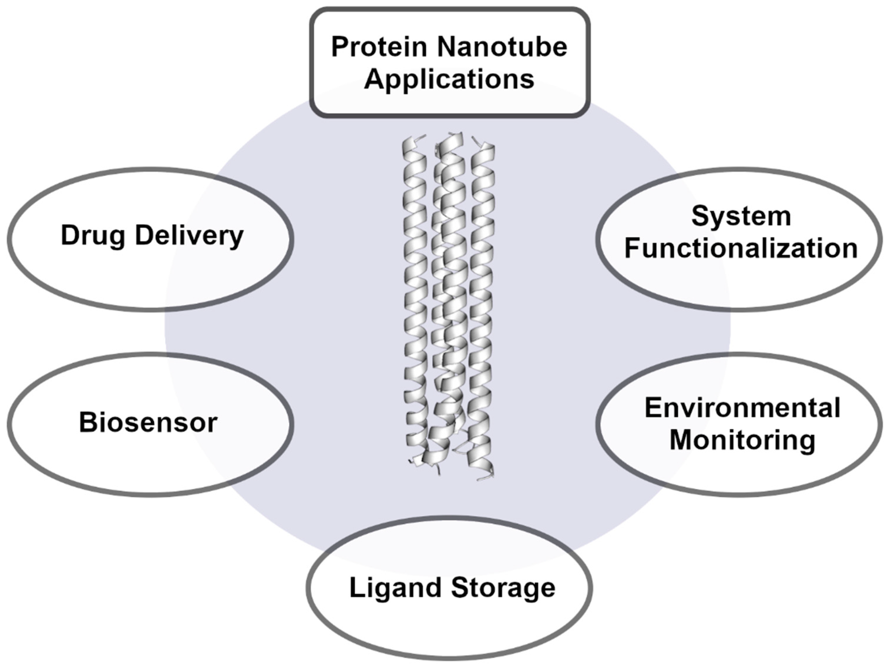
2. Applications for Nanotube Cavities

Even though protein nanotubes are considered a niche material in nanotechnology, they offer unique advantages over common materials such as carbon nanotubes. Carbon nanotubes have been explored for multiple applications including energy storage [20], transistors, sensors [21][22] and drug delivery [23][24]. However, their immediate toxicity and unknown long-term health effects make them mostly unsuitable for biological systems [25][26]. Adding to that are the obstacles of commercial production in high yields and of adequate purity [27]. Protein nanotubes can address these issues and prove to be promising nanomaterials, especially for biotechnological applications (Figure 1). In general, organic polymer-based nanomaterials serve as unique carrier systems that can be engineered to bind ligands of interest and transport them to specific target locations for potential release. Due to their organic composition, they offer unique chemical interactions with biological systems and drugs alike [28]. Protein-based nanomaterials exhibit low direct toxicity [29][30], are biodegradable [31] and are highly stable if designed correctly. Although these are preferred characteristics for efficient drug delivery, issues due to cytotoxic effects have been reported [29][32][33] and can occur readily if the drug carrier nanomaterial interacts with nontarget biological systems. Still, cytotoxic effects are being considered in biotechnological developments, and multiple protein nanoparticle–drug complexes have already been approved for therapeutic use [34][35]. The chemical diversity of the various possible amino acids allows for complex engineering to fulfill various functions. These functions include not only the cavity spaces that can be filled with target ligands but also the possibility of functionalizing the outer nanotube surface to associate with molecular targets. Hence, many recent applications have aimed to develop nanotubes into drug carrier systems that attach to cellular targets.
Figure 1. General applications for protein nanotubes in current use or development. These include but are not limited to drug delivery, biosensor, general ligand storage, environmental monitoring and system functionalization applications.
The repurposing and de novo design of novel nanotubes for certain functions has progressed quickly due to the collective increase in coiled coil and cavity knowledge. The naturally occurring protein nanotube RHCC was found to bind a host of medicinal compelling ligands including cisplatin [36] and ortho-carborane [37]. Both of these drugs are applicable towards cancer treatments but lack adequate solubility. In addition, cisplatin is a highly toxic compound [38]. Upon uptake of the drugs into the central hydrophobic cavities, issues with solubility and toxicity are overcome. The nanotube is highly soluble and can be used for drug delivery; assays confirmed that the nanotube enters cellular targets, and additional mouse studies showed that cytotoxic effects of the isolated nanotube were negligible [29]. Similarly, α-lactalbumin nanotubes have been shown to take up various ligands into their large cavities including capsaicin [39] and lycopene [40]. Whereas capsaicin has antimicrobial and pain relieving properties for medical applications, it causes irritation on mucosal surfaces and is poorly soluble in water, which limits the use of the drug [41]. Comparatively, lycopene is a compelling antioxidant that is also restricted by poor solubility and stability [42]. The uptake of these ligands into protein nanotubes was shown to circumvent solubility issues and improve the general stability of the molecules. In the case of the nanotube–capsaicin complex, effective mucus penetration and retention in gastrointestinal tracts of mice was demonstrated [39]. In short, both of these nanotubes show great potential as repurposed drug delivery nanomaterials.
The intelligent design of peptide sequences to create coiled-coil assemblies that bind ligands for drug delivery has also seen large improvements. Initial studies such as the constructions of tetrameric coiled coils experimented with a and d position substitutions in the heptad repeat to change the physicochemical properties of a central cavity. Various combinations allowed for the uptake of various organic ligands such as adamantane, camphor and corresponding derivatives [43]. The nanomaterial in complex with ligands was shown to be stable, and although subsequent drug delivery was not explored, an early basis for protein nanotube ligand binding was presented. More recent studies have investigated the construction of protein nanotubes around more complex ligands including porphyrin [12] and apixaban [11]. The de novo designs of both nanotubes were directed towards creating a functional ligand-binding protein, which inevitably created intricate cavities. Being able to create nanotubes with binding cavities for target ligands highlights the beneficial modularity of proteins for prospect drug delivery, especially for ligands with difficult pharmacokinetic properties. In contrast, it is also possible to design unspecific cavities that bind a variety of nonpolar molecules. These include novel constructs from a series of coiled-coil nanotubes ranging from trimeric to nonameric assemblies [15][16]. As the number of helices increases, the central cavity space increases in volume, which allowed for the uptake of large nonpolar ligands. In general, nonpolar target molecules show poor pharmacokinetics as they are poorly soluble, resulting in low bioavailability; in certain cases, nonpolar molecules exhibit inherent toxic properties [44][45]. These can easily be overcome by uptake into an appropriately sized, hydrophobic cavity. Still, it should be mentioned that translation from initial nanotube development and the characterization of protein–ligand complexes to a clinical setting has been lacking. This is most likely due to a current emphasis on understanding coiled-coil arrangements and their underlying characteristics. Additionally, although the successful design of novel nanotubes that bind relevant drugs demonstrates the basic understanding, subsequent studies should aim to further establish functional nanotubes as targeted drug-delivery agents.
Even though drug delivery appears to be a prevalent target application for protein nanotubes, other studies have examined potential uses as biosensors. Here, the cavities are filled with ligands that can be detected by absorbance, fluorescence [16], magnetic [19] or even electronic [46] measurements. The essential mechanisms for application development are highly similar to drug-delivery approaches. Herein, however, the presence of ligands is detected using external methods. The central cavities for the nanotube biosensors cover a broad spectrum with varying shapes, sizes and hydrophobicity indices. Upon the uptake of a luminescent ruthenium(II) polypyridyl species into a protein nanocage cavity, the complexes could be detected by spectroscopic methods in cell imaging analyses [47]. Although this complex was not a nanotube material, the cavity was highly reminiscent of hydrophobic cavities in nanotubes where nonpolar side chains stabilize the internalized ligand. Comparatively, nonpolar dyes have been shown to bind Into nanotube cavities nonspecifically, including 1,6-diphenylhexatriene, prodan and β-carotene [16]. These ligands display unique absorbance or fluorescence characteristics that can be taken advantage of for biosensors and diagnostic tools upon targeted delivery by the encapsulating nanotube. Additional imaging applications are possible through the uptake of suitable magnetic resonant compounds. Lanthanides are paramagnetic elements for which a nanotube cavity was designed that coordinates storage with high stability. Multiple Ln3+ atoms such as terbium, cerium, neodymium and europium can be stabilized inside the core by the polar side chains of aspartic acid residues and interhelical water molecules that form a close hydrogen bonding network [19]. These protein–ligand complexes have the potential for use as magnetic resonance imaging (MRI) contrast agents. In general, the lanthanide elements are moderately to highly toxic, which leads to serious side effects upon drug usage [48]. However, these can be circumvented with the use of drug-carrier systems. The recent binding studies of functional metalloprotein complexes show that proteins can be used to effectively stabilize metals of interest over various polar residues. In light of this, other metals have also been placed inside of protein nanotubes and could provide other attractive functions such as centers for catalytic reactions. Copper, nickel, zinc and also lead have been shown to bind into the cavities of de novo protein nanotubes [18][49]. These nanomaterials were designed to coordinate multiple metal atoms, which effectively increases the load capacity per nanotube. These studies not only showed that the intelligent design of metal binding nanotubes is possible but also laid the groundwork for the design of soluble catalytic site nanomaterials.
A somewhat unique application of a protein nanotube is the environmental monitoring of toxic polycyclic aromatic hydrocarbons upon crude oil spillage. A nanotube was able to take up and store these nonpolar compounds up to a specific size limit [17], after which it was used as a suspended medium to detect concentrations in an experimental setting. For these experiments, the protein nanotube medium was placed into lake mesocosms for up to 14 days and showed no signs of degradation by environmental factors [50]. As previously discussed, an appropriately designed nanotube provides a highly stable material for versatile functionality. The nonspecific uptake of nonpolar ligands is an appealing concept, as large enough protein nanotubes can also be used to functionalize carbon nanotubes. Numerous proteins have already been used to coat carbon nanotubes to add the advantageous properties of proteins to carbon nanotube applications [31][51]. Common goals for carbon nanotube functionalization are an increase in solubility [52], a decrease in toxicity [53] and options for biological targeting [54]. A recent functionalization study showed that a heptameric protein nanotube was able to retain a single-walled carbon nanotube in its central channel that effectively solubilized the carbon nanotube [55]. Herein, it was created a novel nanomaterial that combined the internal properties of single-walled carbon nanotubes with the outer surface properties of proteins.
The strength of protein-based materials is their high diversity and modularity due to the many different sequences that can be constructed. The modularity of proteins also allows for the introduction of chemical labels and modifications to outer protein surfaces that can bind specific biological targets or aid in visualization [56][57]. These chemical labels can be as simple as attaching folate ligands via click chemistry, which enhances cancer cell uptake due to the common overexpression of folate receptors [58][59][60]. Outer surfaces of nanotubes have also been shown to include DNA-binding motifs that can be used for targeting [61][62]. Other targeting approaches include more complex methods such as antibody conjugation [63][64] and the construction of larger nanobodies with high load capacities for potential drug delivery [65]. These allow for improved accumulation around target cells. In addition to biological targeting labels, visualization tags such as fluorescent dyes can be covalently and site-specifically linked to protein surfaces [57][66]. Some investigators have employed noncanonical amino acids that contain reactive side chains and expand on the base number of amino acids, which then allowed for site specific labelling [67][68]. Still, the surfaces of protein nanotubes contain functional groups including primary amines and carboxyl groups that are ideal for quick and less-specific chemical modifications. The quick labeling of amino side chains also allows for the crosslinking of coiled coils that can then form an extended protein material. This has recently been shown where a trimeric coiled coil was linked via covalent chemical crosslinkers and electrostatic interactions to form a larger three-dimensional assembly [69]. In short, the general functionalization of proteinaceous nanotubes is straightforward and usually yields stable nanomaterials.
Nevertheless, the applications for protein-based nanotubes are mostly in the developmental stages. This is in part due to the niche nature of these nanomaterials and current issues with the cost-effective production of sufficient quantities. Additionally, laboratory-to-clinical translation for medical purposes is still in its early stages, with few therapeutic applications in clinical development. Although these can potentially be overcome as technologies advance, current limitations prevent the broader expansion of proteins as nanomaterials. However, a collection of nanotubes have progressed past proof-of-concept studies and show great potential across various fields. In particular, protein nanotubes tend to be focused on in biotechnological settings where carbon nanotubes are generally more difficult to employ. The biocompatibility of proteins combined with their modularity and high stability ratify them as suitable carrier systems. If combined with ligand binding cavities, protein nanotubes present themselves as a promising tool that can be directed towards diverse objectives.

References

  1. Liu, X.; Zhao, Y.; Liu, P.; Wang, L.; Lin, J.; Fan, C. Biomimetic DNA Nanotubes: Nanoscale Channel Design and Applications. Angew. Chem. Int. Ed. 2019, 58, 8996–9011.
  2. Chen, Z.; Boyken, S.E.; Jia, M.; Busch, F.; Flores-Solis, D.; Bick, M.J.; Lu, P.; VanAernum, Z.L.; Sahasrabuddhe, A.; Langan, R.A.; et al. Programmable Design of Orthogonal Protein Heterodimers. Nature 2019, 565, 106–111.
  3. Fujita, S.; Matsuura, K. Self-Assembled Artificial Viral Capsids Bearing Coiled-Coils at the Surface. Org. Biomol. Chem. 2017, 15, 5070–5077.
  4. Villegas, J.A.; Sinha, N.J.; Teramoto, N.; Von Bargen, C.D.; Pochan, D.J.; Saven, J.G. Computational Design of Single-Peptide Nanocages with Nanoparticle Templating. Molecules 2022, 27, 1237.
  5. Nambiar, M.; Nepal, M.; Chmielewski, J. Self-Assembling Coiled-Coil Peptide Nanotubes with Biomolecular Cargo Encapsulation. ACS Biomater. Sci. Eng. 2019, 5, 5082–5087.
  6. Hughes, S.A.; Wang, F.; Wang, S.; Kreutzberger, M.A.B.; Osinski, T.; Orlova, A.; Wall, J.S.; Zuo, X.; Egelman, E.H.; Conticello, V.P. Ambidextrous Helical Nanotubes from Self-Assembly of Designed Helical Hairpin Motifs. Proc. Natl. Acad. Sci. USA 2019, 116, 14456–14464.
  7. Mondal, S.; Adler-Abramovich, L.; Lampel, A.; Bram, Y.; Lipstman, S.; Gazit, E. Formation of Functional Super-Helical Assemblies by Constrained Single Heptad Repeat. Nat. Commun. 2015, 6, 8615.
  8. Beesley, J.L.; Woolfson, D.N. The de Novo Design of α-Helical Peptides for Supramolecular Self-Assembly. Curr. Opin. Biotechnol. 2019, 58, 175–182.
  9. Lajoie, M.J.; Boyken, S.E.; Salter, A.I.; Bruffey, J.; Rajan, A.; Langan, R.A.; Olshefsky, A.; Muhunthan, V.; Bick, M.J.; Gewe, M.; et al. Designed Protein Logic to Target Cells with Precise Combinations of Surface Antigens. Science 2020, 369, 1637–1643.
  10. Chen, Y.; Chen, Q.; Liu, H. DEPACT and PACMatch: A Workflow of Designing De Novo Protein Pockets to Bind Small Molecules. J. Chem. Inf. Model. 2022, 62, 971–985.
  11. Polizzi, N.F.; DeGrado, W.F. A Defined Structural Unit Enables de Novo Design of Small-Molecule-Binding Proteins. Science 2020, 369, 1227–1233.
  12. Polizzi, N.F.; Wu, Y.; Lemmin, T.; Maxwell, A.M.; Zhang, S.Q.; Rawson, J.; Beratan, D.N.; Therien, M.J.; DeGrado, W.F. De Novo Design of a Hyperstable Non-Natural Protein-Ligand Complex with Sub-Å Accuracy. Nat. Chem. 2017, 9, 1157–1164.
  13. Naudin, E.A.; Albanese, K.I.; Smith, A.J.; Mylemans, B.; Baker, E.G.; Weiner, O.D.; Andrews, D.M.; Tigue, N.; Savery, N.J.; Woolfson, D.N. From Peptides to Proteins: Coiled-Coil Tetramers to Single-Chain 4-Helix Bundles. Chem. Sci. 2022, 13, 11330–11340.
  14. Boyken, S.E.; Chen, Z.; Groves, B.; Langan, R.A.; Oberdorfer, G.; Ford, A.; Gilmore, J.M.; Xu, C.; Dimaio, F.; Henrique Pereira, J.; et al. De Novo Design of Protein Homo-Oligomers with Modular Hydrogen-Bond Network–Mediated Specificity. Science 2016, 352, 69–72.
  15. Dawson, W.M.; Martin, F.J.O.; Rhys, G.G.; Shelley, K.L.; Brady, R.L.; Woolfson, D.N. Coiled Coils 9-to-5: Rational: De Novo Design of α-Helical Barrels with Tunable Oligomeric States. Chem. Sci. 2021, 12, 6923–6928.
  16. Thomas, F.; Dawson, W.M.; Lang, E.J.M.; Burton, A.J.; Bartlett, G.J.; Rhys, G.G.; Mulholland, A.J.; Woolfson, D.N. De Novo-Designed α-Helical Barrels as Receptors for Small Molecules. ACS Synth. Biol. 2018, 7, 1808–1816.
  17. McDougall, M.; Francisco, O.; Harder-Viddal, C.; Roshko, R.; Heide, F.; Sidhu, S.; Khajehpour, M.; Leslie, J.; Palace, V.; Tomy, G.T.; et al. Proteinaceous Nano Container Encapsulate Polycyclic Aromatic Hydrocarbons. Sci. Rep. 2019, 9, 1058.
  18. Tolbert, A.E.; Ervin, C.S.; Ruckthong, L.; Paul, T.J.; Jayasinghe-Arachchige, V.M.; Neupane, K.P.; Stuckey, J.A.; Prabhakar, R.; Pecoraro, V.L. Heteromeric Three-Stranded Coiled Coils Designed Using a Pb(Ii)(Cys)3 Template Mediated Strategy. Nat. Chem. 2020, 12, 405–411.
  19. Slope, L.N.; Daubney, O.J.; Campbell, H.; White, S.A.; Peacock, A.F.A. Location-Dependent Lanthanide Selectivity Engineered into Structurally Characterized Designed Coiled Coils. Angew. Chem. Int. Ed. 2021, 60, 24473–24477.
  20. Chitranshi, M.; Pujari, A.; Ng, V.; Chen, D.; Chauhan, D.; Hudepohl, R.; Saleminik, M.; Kim, S.Y.; Kubley, A.; Shanov, V.; et al. Carbon Nanotube Sheet-Synthesis and Applications. Nanomaterials 2020, 10, 2023.
  21. Mandeep; Shukla, P. Microbial Nanotechnology for Bioremediation of Industrial Wastewater. Front. Microbiol. 2020, 11, 590631.
  22. Piperopoulos, E.; Calabrese, L.; Khaskhoussi, A.; Proverbio, E.; Milone, C. Thermo-Physical Characterization of Carbon Nanotube Composite Foam for Oil Recovery Applications. Nanomaterials 2020, 10, 86.
  23. Tanaka, M.; Aoki, K.; Haniu, H.; Kamanaka, T.; Takizawa, T.; Sobajima, A.; Yoshida, K.; Okamoto, M.; Kato, H.; Saito, N. Applications of Carbon Nanotubes in Bone Regenerative Medicine. Nanomaterials 2020, 10, 659.
  24. Dehaghani, M.Z.; Yousefi, F.; Seidi, F.; Bagheri, B.; Mashhadzadeh, A.H.; Naderi, G.; Esmaeili, A.; Abida, O.; Habibzadeh, S.; Saeb, M.R.; et al. Encapsulation of an Anticancer Drug Isatin inside a Host Nano-Vehicle SWCNT: A Molecular Dynamics Simulation. Sci. Rep. 2021, 11, 18753.
  25. Zare, H.; Ahmadi, S.; Ghasemi, A.; Ghanbari, M.; Rabiee, N.; Bagherzadeh, M.; Karimi, M.; Webster, T.J.; Hamblin, M.R.; Mostafavi, E. Carbon Nanotubes: Smart Drug/Gene Delivery Carriers. Int. J. Nanomed. 2021, 16, 1681–1706.
  26. Hwang, Y.; Park, S.H.; Lee, J.W. Applications of Functionalized Carbon Nanotubes for the Therapy and Diagnosis of Cancer. Polymers 2017, 9, 13.
  27. Bati, A.S.R.; Yu, L.; Batmunkh, M.; Shapter, J.G. Recent Advances in Applications of Sorted Single-Walled Carbon Nanotubes. Adv. Funct. Mater. 2019, 29, 1902273.
  28. Khan, M.I.; Hossain, M.I.; Hossain, M.K.; Rubel, M.H.K.; Hossain, K.M.; Mahfuz, A.M.U.B.; Anik, M.I. Recent Progress in Nanostructured Smart Drug Delivery Systems for Cancer Therapy: A Review. ACS Appl. Bio Mater. 2022, 5, 971–1012.
  29. Thanasupawat, T.; Bergen, H.; Hombach-Klonisch, S.; Krcek, J.; Ghavami, S.; Del Bigio, M.R.; Krawitz, S.; Stelmack, G.; Halayko, A.; McDougall, M.; et al. Platinum (IV) Coiled Coil Nanotubes Selectively Kill Human Glioblastoma Cells. Nanomed. Nanotechnol. Biol. Med. 2015, 11, 913–925.
  30. Corbo, C.; Molinaro, R.; Parodi, A.; Toledano Furman, N.E.; Salvatore, F.; Tasciotti, E. The Impact of Nanoparticle Protein Corona on Cytotoxicity, Immunotoxicity and Target Drug Delivery. Nanomedicine 2016, 11, 81–100.
  31. Jaiswal, S.; Manhas, A.; Pandey, A.K.; Priya, S.; Sharma, S.K. Engineered Nanoparticle-Protein Interactions Influence Protein Structural Integrity and Biological Significance. Nanomaterials 2022, 12, 1214.
  32. Ren, D.; Dalmau, M.; Randall, A.; Shindel, M.M.; Baldi, P.; Wang, S.W. Biomimetic Design of Protein Nanomaterials for Hydrophobic Molecular Transport. Adv. Funct. Mater. 2012, 22, 3170–3180.
  33. Montes-Fonseca, S.L.; Sánchez-Ramírez, B.; Luna-Velasco, A.; Arzate-Quintana, C.; Silva-Cazares, M.B.; González Horta, C.; Orrantia-Borunda, E. Cytotoxicity of Protein-Carbon Nanotubes on J774 Macrophages Is a Functionalization Grade-Dependent Effect. Biomed. Res. Int. 2015, 2015, 796456.
  34. Kim, M.T.; Chen, Y.; Marhoul, J.; Jacobson, F. Statistical Modeling of the Drug Load Distribution on Trastuzumab Emtansine (Kadcyla), a Lysine-Linked Antibody Drug Conjugate. Bioconjug. Chem. 2014, 25, 1223–1232.
  35. Miele, E.; Spinelli, G.P.; Miele, E.; Tomao, F.; Tomao, S. Albumin-Bound Formulation of Paclitaxel (Abraxane® ABI-007) in the Treatment of Breast Cancer. Int. J. Nanomed. 2009, 4, 99–105.
  36. Eriksson, M.; Hassan, S.; Larsson, R.; Linder, S.; Ramqvist, T.; Lövborg, H.; Vikinge, T.; Figgemeier, E.; Müller, J.; Stetefeld, J.; et al. Utilization of a Right-Handed Coiled-Coil Protein from Archaebacterium Staphylothermus Marinus as a Carrier for Cisplatin. Anticancer Res. 2009, 29, 11–18.
  37. Heide, F.; McDougall, M.; Harder-Viddal, C.; Roshko, R.; Davidson, D.; Wu, J.; Aprosoff, C.; Moya-Torres, A.; Lin, F.; Stetefeld, J. Boron Rich Nanotube Drug Carrier System Is Suited for Boron Neutron Capture Therapy. Sci. Rep. 2021, 11, 1–9.
  38. Scagliotti, G.V.; Park, K.; Patil, S.; Rolski, J.; Goksel, T.; Martins, R.; Gans, S.J.M.; Visseren-Grul, C.; Peterson, P. Survival without Toxicity for Cisplatin plus Pemetrexed versus Cisplatin plus Gemcitabine in Chemonaïve Patients with Advanced Non-Small Cell Lung Cancer: A Risk-Benefit Analysis of a Large Phase III Study. Eur. J. Cancer 2009, 45, 2298–2303.
  39. Yuan, Y.; Liu, Y.; He, Y.; Zhang, B.; Zhao, L.; Tian, S.; Wang, Q.; Chen, S.; Li, Z.; Liang, S.; et al. Intestinal-Targeted Nanotubes-in-Microgels Composite Carriers for Capsaicin Delivery and Their Effect for Alleviation of Salmonella Induced Enteritis. Biomaterials 2022, 287, 121613.
  40. Chang, R.; Liu, B.; Wang, Q.; Zhang, J.; Yuan, F.; Zhang, H.; Chen, S.; Liang, S.; Li, Y. The Encapsulation of Lycopene with α-Lactalbumin Nanotubes to Enhance Their Anti-Oxidant Activity, Viscosity and Colloidal Stability in Dairy Drink. Food Hydrocoll. 2022, 131, 107792.
  41. Merritt, J.C.; Richbart, S.D.; Moles, E.G.; Cox, A.J.; Brown, K.C.; Miles, S.L.; Finch, P.T.; Hess, J.A.; Tirona, M.T.; Valentovic, M.A.; et al. Anti-Cancer Activity of Sustained Release Capsaicin Formulations. Pharmacol. Ther. 2022, 238, 108177.
  42. Youssef, R.B.; Fouad, M.A.; El-Zaher, A.A. Bioanalytical Study of the Effect of Lycopene on the Pharmacokinetics of Theophylline in Rats. Pharm. Chem. J. 2020, 53, 1053–1058.
  43. Mizuno, T.; Hasegawa, C.; Tanabe, Y.; Hamajima, K.; Muto, T.; Nishi, Y.; Oda, M.; Kobayashi, Y.; Tanaka, T. Organic Ligand Binding by a Hydrophobic Cavity in a Designed Tetrameric Coiled-Coil Protein. Chem.–A Eur. J. 2009, 15, 1491–1498.
  44. Chen, Y.-Y.; Kao, T.-W.; Wang, C.-C.; Wu, C.-J.; Zhou, Y.-C.; Chen, W.-L. Association between Polycyclic Aromatic Hydrocarbons Exposure and Bone Turnover in Adults. Eur. J. Endocrinol. 2020, 182, 333–341.
  45. Kim, K.-W.; Won, Y.L.; Park, D.J.; Kim, Y.S.; Jin, E.S.; Lee, S.K. Combined Toxic Effects of Polar and Nonpolar Chemicals on Human Hepatocytes (HepG2) Cells by Quantitative Property—Activity Relationship Modeling. Toxicol. Res. 2016, 32, 337–343.
  46. Mahendran, K.R.; Niitsu, A.; Kong, L.; Thomson, A.R.; Sessions, R.B.; Woolfson, D.N.; Bayley, H. A Monodisperse Transmembrane α-Helical Peptide Barrel. Nat. Chem. 2017, 9, 411–419.
  47. Li, X.; Zhang, Y.; Chen, H.; Sun, J.; Feng, F. Protein Nanocages for Delivery and Release of Luminescent Ruthenium(II) Polypyridyl Complexes. ACS Appl. Mater. Interfaces 2016, 8, 22756–22761.
  48. Bao, G. Lanthanide Complexes for Drug Delivery and Therapeutics. J. Lumin. 2020, 228, 117622.
  49. Boyle, A.L.; Rabe, M.; Crone, N.S.A.; Rhys, G.G.; Soler, N.; Voskamp, P.; Pannu, N.S.; Kros, A. Selective Coordination of Three Transition Metal Ions within a Coiled-Coil Peptide Scaffold. Chem. Sci. 2019, 10, 7456–7465.
  50. Heide, F.; Aprosoff, C.; Peters, L.; Palace, V.; Tomy, G.; Stetefeld, J.; McDougall, M. A Novel Passive Sampling Device for Low Molecular Weight PAHs with a Proteinaceous Medium. Environ. Nanotechnol. Monit. Manag. 2022, 17, 100609.
  51. Breitwieser, A.; Sleytr, U.B.; Pum, D. A New Method for Dispersing Pristine Carbon Nanotubes Using Regularly Arranged S-Layer Proteins. Nanomaterials 2021, 11, 1346.
  52. García-Hevia, L.; Saramiforoshani, M.; Monge, J.; Iturrioz-Rodríguez, N.; Padín-González, E.; González, F.; González-Legarreta, L.; González, J.; Fanarraga, M.L. The Unpredictable Carbon Nanotube Biocorona and a Functionalization Method to Prevent Protein Biofouling. J. Nanobiotechnology 2021, 19, 129.
  53. Martins, C.H.Z.; Côa, F.; Da Silva, G.H.; Bettini, J.; De Farias, M.A.; Portugal, R.V.; de Aragao Umbuzeiro, G.; Alves, O.L.; Martinez, D.S.T. Functionalization of Carbon Nanotubes with Bovine Plasma Biowaste by Forming a Protein Corona Enhances Copper Removal from Water and Ecotoxicity Mitigation. Environ. Sci. Nano 2022, 9, 2887–2905.
  54. Mann, F.A.; Lv, Z.; Großhans, J.; Opazo, F.; Kruss, S. Nanobody-Conjugated Nanotubes for Targeted Near-Infrared In Vivo Imaging and Sensing. Angew. Chem. Int. Ed. 2019, 58, 11469–11473.
  55. Mann, F.A.; Horlebein, J.; Meyer, N.F.; Meyer, D.; Thomas, F.; Kruss, S. Carbon Nanotubes Encapsulated in Coiled-Coil Peptide Barrels. Chem.—A Eur. J. 2018, 24, 12241–12245.
  56. Klermund, L.; Poschenrieder, S.T.; Castiglione, K. Simple Surface Functionalization of Polymersomes Using Non-Antibacterial Peptide Anchors. J. Nanobiotechnol. 2016, 14, 48.
  57. Zhang, Y.; Zang, C.; An, G.; Shang, M.; Cui, Z.; Chen, G.; Xi, Z.; Zhou, C. Cysteine-Specific Protein Multi-Functionalization and Disulfide Bridging Using 3-Bromo-5-Methylene Pyrrolones. Nat. Commun. 2020, 11, 1015.
  58. Farran, B.; Montenegro, R.C.; Kasa, P.; Pavitra, E.; Huh, Y.S.; Han, Y.K.; Kamal, M.A.; Nagaraju, G.P.; Rama Raju, G.S. Folate-Conjugated Nanovehicles: Strategies for Cancer Therapy. Mater. Sci. Eng. C 2020, 107, 110341.
  59. Kwan, H.Y.; Xu, Q.; Gong, R.; Bian, Z.; Chu, C.C. Targeted Chinese Medicine Delivery by A New Family of Biodegradable Pseudo-Protein Nanoparticles for Treating Triple-Negative Breast Cancer: In Vitro and In Vivo Study. Front. Oncol. 2021, 10, 600298.
  60. Loureiro, A.; Bernardes, G.J.L.; Shimanovich, U.; Sárria, M.P.; Nogueira, E.; Preto, A.; Gomes, A.C.; Cavaco-Paulo, A. Folic Acid-Tagged Protein Nanoemulsions Loaded with CORM-2 Enhance the Survival of Mice Bearing Subcutaneous A20 Lymphoma Tumors. Nanomed. Nanotechnol. Biol. Med. 2015, 11, 1077–1083.
  61. Hayashi, I. The C-Terminal Region of the Plasmid Partitioning Protein TubY Is a Tetramer That Can Bind Membranes and DNA. J. Biol. Chem. 2020, 295, 17770–17780.
  62. Murase, S.; Ishino, S.; Ishino, Y.; Tanaka, T. Control of Enzyme Reaction by a Designed Metal-Ion-Dependent α-Helical Coiled-Coil Protein. J. Biol. Inorg. Chem. 2012, 17, 791–799.
  63. Majerle, A.; Hadzi, S.; Aupič, J.; Satler, T.; Lapenta, F.; Strmšek, Ž.; Lah, J.; Loris, R.; Jerala, R. A Nanobody Toolbox Targeting Dimeric Coiled-Coil Modules for Functionalization of Designed Protein Origami Structures. Proc. Natl. Acad. Sci. USA 2021, 118.
  64. Gil-Garcia, M.; Ventura, S. Multifunctional Antibody-Conjugated Coiled-Coil Protein Nanoparticles for Selective Cell Targeting. Acta Biomater. 2021, 131, 472–482.
  65. Ahn, B.; Lee, S.G.; Yoon, H.R.; Lee, J.M.; Oh, H.J.; Kim, H.M.; Jung, Y. Four-Fold Channel-Nicked Human Ferritin Nanocages for Active Drug Loading and PH-Responsive Drug Release. Angew. Chem. Int. Ed. 2018, 57, 2909–2913.
  66. Agrawalla, B.K.; Wang, T.; Riegger, A.; Domogalla, M.P.; Steinbrink, K.; Dörfler, T.; Chen, X.; Boldt, F.; Lamla, M.; Michaelis, J.; et al. Chemoselective Dual Labeling of Native and Recombinant Proteins. Bioconjug. Chem. 2018, 29, 29–34.
  67. Saleh, A.M.; Wilding, K.M.; Calve, S.; Bundy, B.C.; Kinzer-Ursem, T.L. Non-Canonical Amino Acid Labeling in Proteomics and Biotechnology. J. Biol. Eng. 2019, 13, 43.
  68. Dieterich, D.C.; Lee, J.J.; Link, A.J.; Graumann, J.; Tirrell, D.A.; Schuman, E.M. Labeling, Detection and Identification of Newly Synthesized Proteomes with Bioorthogonal Non-Canonical Amino-Acid Tagging. Nat. Protoc. 2007, 2, 532–540.
  69. Jorgensen, M.D.; Chmielewski, J. Reversible Crosslinked Assembly of a Trimeric Coiled-coil Peptide into a Three-dimensional Matrix for Cell Encapsulation and Release. J. Pept. Sci. 2022, 28, e3302.
More
Information
Contributors MDPI registered users' name will be linked to their SciProfiles pages. To register with us, please refer to https://encyclopedia.pub/register : ,
View Times: 328
Revisions: 2 times (View History)
Update Date: 24 Nov 2022
1000/1000
Video Production Service