Submitted Successfully!
To reward your contribution, here is a gift for you: A free trial for our video production service.
Thank you for your contribution! You can also upload a video entry or images related to this topic.
Version Summary Created by Modification Content Size Created at Operation
1 -- 2254 2022-11-07 09:29:39 |
2 format correct Meta information modification 2254 2022-11-08 04:41:15 | |
3 format correct + 5 word(s) 2259 2022-11-08 04:48:04 | |
4 format correct + 2 word(s) 2261 2022-11-08 04:49:21 | |
5 format correct Meta information modification 2261 2022-11-08 04:50:35 |

Video Upload Options

Do you have a full video?

Confirm

Are you sure to Delete?
Cite
If you have any further questions, please contact Encyclopedia Editorial Office.
Clergue-Duval, V.;  Coulbault, L.;  Questel, F.;  Cabé, N.;  Laniepce, A.;  Delage, C.;  Boudehent, C.;  Bloch, V.;  Segobin, S.;  Naassila, M.; et al. Alcohol-Induced Oxidative Stress for the Brain. Encyclopedia. Available online: https://encyclopedia.pub/entry/33280 (accessed on 28 April 2024).
Clergue-Duval V,  Coulbault L,  Questel F,  Cabé N,  Laniepce A,  Delage C, et al. Alcohol-Induced Oxidative Stress for the Brain. Encyclopedia. Available at: https://encyclopedia.pub/entry/33280. Accessed April 28, 2024.
Clergue-Duval, Virgile, Laurent Coulbault, Frank Questel, Nicolas Cabé, Alice Laniepce, Clément Delage, Céline Boudehent, Vanessa Bloch, Shailendra Segobin, Mickael Naassila, et al. "Alcohol-Induced Oxidative Stress for the Brain" Encyclopedia, https://encyclopedia.pub/entry/33280 (accessed April 28, 2024).
Clergue-Duval, V.,  Coulbault, L.,  Questel, F.,  Cabé, N.,  Laniepce, A.,  Delage, C.,  Boudehent, C.,  Bloch, V.,  Segobin, S.,  Naassila, M.,  Pitel, A., & Vorspan, F. (2022, November 07). Alcohol-Induced Oxidative Stress for the Brain. In Encyclopedia. https://encyclopedia.pub/entry/33280
Clergue-Duval, Virgile, et al. "Alcohol-Induced Oxidative Stress for the Brain." Encyclopedia. Web. 07 November, 2022.
Alcohol-Induced Oxidative Stress for the Brain
Edit

Alcohol use disorders (AUD) is defined by the loss of control over alcohol intake and chronic, compulsive, heavy alcohol use despite adverse consequences. Among patients seeking treatment for AUD, the proportion of patients at treatment entry endorsing the criteria for pharmacological dependence was 63% for tolerance and 14% for withdrawal. Alcohol withdrawal (AW) syndrome is the combination of signs and symptoms occurring as soon as three to six hours after the last intake of alcohol in subjects with pharmacological dependence. The classical symptoms are tremor, perspiration, anxiety and adrenergic signs (hypertension, tachycardia). Untreated AW can lead to specific complications: delirium tremens (DT) and seizure. Several indirect complications of the adrenergic syndrome may also occur during an untreated AW syndrome as dehydration, cardiac failure or renal failure. Mortality reaches 8% in patients with AW syndrome hospitalized in intensive care units, because of any or the combination of those multiple organs complications. AW is still considered as a dangerous complication of undetected AUD during any surgery or medical inpatient treatment. 

oxidative stress AUD DT alcohol cognitive impairment

1. Alcohol Withdrawal as an Oxidative Stress Challenge for the Brain

As ethanol is a small molecule that easily passes the blood–brain barrier and is ubiquitously distributed in the brain, chronic alcohol use induces neuroadaptation [1][2]. This neuroadaptation is obviously revealed in cases of observable pharmacological dependence, characterized by the phenomenon of tolerance and the presence of signs of withdrawal when alcohol use ceases. Thus, at the brutal alcohol cessation, the equilibrium resulting from this neuroadaptation is disrupted and it will take several days to restore the balance. What occurs at the whole brain level is mainly an increase in glutamate and norepinephrine, a decrease in GABA and an increase in intracellular calcium concentration [1][3][4]. This hyperadrenergic state induces the physical signs of alcohol withdrawal (AW), but in actual practice, no direct clinical evaluation of the glutamatergic surge exists. As the result of this hyperglutamatergic state, a central nervous system hyperexcitability and an autonomic hyperactivity occurs. Then, a vicious cycle of AW exacerbating both central nervous system hyperexcitability and an autonomic hyperactivity may start. This leads to an intense oxidative stress and reactive oxygen species (ROS) production during early alcohol withdrawal [5][6], via the increase in excitatory neurotransmitters and intracellular calcium concentration [2]. At present, it is not known whether these changes in oxidative brain status persist after the initial period of AW [2]. Interestingly, the antioxidant N-acetylcysteine has been shown to be effective in reducing alcohol drinking and relapse in chronic alcohol consuming rats, and its efficacy may come from its ability to normalize glutamatergic homeostasis but also from its antioxidative properties [7][8][9]. However, the specific impact of this oxidative stress during AW has not been established per se [2], but it is a strong candidate mechanism that could contribute to the development of brain injuries and alcohol-related neurocognitive deficits.

1.1. Epilepsy, Delirium Tremens as  Alcohol Withdrawal Neurological Complications: The Role of Oxidative Stress

The two major and well-known complications of AW, occurring in case of absence or inadequate medical management of AW symptoms, are epileptic seizure and DT. Seizures are provoked by the hyperexcitability of neurons and intracellular calcium dysregulation [4], associated with neuroinflammation and an excessive production of ROS. They induce a synchronous depolarization of neurons, itself increasing the oxygen consumption by neurons and the production of ROS, maintaining and spreading the seizure [10]. The development of innovative treatment for epilepsy includes, amongst other pathways, molecules targeting the production of ROS [11]. DT, also known as AW delirium, is a complication of AW constituted by an acute confusion state in addition to the signs and symptoms of AW (tremor, perspiration and hyperadrenergic signs). It is a life-endangering condition requiring intensive care. Consequently, experimental studies in humans trying to link DT with oxidative stress are scarce. One study assessed 8-hydroxy-2′-deoxyguanosine (8-OHdG), a peripheral blood biomarker of oxidative DNA damage, in patients with pharmacological dependence undergoing AW with and without DT [12]. The 8-OhdG level was higher in the DT group, but some patients of the AW without DT group also had elevated levels, suggesting the hyperexcitability of AW is sufficient to induce an oxidative stress. DT may not be categorically different from AW, but only dimensionally a more severe form of AW.

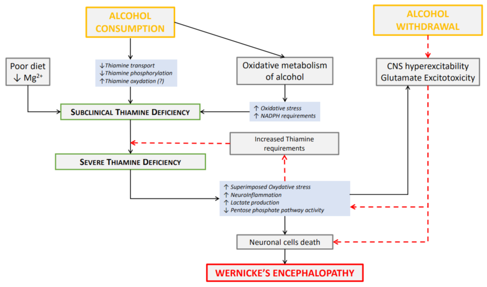
1.2.  Alcohol Withdrawal as a Vulnerability Period for Wernicke’s Encephalopathy: The Role of Oxidative Stress

Wernicke’s encephalopathy (WE) is caused by the brain toxicity related to an acute thiamine diphosphate deficiency (also named thiamine pyrophosphate) or an altered thiamine metabolism and distribution [13][14][15][16][17][18][19]. Even though WE is not the direct consequence of AW only, this particular timeframe of “periwithdrawal” should be considered as a vulnerability period for the occurrence of WE.

1.2.1. Wernicke’s Encephalopathy Occurrence

WE is frequent in AUD patients [19]. It is estimated that it affects between 10 and 35% of patients with AUD assessed when they are hospitalized for AW [19][20]; however, the prevalence is not known outside of the withdrawal period in AUD patients with active chronic alcohol use. In the general population, the prevalence of WE is much lower, and would be 0.4 to 2.8% [18]. As with the other complication of the thiamine deficiency spectrum [21], WE also appears in non-AUD patients during events of subacute (or more often acute) undernutrition, such as starvation, anorexia and food refusal, episodes of severe and repeated vomiting, malabsorption phenomena (exacerbation of inflammatory bowel disease or after bariatric surgery), but also in the context of hypercatabolism (neoplasia, sepsis, malaria, thyrotoxicosis) [22][23][24][25]. The prevalence of WE in those conditions is not precisely known, but the phenomenon is rare, and does not compare to the 10 and 35% of patients with AUD assessed when they are hospitalized for AW [19][20].

1.2.2. Wernicke’s Encephalopathy Diagnosis

The clinical Caine’s criteria for the identification of WE are the presence of two criteria among: ataxia, oculomotor dysfunction, confusion and dietary deficiency [26]. However, the clinical triad of the neurologic symptoms (ataxia, oculomotor dysfunction and confusion) is often incomplete [16][18]. The criteria, especially when they are subtle, are often difficult to identify in chronic alcohol users because there may be ambiguity between WE and signs of acute alcohol intoxication, or in non-alcohol users, because of a lack of knowledge of this symptomatology and a poor assessment of the risk of malnutrition, leading to an underdiagnosis [16][18][20].
Regarding the imaging diagnosis of WE, brain magnetic resonance imaging (MRI) has a good specificity but a low sensitivity in clinical practice. It shows bilateral FLAIR hyperintensities in specific regions, such as the mammillary bodies, thalamus, hypothalamus, periaqueductal region and floor of the fourth ventricle [18]. In the context of this low sensitivity, it is actually the case that the value of MRI and imaging in clinical practice is mainly to rule out differential diagnoses [27].
Regarding blood assays, free thiamine (the inactive form) represents a small part of the total thiamine. It is rarely impaired and is not an indicator of thiamine status [17][28]. To objectify thiamine deficiency, the esters should be assessed (only thiamine diphosphate is assessed in clinical practice) by high-performance liquid chromatography or indirectly assessed by measuring the erythrocyte transketolase activity [17][28][29]. These assays are not performed in routine practice and are highly heterogeneous between laboratories [28]. In addition, the phosphorylation capacity and the active passage of the blood–brain barrier contribute to the cerebral availability of thiamine [17][30][31][32]. As a result, the value of thiamine blood measurement is relatively poor, and it is not recommended in clinical practice. Thiamine blood measurement strategies are not shown to be effective in comparison with systematic supplementation in AUD patients hospitalized for AW [33]. Because the diagnosis of WE in non-AUD patients can occur during various medical conditions leading to thiamine depletion, the blood quantification is relevant to provide an etiological diagnostic. Indeed, low blood concentration in free thiamine or thiamine diphosphate during a comprehensive confusion investigation or in case of suspicion of thiamine deficiency confirms the diagnosis [21].

1.2.3. Systematic Thiamine Supplementation in Alcohol Use Disorder Patients

In clinical addiction medicine practice, given the prevalence of WE in hospitalized AUD patients, the absence of and the low sensitivity of the MRI or thiamine blood measurements, the majority of international medical societies recommend to systematically prescribe thiamine to patients displaying signs of AW [34]. Some recommendations advise prescribing according to the individual risk of WE notably based on the nutritional status, or the severity of the AW (delirium, intensity of withdrawal, signs of malnutrition, liver disease) [34]. Only the Australian recommendation indicates that thiamine needs to be continued indefinitely in AUD patients when alcohol consumption continues [35]. The other official recommendations do not refer to the appropriate management outside the period of acute AW [34], although thiamine prescription is a common practice.

1.2.4. Wernicke’s Encephalopathy and Alcohol Withdrawal

Animal models of alcohol-related brain damage display similar brain and cognitive alterations to what is seen in AUD patients. Numerous studies have shown that thiamine deficiency [36], or even subclinical thiamine deficiency during chronic heavy alcohol consumption, is critical for the development of significant cognitive alterations affecting spatial memory and cognitive flexibility [37] associated with neuronal and neurotrophin loss.
However, only a few studies have investigated the interaction and the synergistic effects of both ethanol toxicity and thiamine deficiency [38][39][40] to induce brain lesions.
While clinical recommendations regarding the timing of preventive management and thiamine administration acknowledge a specific risk period during AW, the link between the physiological event of AW and the occurrence of WE is only suspected but not currently demonstrated [14][15].
In clinical practice, assessing the time course of WE as a consequence of AW or AW occurring in patients with subtle symptoms of WE is highly difficult. The difficulties include the absence of easily accessible plasma thiamine measurements [28][33], the frequent absence of any prior medical consultation enabling a comprehensive premorbid clinical evaluation, and the low sensitivity of imaging. Furthermore, the symptoms of WE and AW are difficult to disentangle. Here researchers will develop the pathophysiological data supporting researchers' core hypothesis of AW favoring the occurrence of WE.

1.2.5. Wernicke’s Encephalopathy and Oxidative Stress

The pathophysiology of WE is complex and multi-factorial, including glutamatergic excitotoxicity, oxidative stress, lactic acidosis and blood–brain barrier disruption [13] and cannot be restricted to the sole well-known thiamine deficiency.
Thiamine is an essential water-soluble vitamin required for the Krebs cycle and the production of adenosine triphosphate in the mitochondria [13][18]. Thiamine itself has antioxidant properties (Huang et al. 2010). Its deficiency leads to the depletion of adenosine triphosphate and the increase in lactates production [2][13][18].
During AW, by increasing catabolism, withdrawal symptoms could increase the consumption of thiamine in the Krebs cycle, deplete its reserves and induce a further increase in lactates. Thiamine is also needed for the pentose phosphate pathway and the production of nicotinamide adenine dinucleotide phosphate, which helps to eliminate ROS and to reduce lactic acidosis [13][18]. This role contributes to the increase in oxidative stress and the disruption of the homeostasis of cellular electrolytes [13][18]. In the brain, astrocytes are the main source of lactate production [13].
In addition, astrocytic dysfunction, induced by the thiamine deficiency and this increase in oxidative stress, alters its function of reuptake and metabolism of extracellular glutamate, especially with the loss of the glutamate transporters [13][41][42][43]. Using an animal model of Wernicke–Korsakoff syndrome, in which rats were submitted to a chronic ethanol treatment with or without a thiamine deficiency episode, the glutamate uptake was found to be reduced in the prefrontal cortex by thiamine deficiency, but not by chronic ethanol intake [44].
Confronted with this decreased capacity of metabolism by astrocytes, the hyperglutamatergia of AW could exacerbate the excitotoxicity caused by thiamine deficiency and worsen the induced neuronal deaths. This hyperglutamatergia contributes, notably in a context of calcium channels dysregulation by chronic alcohol use [4], to an increase in intracellular calcium concentration in neuronal and glial cells [1][43] and increases the production of ROS [2]. In practice, the hyperglutamatergic excitotoxicity is this major pathophysiological mechanism inducing the cerebral cells death and the histological lesions observed in WE [13]. Thus, researchers suggest a synergistic effect of the two excitotoxic phenomena, acute thiamine deficiency and AW that is summarized in Figure 1.
Figure 1. Relationships between alcohol consumption, alcohol withdrawal and Wernicke’s encephalopathy. Alcohol consumption may be responsible for subclinical thiamine deficiency due to elevated thiamine requirements for energy metabolism and oxidative alcohol metabolism, impaired thiamine distribution and metabolism, and impaired nutritional status. Red dotted arrows indicate the effect of alcohol withdrawal that induces central nervous system (CNS) hyperexcitability, glutamate excitotoxicity, and exacerbates oxidative stress, neuroinflammation and thiamine requirements leading in some cases to severe thiamine deficiency, synchronous neuronal cell death, including localized thalamic and mamillary bodies neuron death responsible for Wernicke’s encephalopathy. The «(?)» corresponds to a hypothesis.

2. What Makes the Alcohol Withdrawal Period at High Risk for Oxidative Brain Damage?

AW occurs in subjects with severe AUD and pharmacological dependence. Those patients, by the combined effects of high chronic alcohol intake, poor diet, modified intestinal absorption and social disadvantages display a high rate of nutritional depletions [17][45][46][47][48]. Nutritional depletion puts them at high risk for brain suffering during the hyperglutamatergic and hyperadrenergic state induced by AW, potentially leading to WE in patients with subclinical thiamine deficiency or an individual genetic predisposition to thiamine deficiency [49]. Nutritional deficits include, apart from the previously discussed thiamine, magnesium and ascorbic acid (Vitamin C). Magnesium is notably a thiamine cofactor and also a glutamate NMDA receptor channel blocker. Beyond targeted correction in deficient patients, the relevance of its supplementation to reduce the intensity of AW and its consequences are discussed [50][51][52], although not recommended today in clinical practice. In addition, other nutritional interventions are discussed for the reduction in the oxidative stress and the prevention of neurotoxicity in AW, as ascorbic acid [46], nutritional ketosis [53] or omega-3 fatty acid treatments [54]. Those data suggest that a comprehensive nutritional assessment and/or supplementation should be developed for the peri-withdrawal period to prevent WE.

References

  1. Becker, H.C.; Mulholland, P.J. Neurochemical mechanisms of alcohol withdrawal. Handb. Clin. Neurol. 2014, 125, 133–156.
  2. Jung, M.E.; Metzger, D.B. Alcohol Withdrawal and Brain Injuries: Beyond Classical Mechanisms. Molecules 2010, 15, 4984–5011.
  3. Brousse, G.; Arnaud, B.; Vorspan, F.; Richard, D.; Dissard, A.; Dubois, M.; Pic, D.; Geneste, J.; Xavier, L.; Authier, N.; et al. Alteration of Glutamate/GABA Balance During Acute Alcohol Withdrawal in Emergency Department: A Prospective Analysis. Alcohol Alcohol. 2012, 47, 501–508.
  4. N’Gouemo, P. Voltage-Sensitive Calcium Channels in the Brain: Relevance to Alcohol Intoxication and Withdrawal. Handb. Exp. Pharm. 2018, 248, 263–280.
  5. Huang, M.-C.; Chen, C.-H.; Peng, F.-C.; Tang, S.-H.; Chen, C.-C. Alterations in Oxidative Stress Status During Early Alcohol Withdrawal in Alcoholic Patients. J. Med. Assoc. 2009, 108, 560–569.
  6. Jung, M.E.; Yan, L.-J.; Forster, M.J.; Simpkins, J.W. Ethanol withdrawal provokes mitochondrial injury in an estrogen preventable manner. J. Bioenerg. Biomembr. 2008, 40, 35–44.
  7. Lebourgeois, S.; González-Marín, M.C.; Jeanblanc, J.; Naassila, M.; Vilpoux, C. Effect of N-acetylcysteine on motivation, seeking and relapse to ethanol self-administration: NAC reduces ethanol seeking and relapse in rats. Addict. Biol. 2018, 23, 643–652.
  8. Lebourgeois, S.; González-Marín, M.C.; Antol, J.; Naassila, M.; Vilpoux, C. Evaluation of N-acetylcysteine on ethanol self-administration in ethanol-dependent rats. Neuropharmacology 2019, 150, 112–120.
  9. Quintanilla, M.E.; Ezquer, F.; Morales, P.; Ezquer, M.; Olivares, B.; Santapau, D.; Herrera-Marschitz, M.; Israel, Y. N-Acetylcysteine and Acetylsalicylic Acid Inhibit Alcohol Consumption by Different Mechanisms: Combined Protection. Front. Behav. Neurosci. 2020, 14, 122.
  10. Parsons, A.L.M.; Bucknor, E.M.V.; Castroflorio, E.; Soares, T.R.; Oliver, P.L.; Rial, D. The Interconnected Mechanisms of Oxidative Stress and Neuroinflammation in Epilepsy. Antioxidants 2022, 11, 157.
  11. Lee, S.H.; Lee, M.; Ko, D.G.; Choi, B.Y.; Suh, S.W. The Role of NADPH Oxidase in Neuronal Death and Neurogenesis after Acute Neurological Disorders. Antioxidants 2021, 10, 739.
  12. Huang, M.-C.; Chen, C.-C.; Pan, C.-H.; Chen, C.-H. Comparison of Oxidative DNA Damage Between Alcohol-Dependent Patients with and Without Delirium Tremens. Alcohol. Clin. Exp. Res. 2014, 38, 2523–2528.
  13. Abdou, E.; Hazell, A.S. Thiamine Deficiency: An Update of Pathophysiologic Mechanisms and Future Therapeutic Considerations. Neurochem. Res. 2015, 40, 353–361.
  14. Arts, N.J.M.; Walvoort, S.J.; Kessels, R.P. Korsakoff’s syndrome: A critical review. Neuropsychiatr. Dis. Treat. 2017, 13, 2875–2890.
  15. Arts, N.J.M.; Pitel, A.-L.; Kessels, R.P.C. The contribution of mamillary body damage to Wernicke’s encephalopathy and Korsakoff’s syndrome. Handb. Clin. Neurol. 2021, 180, 455–475.
  16. Chandrakumar, A.; Bhardwaj, A.; W’t Jong, G.W. Review of thiamine deficiency disorders: Wernicke encephalopathy and Korsakoff psychosis. J. Basic. Clin. Physiol. Pharm. 2019, 30, 153–162.
  17. Coulbault, L.; Ritz, L.; Vabret, F.; Lannuzel, C.; Boudehent, C.; Nowoczyn, M.; Beaunieux, H.; Pitel, A.L. Thiamine and phosphate esters concentrations in whole blood and serum of patients with alcohol use disorder: A relation with cognitive deficits. Nutr. Neurosci. 2021, 24, 530–541.
  18. Ota, Y.; Capizzano, A.A.; Moritani, T.; Naganawa, S.; Kurokawa, R.; Srinivasan, A. Comprehensive review of Wernicke encephalopathy: Pathophysiology, clinical symptoms and imaging findings. Jpn. J. Radiol. 2020, 38, 809–820.
  19. Thomson, A.D.; Marshall, E.J. The natural history and pathophysiology of Wernicke’s Encephalopathy and Korsakoff’s Psychosis. Alcohol Alcohol. 2006, 41, 151–158.
  20. Thomson, A.D.; Marshall, E.J. The treatment of patients at risk of developing wernicke’s encephalopathy in the community. Alcohol Alcohol. 2006, 41, 159–167.
  21. Smith, T.J.; Johnson, C.R.; Koshy, R.; Hess, S.Y.; Qureshi, U.A.; Mynak, M.L.; Fischer, P.R. Thiamine deficiency disorders: A clinical perspective. Ann. N. Y. Acad. Sci. 2021, 1498, 9–28.
  22. Gomes, F.; Bergeron, G.; Bourassa, M.W.; Fischer, P.R. Thiamine deficiency unrelated to alcohol consumption in high-income countries: A literature review. Ann. N. Y. Acad. Sci. 2021, 1498, 46–56.
  23. Oudman, E.; Wijnia, J.W.; Oey, M.J.; van Dam, M.; Postma, A. Wernicke-Korsakoff syndrome despite no alcohol abuse: A summary of systematic reports. J. Neurol. Sci. 2021, 426, 117482.
  24. Scalzo, S.J.; Bowden, S.C.; Ambrose, M.L.; Whelan, G.; Cook, M.J. Wernicke-Korsakoff syndrome not related to alcohol use: A systematic review. J. Neurol. Neurosurg. Psychiatry 2015, 86, 1362–1368.
  25. Wijnia, J.W.; Oudman, E.; van Gool, W.A.; Wierdsma, A.I.; Bresser, E.L.; Bakker, J.; van de Wiel, A.; Mulder, C.L. Severe Infections are Common in Thiamine Deficiency and May be Related to Cognitive Outcomes: A Cohort Study of 68 Patients With Wernicke-Korsakoff Syndrome. Psychosomatics 2016, 57, 624–633.
  26. Caine, D.; Halliday, G.M.; Kril, J.J.; Harper, C.G. Operational criteria for the classification of chronic alcoholics: Identification of Wernicke’s encephalopathy. J. Neurol. Neurosurg. Psychiatry 1997, 62, 51–60.
  27. Société Française d’Alcoologie. Mésusage de l’alcool: Dépistage, diagnostic et traitement. Recommandation de bonne pratique. Alcoologie Addictologie 2015, 37, 5–84.
  28. Whitfield, K.C.; Bourassa, M.W.; Adamolekun, B.; Bergeron, G.; Bettendorff, L.; Brown, K.H.; Cox, L.; Fattal-Valevski, A.; Fischer, P.R.; Frank, E.L.; et al. Thiamine deficiency disorders: Diagnosis, prevalence, and a roadmap for global control programs. Ann. N. Y. Acad. Sci. 2018, 1430, 3–43.
  29. Jones, K.S.; Parkington, D.A.; Cox, L.J.; Koulman, A. Erythrocyte transketolase activity coefficient (ETKAC) assay protocol for the assessment of thiamine status. Ann. N. Y. Acad. Sci. 2021, 1498, 77–84.
  30. Ganapathy, V.; Smith, S.B.; Prasad, P.D. SLC19: The folate/thiamine transporter family. Pflügers Arch. 2004, 447, 641–646.
  31. Lockman, P.R.; McAfee, J.H.; Geldenhuys, W.J.; Allen, D.D. Cation Transport Specificity at the Blood-Brain Barrier. Neurochem. Res. 2004, 29, 2245–2250.
  32. Thomson, A.D.; Guerrini, I.; Marshall, E.J. The Evolution and Treatment of Korsakoff’s Syndrome: Out of Sight, Out of Mind? Neuropsychol. Rev. 2012, 22, 81–92.
  33. Galvin, R.; Bråthen, G.; Ivashynka, A.; Hillbom, M.; Tanasescu, R.; Leone, M.A. EFNS guidelines for diagnosis, therapy and prevention of Wernicke encephalopathy: EFNS guidelines for Wernicke encephalopathy. Eur. J. Neurol. 2010, 17, 1408–1418.
  34. Pruckner, N.; Baumgartner, J.; Hinterbuchinger, B.; Glahn, A.; Vyssoki, S.; Vyssoki, B. Thiamine Substitution in Alcohol Use Disorder: A Narrative Review of Medical Guidelines. Eur. Addict. Res. 2019, 25, 103–110.
  35. Haber, P.S.; Riordan, B.C.; Winter, D.T.; Barrett, L.; Saunders, J.; Hides, L.; Gullo, M.; Manning, V.; Day, C.A.; Bonomo, Y.; et al. New Australian guidelines for the treatment of alcohol problems: An overview of recommendations. Med. J. Aust. 2021, 215, S3–S32.
  36. Vetreno, R.P.; Ramos, R.L.; Anzalone, S.; Savage, L.M. Brain and behavioral pathology in an animal model of Wernicke’s encephalopathy and Wernicke-Korsakoff Syndrome. Brain Res. 2012, 1436, 178–192.
  37. Vedder, L.C.; Hall, J.M.; Jabrouin, K.R.; Savage, L.M. Interactions Between Chronic Ethanol Consumption and Thiamine Deficiency on Neural Plasticity, Spatial Memory, and Cognitive Flexibility. Alcohol. Clin. Exp. Res. 2015, 39, 2143–2153.
  38. Ciccia, R.M.; Langlais, P.J. An examination of the synergistic interaction of ethanol and thiamine deficiency in the development of neurological signs and long-term cognitive and memory impairments. Alcohol. Clin. Exp. Res. 2000, 24, 622–634.
  39. Homewood, J.; Bond, N.W.; Mackenzie, A. The effects of single and repeated episodes of thiamin deficiency on memory in alcohol-consuming rats. Alcohol 1997, 14, 81–91.
  40. Palencia, G.; Teixeira, F.; Ortiz, A.; Perez, R.; Rios, C.; Sotelo, J. Detrimental effects of malnutrition on the damage induced by alcoholism: A study of animal models that simulate chronic alcoholism and malnutrition of large human groups. J. Stud. Alcohol 1994, 55, 113–120.
  41. Hazell, A.S. Astrocytes are a major target in thiamine deficiency and Wernicke’s encephalopathy. Neurochem. Int. 2009, 55, 129–135.
  42. Hazell, A.S.; Sheedy, D.; Oanea, R.; Aghourian, M.; Sun, S.; Jung, J.Y.; Wang, D.; Wang, C. Loss of astrocytic glutamate transporters in Wernicke encephalopathy. Glia 2010, 58, 148–156.
  43. Jhala, S.S.; Hazell, A.S. Modeling neurodegenerative disease pathophysiology in thiamine deficiency: Consequences of impaired oxidative metabolism. Neurochem. Int. 2011, 58, 248–260.
  44. Carvalho, F.; Pereira, S.; Pires, R.; Ferraz, V.; Romanosilva, M.; Oliveirasilva, I.; Ribeiro, A. Thiamine deficiency decreases glutamate uptake in the prefrontal cortex and impairs spatial memory performance in a water maze test. Pharmacol. Biochem. Behav. 2006, 83, 481–489.
  45. McLean, C.; Tapsell, L.; Grafenauer, S.; McMahon, A. Systematic review of nutritional interventions for people admitted to hospital for alcohol withdrawal. Nutr. Diet. 2020, 77, 76–89.
  46. Clergue-Duval, V.; Azuar, J.; Fonsart, J.; Delage, C.; Rollet, D.; Amami, J.; Frapsauce, A.; Gautron, M.-A.; Hispard, E.; Bellivier, F.; et al. Ascorbic Acid Deficiency Prevalence and Associated Cognitive Impairment in Alcohol Detoxification Inpatients: A Pilot Study. Antioxidants 2021, 10, 1892.
  47. Gautron, M.-A.; Questel, F.; Lejoyeux, M.; Bellivier, F.; Vorspan, F. Nutritional Status During Inpatient Alcohol Detoxification. Alcohol Alcohol. 2018, 53, 64–70.
  48. Girre, C.; Hispard, E.; Therond, P.; Guedj, S.; Bourdon, R.; Dally, S. Effect of Abstinence from Alcohol on the Depression of Glutathione Peroxidase Activity and Selenium and Vitamin E Levels in Chronic Alcoholic Patients. Alcohol. Clin. Exp. Res. 1990, 14, 909–912.
  49. O’Brien, N.L.; Quadri, G.; Lightley, I.; Sharp, S.I.; Guerrini, I.; Smith, I.; Heydtmann, M.; Morgan, M.Y.; Thomson, A.D.; Bass, N.J.; et al. SLC19A1 Genetic Variation Leads to Altered Thiamine Diphosphate Transport: Implications for the Risk of Developing Wernicke-Korsakoff’s Syndrome. Alcohol Alcohol. 2022, 57, 581–588.
  50. Airagnes, G.; Ducoutumany, G.; Laffy-Beaufils, B.; Le Faou, A.-L.; Limosin, F. Alcohol withdrawal syndrome management: Is there anything new? Rev. Med. Interne 2019, 40, 373–379.
  51. Maguire, D.; Burns, A.; Talwar, D.; Catchpole, A.; Stefanowicz, F.; Ross, D.P.; Galloway, P.; Ireland, A.; Robson, G.; Adamson, M.; et al. Randomised trial of intravenous thiamine and/or magnesium sulphate administration on erythrocyte transketolase activity, lactate concentrations and alcohol withdrawal scores. Sci. Rep. 2022, 12, 6941.
  52. Sarai, M.; Tejani, A.M.; Chan, A.H.W.; Kuo, I.F.; Li, J. Magnesium for alcohol withdrawal. Cochrane Database Syst. Rev. 2013, CD008358.
  53. Mahajan, V.R.; Elvig, S.K.; Vendruscolo, L.F.; Koob, G.F.; Darcey, V.L.; King, M.T.; Kranzler, H.R.; Volkow, N.D.; Wiers, C.E. Nutritional Ketosis as a Potential Treatment for Alcohol Use Disorder. Front. Psychiatry 2021, 12, 781668.
  54. Shi, Z.; Xie, Y.; Ren, H.; He, B.; Wang, M.; Wan, J.; Yuan, T.; Yao, X.; Su, H. Fish oil treatment reduces chronic alcohol exposure induced synaptic changes. Addict. Biol. 2019, 24, 577–589.
More
Information
Subjects: Neurosciences
Contributors MDPI registered users' name will be linked to their SciProfiles pages. To register with us, please refer to https://encyclopedia.pub/register : , , , , , , , , , , ,
View Times: 459
Entry Collection: Neurodegeneration
Revisions: 5 times (View History)
Update Date: 08 Nov 2022
1000/1000