Submitted Successfully!
To reward your contribution, here is a gift for you: A free trial for our video production service.
Thank you for your contribution! You can also upload a video entry or images related to this topic.
Version Summary Created by Modification Content Size Created at Operation
1 -- 1832 2022-10-04 16:04:33 |
2 format corrected. -5 word(s) 1827 2022-10-04 16:16:05 |

Video Upload Options

Do you have a full video?

Confirm

Are you sure to Delete?
Cite
If you have any further questions, please contact Encyclopedia Editorial Office.
Alberio, T.;  Brughera, M.;  Lualdi, M. Neurodegeneration by the Italian Proteomics Community. Encyclopedia. Available online: https://encyclopedia.pub/entry/28246 (accessed on 21 July 2024).
Alberio T,  Brughera M,  Lualdi M. Neurodegeneration by the Italian Proteomics Community. Encyclopedia. Available at: https://encyclopedia.pub/entry/28246. Accessed July 21, 2024.
Alberio, Tiziana, Martina Brughera, Marta Lualdi. "Neurodegeneration by the Italian Proteomics Community" Encyclopedia, https://encyclopedia.pub/entry/28246 (accessed July 21, 2024).
Alberio, T.,  Brughera, M., & Lualdi, M. (2022, October 04). Neurodegeneration by the Italian Proteomics Community. In Encyclopedia. https://encyclopedia.pub/entry/28246
Alberio, Tiziana, et al. "Neurodegeneration by the Italian Proteomics Community." Encyclopedia. Web. 04 October, 2022.
Neurodegeneration by the Italian Proteomics Community
Edit

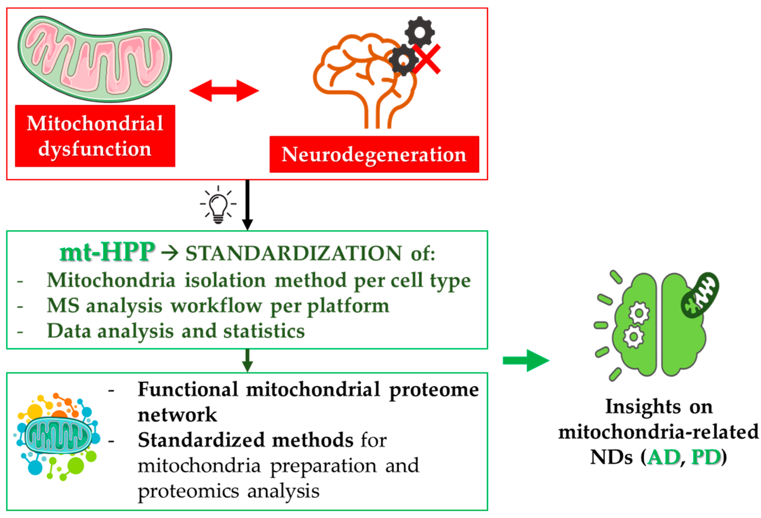
The growing number of patients affected by neurodegenerative disorders represents a huge problem for healthcare systems, human society, and economics. In this context, omics strategies are crucial for the identification of molecular factors involved in disease pathobiology, and for the discovery of biomarkers that allow early diagnosis, patients’ stratification, and treatment response prediction. The integration of different omics data is a required step towards the goal of personalized medicine. The Italian proteomics community is actively developing and applying proteomics approaches to the study of neurodegenerative disorders; moreover, it is leading the mitochondria-focused initiative of the Human Proteome Project, which is particularly important given the central role of mitochondrial impairment in neurodegeneration. 

neurodegeneration proteomics Italy integrated omics systems biology mitochondria

1. Introduction

Neurodegenerative disorders (NDs) are complex diseases affecting a large part of the elderly population worldwide. As the average human lifespan is continuously increasing, the number of patients is expected to dramatically grow in the coming decades. Thus, studying the mechanisms involved in the pathobiology of NDs, for the development of novel disease-modifying therapies, represents an urgent unmet need, especially in terms of the impact on human society and economics.
The majority of NDs involve both genetic and environmental factors [1]. Familial forms—characterized by a known causative genetic alteration with classical mendelian inheritance—usually represent a small percentage (5–15%) of the total number of cases, while sporadic forms represent the majority of them. The latter often display high inter-individual variability, in terms of clinical manifestations, severity, and response to therapeutic treatments.
In this context, omics strategies are of pivotal importance for the identification of novel molecular factors involved in disease pathobiology, and for the discovery of disease biomarkers for early diagnosis, patients’ stratification, prognosis, and prediction of response to treatments [2][3]. These objectives are difficult to achieve, especially in the context of neurodegeneration research, due to many overlapping factors contributing to more than one disease, or simply related to ageing. The integration of omics data (genomics, transcriptomics, proteomics, metabolomics, meta-omics) represents the most important step towards the goal of personalized medicine.
The Italian proteomics community has been actively involved in the study of neurodegenerative disorders, by developing and using several proteomics approaches, which have contributed to changing the treatment of many complex neurological disorders during the last decade. Moreover, Italy is leading the Human Proteome Project initiative, which is dedicated to studying mitochondria as part of both chromosome-centric (c-HPP) and Biology/Disease (B/D-HPP) programs [4]. This is particularly important in view of the central role of the mitochondrial impairment in the pathogenesis of several NDs.

2. Mitochondrial Proteomics and Neurodegeneration

Neurons are terminally differentiated cells which fully rely on oxidative phosphorylation for their energy supply. Healthy mitochondria guarantee not only ATP production, but also the maintenance of a correct metabolic homeostasis, the regulation of Ca2+ fluxes and redox signaling, and the arbitration of synaptic activity and cell survival [5].
Mitochondria are unique organelles endowed with their own genome, the mitochondrial DNA (mtDNA), encoding thirteen mitochondrial proteins of the electron transport chain (ETC) [6]; thus, mitochondrial activities are orchestrated by both nuclear and mitochondrial genes. Moreover, mitochondria are not standalone entities; rather, they are organized in complex, continuously remodeled networks, that allow them to exchange components and to dispose dysfunctional organelles. Network dynamics are mainly regulated by fusion and fission processes, which collectively represent the mitochondrial quality-control machinery [7][8][9]. When the mitochondrial quality control fails, and dysfunctional mitochondria are not properly eliminated, low ATP production, in combination with the generation of reactive oxygen species, leads to increased oxidative stress. This causes dramatic changes in cellular homeostasis, which can trigger cell death. In neurons, these mechanisms initiate and/or foster neurodegeneration. As demonstrated by a literature meta-analysis [10], the degeneration of synapses, led by mitochondrial impairment and metabolic failure, can be the starting event in these neurodegenerative processes. For this reason, mitochondrial dysfunction is well-recognized, and has been deeply investigated as an early event in the pathogenesis of several NDs, especially AD and PD [11][12]. In this frame, the efforts of the Italian mt-HPP initiative helped the identification of novel mechanisms in the crosstalk between mitochondrial dysfunction and neuronal cell death in NDs (Figure 1).
Figure 1. Mitochondria and neurodegeneration. Mitochondrial dysfunction represents one of the most frequent pathological alterations in NDs. Indeed, the failure of the tightly regulated mitochondrial quality control contributes to neuron death. The mitochondria-centered initiative of the Human Proteome Project (mtHPP), which aims at the in-depth investigation of the mitochondrial proteome in health and disease, has enabled the standardization of procedures related to mitochondria isolation and quantitative analysis of mitochondrial proteins. The fulfillment of the objectives of this project will achieve important advances in the understanding of several NDs, especially AD and PD .

2.1. The Italian mt-HPP Initiative

As mentioned above, the mitochondrial Human Proteome Project (mt-HPP) is a Human Proteome Organization (HUPO) initiative led by the Italian Proteomics Association (ItPA) (https://hupo.org/mitochondria). The main goal of this effort is to deepen the integrative role of proteins acting at the mitochondrial level, considering both those coded by the mt-DNA and those coded by the nuclear genome. The specific aims of this initiative have been: (i) the definition of the mitochondrial proteome by the characterization of all mitochondrial or mitochondria-associated proteins and their interactions in health and disease [13]; (ii) the definition of a functional mitochondrial proteome network; (iii) the development of standardized methods for mitochondria preparation and proteomics analysis; (iv) the characterization of the mitochondrial proteome/interactome in both neurodegenerative and metabolic diseases; (v) the identification of three missing proteins in the mitochondrial proteome. The achievement of these aims will dramatically increase the researchers' knowledge of mitochondrial biology, in both physiological conditions and disease states.
In the context of the mt-HPP, the Italian proteomics community has contributed to investigating the role played by mitochondrial proteins in the initiation and progression of neurodegenerative processes: multicentered experiments have been performed, using different MS platforms and different cellular models, to establish standardized protocols for mitochondria proteome analysis [14].
Furthermore, novel strategies have been proposed and optimized for the characterization of the mitochondrial proteome. The most important example is redox proteomics, which is that branch of proteomics used to identify oxidized proteins, and to determine the extent and location of oxidative modifications in proteins of interest. The steep increase in the development of redox proteomics strategies, pioneered by Perluigi and co-workers, has allowed researchers to unveil neurodegeneration-related changes in protein expression linked to protein oxidation levels [15][16]. This kind of approach is especially important in the study of NDs, where protein oxidation is a frequently observed pathobiological event.

2.2. Mitochondrial Proteomics in Alzheimer’s Disease

The seminal work of Castegna and co-workers suggested, for the first time, a relationship between protein oxidation and protein abundance, thanks to redox proteomics studies in AD brains [17]. They demonstrated that the targets of protein oxidation tend to precipitate as insoluble matter, suggesting an inverse relationship between protein oxidation and the amount of the soluble protein. It is notable that glyceraldehyde 3-phosphate dehydrogenase (GAPDH), aconitase, voltage-dependent anion channels (VDACs), the ATP synthase alpha-chain, lactate dehydrogenase (LDH), beta-actin, and alpha-tubulin—which are either mitochondrial proteins or are known to interact with mitochondria—have been identified as oxidation targets in AD brains. This indicates that protein abundance may discriminate which protein targets are more susceptible to oxidative modifications. In other words, the decrease in the protein levels of several mitochondrial proteins in NDs may be due to their oxidation and precipitation in insoluble protein aggregates [18][19]. Again in the context of AD, the proteomics analysis of mitochondria isolated from peripheral blood lymphocytes has highlighted several proteins altered between controls and AD patients, grouped into four categories: (i) cellular energetics, including GADPH, LDH B-chain, and ATP synthase subunit beta; (ii) structural proteins, including annexin, beta-centractin, and myosin light polypeptide 6; (iii) cell signaling, including Rho GDP-dissociation inhibitor 2 (RhoGDI); and (iv) cellular defense, including thioredoxin-dependent peroxide reductase/peroxiredoxin III (PDXIII) [20]. In addition, reduced OXPHOS complexes I, III, and IV activity has been reported in platelets from AD patients and in postmortem brains [21].

2.3. Mitochondrial Proteomics in Parkinson’s Disease

The Italian proteomics community has also, in the context of PD, significantly contributed to the identification of mitochondrial pathways involved in the pathogenesis. The central role of mitochondrial dysfunction in PD pathobiology was first suggested by the discovery that mutation in genes encoding mitochondrial proteins is a cause of familial forms of PD [22][23]. Following this, a strong body of evidence was collected, supporting the role of mitochondrial dysfunction in the early pathogenesis of the sporadic forms of the disease also, which represent the majority of the cases. In addition to this, mitochondria-targeted toxins (e.g., MPTP and rotenone) were shown to cause Parkinsonian symptoms in humans. Based on these observations, PD animal models have been generated through either the specific knock-out or the pharmacological targeting of ETC complexes [24][25].
In a pioneering paper, Basso and co-workers compared protein extract from the substantia nigra of PD patients and controls, and identified forty-four proteins expressed in this mid-brain region by peptide mass fingerprinting, among which, nine showed changes in their abundance: L and M neurofilament chains were less abundant in PD, whereas peroxiredoxin II, mitochondrial complex III, the ATP synthase D-chain, complexin I, profilin, the L-type calcium channel delta-subunit, and the fatty-acid binding protein were more present in PD samples than controls [26]. These results were the first to suggest a possible potentiation mechanism of afferent signals to SN, upon degeneration of dopaminergic neurons.
De Iuliis and co-workers first highlighted common neurodegeneration mechanisms in PD and AD, related to mitochondrial function, by the analysis of SN from hemiparkinsonian rats, a PD animal model obtained through unilateral intranigral injection of 6-hydroxydopamine (6-OHDA) [27]. After protein separation and quantification by 2DE, they identified alpha-enolase and beta-actin as differentially expressed proteins in lesioned SN, compared to controls. These proteins were also found to be oxidized and modulated in AD.
Alberio and co-workers deeply investigated the alterations in the mitochondrial proteome due to dopamine exposure in the SH-SY5Y human neuroblastoma cell line, by two-dimensional electrophoresis and by shotgun proteomics [28]. The results highlighted the fragmentation of some mitochondrial proteins, suggesting a possible alteration of the activity of mitochondrial proteases, which are key enzymes involved in mitochondrial quality control. The possible aberrant activation of mitochondrial proteases in conditions of altered dopamine homeostasis was further investigated, using a degradomics approach, by Lualdi and co-workers, who first proposed neprilysin as a mitochondria-localized protease aberrantly activated by dopamine treatment in SH-SY5Y cells [29]. The recent development of terminomics approaches in proteomics, which allow the quantitative assessment of protease-generated fragments by their labeling, enrichment, and LC–MS/MS analysis, greatly improved the researchers' knowledge about mitochondrial proteases, their targets, and their role in NDs [30][31]. Indeed, mitochondrial proteases perform quality-control surveillance, by degrading misfolded and non-assembled polypeptides, and regulate the activity of specific substrates by mediating their processing steps. They are also directly involved in NDs (as shown for the m-AAA protease), and may regulate mitochondrial molecules, such as OPA1, which in turn are implicated in the pathogenesis of NDs [32].

References

  1. Armstrong, R. What Causes Neurodegenerative Disease? Folia Neuropathol. 2020, 58, 93–112.
  2. Ruffini, N.; Klingenberg, S.; Schweiger, S.; Gerber, S. Common Factors in Neurodegeneration: A Meta-Study Revealing Shared Patterns on a Multi-Omics Scale. Cells 2020, 9, 2642.
  3. Manzoni, C.; Lewis, P.A.; Ferrari, R. Network Analysis for Complex Neurodegenerative Diseases. Curr. Genet. Med. Rep. 2020, 8, 17–25.
  4. Urbani, A.; De Canio, M.; Palmieri, F.; Sechi, S.; Bini, L.; Castagnola, M.; Fasano, M.; Modesti, A.; Roncada, P.; Timperio, A.M.; et al. The Mitochondrial Italian Human Proteome Project Initiative (Mt-HPP). Mol. Biosyst. 2013, 9, 1984–1992.
  5. Chan, D.C. Mitochondrial Dynamics and Its Involvement in Disease. Annu. Rev. Pathol. 2020, 15, 235–259.
  6. Chinnery, P.F.; Hudson, G. Mitochondrial Genetics. Br. Med. Bull. 2013, 106, 135–159.
  7. Tatsuta, T.; Langer, T. Quality Control of Mitochondria: Protection against Neurodegeneration and Ageing. EMBO J. 2008, 27, 306–314.
  8. Ng, M.Y.W.; Wai, T.; Simonsen, A. Quality Control of the Mitochondrion. Dev. Cell 2021, 56, 881–905.
  9. Stotland, A.; Gottlieb, R.A. Mitochondrial Quality Control: Easy Come, Easy Go. Biochim. Biophys. Acta (BBA)-Mol. Cell Res. 2015, 1853, 2802–2811.
  10. Monti, C.; Colugnat, I.; Lopiano, L.; Chiò, A.; Alberio, T. Network Analysis Identifies Disease-Specific Pathways for Parkinson’s Disease. Mol. Neurobiol. 2018, 55, 370–381.
  11. Yang, D.; Ying, J.; Wang, X.; Zhao, T.; Yoon, S.; Fang, Y.; Zheng, Q.; Liu, X.; Yu, W.; Hua, F. Mitochondrial Dynamics: A Key Role in Neurodegeneration and a Potential Target for Neurodegenerative Disease. Front. Neurosci. 2021, 15, 654785.
  12. Lezi, E.; Swerdlow, R.H. Mitochondria in Neurodegeneration. Adv. Exp. Med. Biol. 2012, 942, 269–286.
  13. Pagliarini, D.J.; Calvo, S.E.; Chang, B.; Sheth, S.A.; Vafai, S.B.; Ong, S.-E.; Walford, G.A.; Sugiana, C.; Boneh, A.; Chen, W.K.; et al. A Mitochondrial Protein Compendium Elucidates Complex I Disease Biology. Cell 2008, 134, 112–123.
  14. Alberio, T.; Pieroni, L.; Ronci, M.; Banfi, C.; Bongarzone, I.; Bottoni, P.; Brioschi, M.; Caterino, M.; Chinello, C.; Cormio, A.; et al. Toward the Standardization of Mitochondrial Proteomics: The Italian Mitochondrial Human Proteome Project Initiative. J. Proteome Res. 2017, 16, 4319–4329.
  15. Butterfield, D.A.; Perluigi, M.; Reed, T.; Muharib, T.; Hughes, C.P.; Robinson, R.A.S.; Sultana, R. Redox Proteomics in Selected Neurodegenerative Disorders: From Its Infancy to Future Applications. Antioxid. Redox Signal. 2012, 17, 1610–1655.
  16. Butterfield, D.A.; Perluigi, M. Redox Proteomics: A Key Tool for New Insights into Protein Modification with Relevance to Disease. Antioxid. Redox Signal. 2017, 26, 277–279.
  17. Castegna, A.; Aksenov, M.; Aksenova, M.; Thongboonkerd, V.; Klein, J.B.; Pierce, W.M.; Booze, R.; Markesbery, W.R.; Butterfield, D.A. Proteomic Identification of Oxidatively Modified Proteins in Alzheimer’s Disease Brain. Part I: Creatine Kinase BB, Glutamine Synthase, and Ubiquitin Carboxy-Terminal Hydrolase L-1. Free Radic. Biol. Med. 2002, 33, 562–571.
  18. Celi, P.; Gabai, G. Oxidant/Antioxidant Balance in Animal Nutrition and Health: The Role of Protein Oxidation. Front. Vet. Sci. 2015, 2, 48.
  19. Butterfield, D.A.; Palmieri, E.M.; Castegna, A. Clinical Implications from Proteomic Studies in Neurodegenerative Diseases: Lessons from Mitochondrial Proteins. Expert Rev. Proteom. 2016, 13, 259–274.
  20. Sultana, R.; Baglioni, M.; Cecchetti, R.; Cai, J.; Klein, J.B.; Bastiani, P.; Ruggiero, C.; Mecocci, P.; Butterfield, D.A. Lymphocyte Mitochondria: Towards Identification of Peripheral Biomarkers in Progression of Alzheimer Disease. Free Radic. Biol. Med. 2013, 65, 595–606.
  21. Bosetti, F.; Brizzi, F.; Barogi, S.; Mancuso, M.; Siciliano, G.; Tendi, E.A.; Murri, L.; Rapoport, S.I.; Solaini, G. Cytochrome c Oxidase and Mitochondrial F1F0-ATPase (ATP Synthase) Activities in Platelets and Brain from Patients with Alzheimer’s Disease. Neurobiol. Aging 2002, 23, 371–376.
  22. Shulman, J.M.; De Jager, P.L.; Feany, M.B. Parkinson’s Disease: Genetics and Pathogenesis. Annu. Rev. Pathol. 2011, 6, 193–222.
  23. Deng, H.; Wang, P.; Jankovic, J. The Genetics of Parkinson Disease. Ageing Res. Rev. 2018, 42, 72–85.
  24. Konnova, E.A.; Swanberg, M. Animal Models of Parkinson’s Disease. In Parkinson’s Disease: Pathogenesis and Clinical Aspects; Stoker, T.B., Greenland, J.C., Eds.; Codon Publications: Brisbane, Australia, 2018; ISBN 978-0-9944381-6-4.
  25. Pingale, T.; Gupta, G.L. Classic and Evolving Animal Models in Parkinson’s Disease. Pharmacol. Biochem. Behav. 2020, 199, 173060.
  26. Basso, M.; Giraudo, S.; Corpillo, D.; Bergamasco, B.; Lopiano, L.; Fasano, M. Proteome Analysis of Human Substantia Nigra in Parkinson’s Disease. Proteomics 2004, 4, 3943–3952.
  27. De Iuliis, A.; Grigoletto, J.; Recchia, A.; Giusti, P.; Arslan, P. A Proteomic Approach in the Study of an Animal Model of Parkinson’s Disease. Clin. Chim. Acta 2005, 357, 202–209.
  28. Alberio, T.; Bondi, H.; Colombo, F.; Alloggio, I.; Pieroni, L.; Urbani, A.; Fasano, M. Mitochondrial Proteomics Investigation of a Cellular Model of Impaired Dopamine Homeostasis, an Early Step in Parkinson’s Disease Pathogenesis. Mol. Biosyst. 2014, 10, 1332–1344.
  29. Lualdi, M.; Ronci, M.; Zilocchi, M.; Corno, F.; Turilli, E.S.; Sponchiado, M.; Aceto, A.; Alberio, T.; Fasano, M. Exploring the Mitochondrial Degradome by the TAILS Proteomics Approach in a Cellular Model of Parkinson’s Disease. Front. Aging Neurosci. 2019, 11, 195.
  30. Kleifeld, O.; Doucet, A.; Prudova, A.; auf dem Keller, U.; Gioia, M.; Kizhakkedathu, J.N.; Overall, C.M. Identifying and Quantifying Proteolytic Events and the Natural N Terminome by Terminal Amine Isotopic Labeling of Substrates. Nat. Protoc. 2011, 6, 1578–1611.
  31. Marshall, N.C.; Klein, T.; Thejoe, M.; von Krosigk, N.; Kizhakkedathu, J.; Finlay, B.B.; Overall, C.M. Global Profiling of Proteolysis from the Mitochondrial Amino Terminome during Early Intrinsic Apoptosis Prior to Caspase-3 Activation. J. Proteome Res. 2018, 17, 4279–4296.
  32. Martinelli, P.; Rugarli, E.I. Emerging Roles of Mitochondrial Proteases in Neurodegeneration. Biochim. Biophys. Acta (BBA)-Bioenerg. 2010, 1797, 1–10.
More
Information
Subjects: Neurosciences
Contributors MDPI registered users' name will be linked to their SciProfiles pages. To register with us, please refer to https://encyclopedia.pub/register : , ,
View Times: 460
Entry Collection: Neurodegeneration
Revisions: 2 times (View History)
Update Date: 04 Oct 2022
1000/1000
Video Production Service