Submitted Successfully!
To reward your contribution, here is a gift for you: A free trial for our video production service.
Thank you for your contribution! You can also upload a video entry or images related to this topic.
Version Summary Created by Modification Content Size Created at Operation
1 -- 2473 2022-09-27 15:31:37 |
2 Reference format revised. Meta information modification 2473 2022-09-28 02:59:29 |

Video Upload Options

Do you have a full video?

Confirm

Are you sure to Delete?
Cite
If you have any further questions, please contact Encyclopedia Editorial Office.
Catalani, E.;  Cherubini, A.;  Quondam, S.D.;  Cervia, D. Alternative Organism Models for Retina Neuroregeneration. Encyclopedia. Available online: https://encyclopedia.pub/entry/27718 (accessed on 03 May 2024).
Catalani E,  Cherubini A,  Quondam SD,  Cervia D. Alternative Organism Models for Retina Neuroregeneration. Encyclopedia. Available at: https://encyclopedia.pub/entry/27718. Accessed May 03, 2024.
Catalani, Elisabetta, Agnese Cherubini, Simona Del Quondam, Davide Cervia. "Alternative Organism Models for Retina Neuroregeneration" Encyclopedia, https://encyclopedia.pub/entry/27718 (accessed May 03, 2024).
Catalani, E.,  Cherubini, A.,  Quondam, S.D., & Cervia, D. (2022, September 27). Alternative Organism Models for Retina Neuroregeneration. In Encyclopedia. https://encyclopedia.pub/entry/27718
Catalani, Elisabetta, et al. "Alternative Organism Models for Retina Neuroregeneration." Encyclopedia. Web. 27 September, 2022.
Alternative Organism Models for Retina Neuroregeneration
Edit

Unlike in vitro cell cultures that cannot mimic tissue homeostasis and physiology, 3D retinal organoids are relatively cheap models and have an undeniable complexity rate. However, they are challenging to isolate and maintain long enough to investigate complex processes such as inflammation and neovascularization. These disadvantages are exacerbated considering the retina, which is mainly due to the global complexity of this tissue. Several classic diagnostic techniques could be applied to retinal organ cultures, such as optical coherence tomography, which explores the morphological aspect of the retinal architecture, electroretinograms that record the electrical response of retinal cells, and microelectrode array recording, which stimulates and records the electrical activity of RGC. Several mammalian retinal organ cultures as alternative models are currently available and well established, including those derived from mice, rats, rabbits, cats, dogs, non-human primates, bovines, and pigs. They are excellent samples for the preliminary phase before the in vivo step and for therapy tests, although organ cultures for the study of complex retinal neurodegenerative pathologies such as diabetic retinopathy (DR), retinitis pigmentosa (RP), age-related macular degeneration, and glaucoma are not entirely reproducing the human condition. Although all the events occurring during the various steps of retinal neurodegenerative diseases, including the clinical progression, are not fully mimicked by a single animal, preclinical in vivo models provide important information on the molecular and cellular mechanisms at the basis of the neuronal impairment. Thus, multiple organisms, including non-mammalian ones, are crucial for validating the mechanisms involved in retinal pathologies and developing new therapeutic options.

neurodegeneration neuronal regeneration retina zebrafish Drosophila melanogaster

1. Introduction

Cell death, inflammation and oxidative stress are the foremost common mechanisms occurring in degenerative diseases of the central nervous system (CNS), including those regarding the retina and the visual system [1][2]. New strategies and approaches targeting these general pathological aspects are continuously evaluated, and particular attention is paid to early biomarkers [2]. Retinal neurodegenerative diseases are the principal cause of vision impairment and vision loss, affecting people globally. The retina, anatomically and developmentally, is known as an extension of the CNS and displays similarities to the brain and spinal cord also in terms of response to insult, immunology, and neurodegenerative manifestations [3][4].
Establishing links between the retina impairments and neurodegenerative diseases appears particularly challenging for clinical strategy’s future development, and there is an urgent need for innovative technologies and standardized methodologies. It is noteworthy that a detailed knowledge of the status of the retina in neurodegenerative conditions is a crucial point to translate eye research to CNS disease. It is well established that neurodegeneration of the visual system reflects a complex scenario as several components contribute, i.e., neurons, vessels, inflammatory cells, immunological features, and biomechanical impairments [2]. Environmental factors, metabolic stress, neurovascular coupling, and genetic backgrounds may also play a fundamental role in retinal neurogenerative disorders, thus representing additional target areas to study for therapeutic interventions [2]. Many possible treatments are under evaluation for retina neurodegeneration, including gene therapy, antioxidants, anti-inflammatory, and antiapoptotic substances, alone or in combination [1]. An intriguing perspective is the possibility of regeneration after injuries [2].

2. Alternative Organism Models for Retina Neuroregeneration

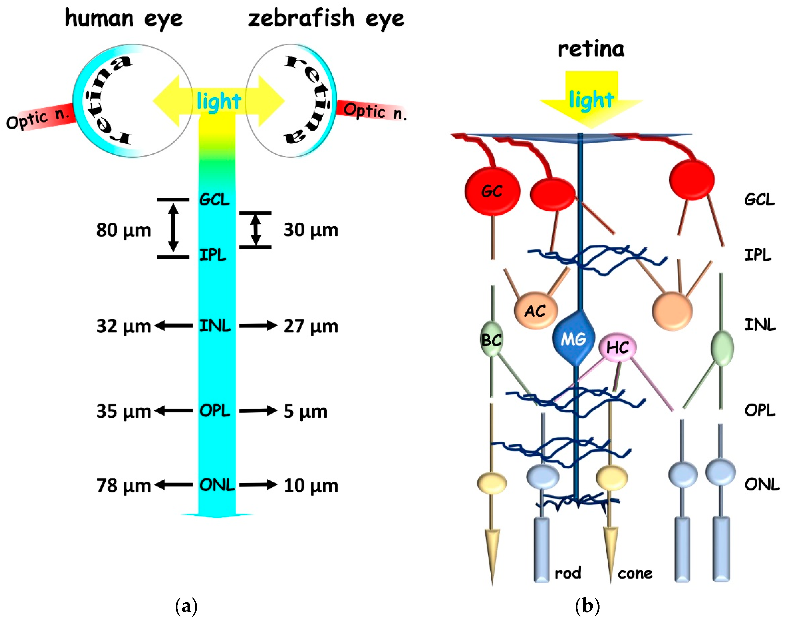
2.1. Zebrafish to Gain Insight in Vertebrate Retina

Zebrafish retina can regenerate after injury and is considered an ideal model for dissecting mechanisms relevant to retinal disease management [5]. Compared to humans, the zebrafish vision system shares structural and functional similarities (Figure 1) [6]; for example, it is cone-dominated, since zebrafish is a diurnal animal. Like in humans, the zebrafish retina consists of three nuclear layers (outer, inner, and RGC layer) containing neuronal soma separated by two plexiform layers (inner and outer) where synapsis takes place [7]. Photoreceptors consist of one rod cell type and blue and red–green cone types. In addition, zebrafish retina contains UV-sensitive cones, which are missing in humans. Zebrafish is a vertebrate with good color vision and high visual acuity [8]. RGC bodies are located in the RGC layer, while inner neurons consist of amacrine, horizontal, and Müller glial (MG) cell bodies, which are a type of retinal stem cell responsible for regenerative responses [9]. MG are radial glial cells in the inner vertebrate retina, which have a cylindrical, fiber-like shape. After an injury, MG de-differentiate and start asymmetric divisions that lead to the production of cells with glial properties and neuronal progenitor cells that proliferate, migrate and differentiate into new neuronal cell types [9]. Zebrafish possess tremendous intrinsic regenerative potential in ocular tissues, including the retinal pigment epithelium (RPE), while mammalian RPE is limited in its regenerative capacity. RPE inflammatory events highly participate in neurodegenerative progress, and RPE dysfunction or disease can lead to blindness in humans [10].
Figure 1. Schematic representation of human and zebrafish retina. (a) Retinal layers thickness. (b) Retinal layers and the main neuronal types. GCL (ganglion cellular layer); IPL (inner plexiform layer); INL (inner nuclear layer); OPL (outer plexiform layer); ONL (outer nuclear layer); GC (ganglion cell; red); AC (amacrine cell; orange); BC (bipolar cell; green); MG (Müller glia; blue); HC (horizontal cell; pink); rod (light blue), cone (yellow), optic n. (optic nerve).
Among the numerous signaling pathways involved in the regenerative potential of the zebrafish retinal neurons, the upregulation of transcription factor achaete-scute complex-like homolog 1 (ASCL1) appeared crucial in MG activation and reprogramming [11]. Indeed, it regulates several regeneration-associated signaling pathways strictly involved in key reprogramming/regenerative retinal steps, including the transcriptional regulator Notch, the transforming growth factor beta (TGF-β), and Wnt/B-catenin. It is noteworthy that Notch and (TGF-β) signaling are negative regulators of reprogramming/regeneration of the MG. Notch maintains MG in a quiescent state around the damaged area to prevent excessive proliferation and block differentiation into new neurons. On the contrary, Wnt/B-catenin signaling is crucial for retinal regenerative processes since it promotes the expression of ASCL1 and the subsequent regeneration response cascade. Furthermore, several other factors activate signaling cascades that lead to the MG reprogramming and regeneration of the injured zebrafish retina. Among them, the hedgehog family (the major regulator for cell differentiation and cell proliferation, including Shh which is critical for retinal regeneration following injury), STAT3 (the signal transducer and activator of transcription 3), α1-tubulin (a neuron-specific microtubule protein), FOXN4 (the forkhead box N4), ZIC2 (the zic family member 2), the transcriptional repressor insm1a, Apobec2b (the apolipoprotein B MRNA Editing Enzyme Catalytic Subunit 2), MAPK (the mitogen-activated protein kinase), PI3K/AKT (the phosphatidylinositol 3-kinase/protein kinase B), PAX6 (a member of the paired box gene family), SOX2 (the sex determining region Y-box 2), MYC (the myelocytomatosis oncogene), OCT4 (octamer-binding transcription factor 4), and RNA-binding protein LIN28, this latter being crucial in inducing pluripotent stem cells [11]. In addition, changes in epigenetics such as DNA methylation, histone modification, and miRNA-mediated degradation of mRNA concur with MG functionality [12], as well as immune response and microglia contribute to retinal regenerative progress mediated by MG [13]. Indeed, microglia cells respond quickly to an injury, which induces an inflammatory reaction promoting MG reprogramming, likely through mTOR signaling.
The stimulation of MG to regenerate injured neurons provides an excellent opportunity to repair degeneration of the retina, which is also associated with aging. In this respect, wild-type zebrafish can live up to 3.5 years in laboratory conditions, and they can accumulate the classic hallmarks of human retinal aging, such as DNA damage, shorter telomeres, and vision decline [14]. Furthermore, retinas of old zebrafish undergo tissue thinning, photoreceptor disorganization and neuronal loss, including RGC and bipolar cells. These morphological alterations occur independently, at least in part, from telomerase, since both wild-type and prematurely aged mutant tert−/− displayed the same scenario. Interestingly, a reduced expression of the crucial molecules is related to the regenerative process and coupled to the altered morphology of MG in aged retinas. In addition, when acute damages occur, aged retinas retained their ability to proliferate into new neurons [14]. These observations suggest that a certain level of key signals, reduced by aging, is necessary for the regenerative processes and manipulating these targets may improve neuroregeneration after injuries as well as in old age, when the already low ability to repair neurons is even more reduced. The main limitations of neural stem cell transplant usage in CNS are the risk of tumors caused by gene mutations and the change of the surrounding environment [15][16].

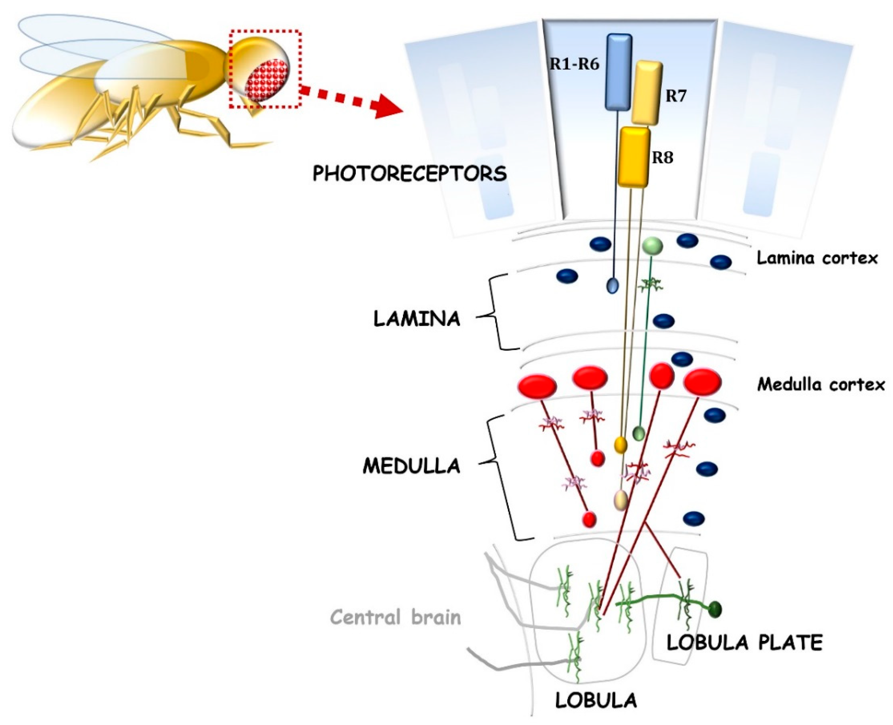
2.2. The Opportunity of D. melanogaster for Neuroregenerative Strategies

Although the drosophila visual system is morphologically and structurally different from the vertebrate one, many parallels can be described (Figure 2). Drosophila captures visual information by the retina and processes it through the optic lobes [17][18]. Each optic lobe consists of four distinct neuropiles: Lamina, Medulla, Lobula, and Lobula plate. The fly retina contains photoreceptors (R1–R8) that project their axons into the optic lobes. In particular, R1–R6 synapses with interneurons in the lamina, while R7 and R8 project to the medulla. These events resemble vertebrate photoreceptors’ synapsis with bipolar cells. In drosophila lamina and medulla, several cell types integrate signals as horizontal and amacrine cells in vertebrates. Furthermore, Lobula cells, such as RGC, send their axons to high-order neurons in the brain. These features suggest that the relevant mechanisms involved in the homeostasis of the retina neurons are well conserved. Remarkably, optic lobes, which project to the central brain, represent more than 60% of the brain. Therefore, in drosophila, a large part of the whole brain is dedicated to sight.
Figure 2. Schematic representation of the drosophila visual system, including the retina and optic neuropils: lamina, medulla, lobula, and lobula plate. Photoreceptors R1–R6 (blue bars) innervate the lamina, and R7–R8 (yellow bars) innervate the medulla. Green, red and blue cells represent lamina neuron, various medullary neurons, and glial cell types, respectively.
The human CNS cannot establish de novo neurogenesis after injury, but it can probably be induced by manipulating a specific molecular target. Since many basic biological, physiological, and neurological properties are extraordinarily conserved between mammals and D. melanogaster, flies represent a well-established system to understand and manage neurogenesis in the mammalian CNS [19], also representing a robust experimental in vivo tool for studying retinal dysfunctions [20].
D. melanogaster possesses glial cells in the visual system, which are also present in the entire nervous system, [21]. Like in mammals, glia plays a complex homeostatic role in the nervous system of flies, being in close morphological and functional connection with neurons. During the visual system development of drosophila, glial cells are crucial in mediating neural circuit assembly and forming boundaries [18][22], while in the mature visual system, they have a pivotal role in synaptic transmission and visual processing [21]. In flies, non-neuronal ommatidial cone cells (CC) support retinal neuronal cells and share structural, molecular, and functional aspects with vertebrate MG [23]. CC express specific conserved effector genes of the glial cells, including the pump Na/K-ATPase, the K-inward rectifying channels (Kir channels), the excitatory amino acid transporters (EAAT1), the glucose transporter 1, and the lactate dehydrogenase (LDH). Moreover, CC express prospero and PAX2 transcription factors related to glia functions.
Critical aspects of regeneration have been studied in the imaginal disc of flies, which has high regeneration ability after injuries [19]. For instance, oxidative stress occurrence in an injured imaginal disc has been observed under several different signaling, including Nox/Duox NADPH- oxidases [24]. In addition, reactive oxygen species activate and regulate c-Jun N-terminal kinases (JNK), p38 stress-activated MAPK, and the JAK/STAT signaling pathway that could drive the proliferative rate of the environment surrounding the wound, also stimulating drosophila insulin-like peptide (dilp8). Dilp8 is crucial in balancing the developmental delay and the growth of healthy and damaged tissue. JNK signaling was shown to target wg (Wnt1 homolog), which is involved in regeneration, dpp (bone morphogenetic protein decapentaplegic), taking part in growth during imaginal disc development, and hippo signaling [24].
Drosophila has been demonstrated to display neuroregenerative ability after penetrating traumatic brain injury, which is more significant in young when compared with aged flies [25]. In this model [26], the new neurons and glia appeared functional and well connected; indeed, there is a recovery of locomotion within 14 days after injury. Remarkably, the neurogenesis in the central brain differs from that in the optic lobe, where there is no proliferation of glia [27].
Undeniably, regeneration is a complex process that depends on injured/damaged tissue/organ, consisting of different stages. Photocontrol of specific engineering neurons by optogenetics enables the development of promising clinical neuroregenerative strategies for replacing degenerated functions or delivering pro-survival signals, since optogenetics can mix spatial and temporal light stimulation with genetic engineering to stimulate cells or tissues during a specific development phase of degenerative disease [28]. In this respect, genetically modified damaged neurons of drosophila have been shown to undertake their regeneration pathways and regulate their growth direction after stimulation with blue light [29]. In particular, the optogenetic activation of both the Raf/MEK/ERK (optoRaf) and AKT (optoAKT) signaling enhanced axon regeneration in injured neurons, but only optoRaf improved dendritic branching in CNS and the peripheral nervous system. Accordingly, it was recently reported that the optogenetics approach may induce beneficial trophic effects in a fly genetic model for parkinsonism [30]. Therefore, because of its simple genetic manipulation, D. melanogaster represents an ideal animal model to expand research in optogenetics and provide proof-of-concept studies.

3. Conclusions

Expanding new strategies in the pathophysiology of CNS is a daily challenge. Certainly, the translation of eye research to CNS and deciphering the role of immune cells in these two systems could improve the understanding and, potentially, the treatment of CNS diseases. Evaluating the impairment of the visual system at early stages to provide biomarkers of neurodegeneration is gaining attention, since it could help to test the efficacy of neuroprotective treatments and identify possible therapeutic strategies. In this context, manipulating signaling pathways that lead to neuronal regeneration offers an excellent opportunity to substitute damaged cells and, thus, restore the CNS functionality. The regenerative ability of vertebrate models, such as zebrafish, is particularly appealing. In addition, the fruit fly is an ideal and alternative animal model for regenerative studies due to its high degree of conservation with vertebrates and the broad spectrum of genetic variants achievable. Furthermore, a large part of the drosophila brain is dedicated to sight, thus offering the possibility of studying common mechanisms of the visual system and the brain at once. On the other side, zebrafish and drosophila are evolutionarily distant from mammals, and far from human complexity, representing the most significant limitation in their use. CNS anatomy differences, the less complex immune system, and the possibility that they could have a different response to stimulating-regeneration drugs imply that results should be verified on more biologically complex organisms. Indeed, the neuroregeneration pathways that work in other species, but not in mammals, may also represent a key confounding factor. In this respect, the validation of proof-of-concept results for future therapeutics would need, for instance, the comparison of treatment responses between fish, flies, and humans and/or further studies about translational biomarkers that bridge these different species. However, undeniably, the knowledge acquired from these alternative models may offer several starting points to manipulate specific well-conserved signal pathways of interest in human regeneration after injuries or during pathologies.

References

  1. Maneu, V.; Lax, P.; Cuenca, N. Current and future therapeutic strategies for the treatment of retinal neurodegenerative diseases. Neural Regen. Res. 2022, 17, 103–104.
  2. Wareham, L.K.; Liddelow, S.A.; Temple, S.; Benowitz, L.I.; Di Polo, A.; Wellington, C.; Goldberg, J.L.; He, Z.; Duan, X.; Bu, G.; et al. Solving neurodegeneration: Common mechanisms and strategies for new treatments. Mol. Neurodegener. 2022, 17, 23.
  3. London, A.; Benhar, I.; Schwartz, M. The retina as a window to the brain-from eye research to CNS disorders. Nat. Rev. Neurol. 2013, 9, 44–53.
  4. Sharma, S.; You, Y. Editorial: Retinal Changes in Neurological Diseases. Front. Neurosci. 2021, 15, 813044.
  5. Chhetri, J.; Jacobson, G.; Gueven, N. Zebrafish—On the move towards ophthalmological research. Eye 2014, 28, 367–380.
  6. Stella, S.L., Jr.; Geathers, J.S.; Weber, S.R.; Grillo, M.A.; Barber, A.J.; Sundstrom, J.M.; Grillo, S.L. Neurodegeneration, Neuroprotection and Regeneration in the Zebrafish Retina. Cells 2021, 10, 633.
  7. Richardson, R.; Tracey-White, D.; Webster, A.; Moosajee, M. The zebrafish eye-a paradigm for investigating human ocular genetics. Eye 2017, 31, 68–86.
  8. Baden, T.; Euler, T.; Berens, P. Understanding the retinal basis of vision across species. Nat. Rev. Neurosci. 2020, 21, 5–20.
  9. Gallina, D.; Todd, L.; Fischer, A.J. A comparative analysis of Muller glia-mediated regeneration in the vertebrate retina. Exp. Eye Res. 2014, 123, 121–130.
  10. Kaur, G.; Singh, N.K. The Role of Inflammation in Retinal Neurodegeneration and Degenerative Diseases. Int. J. Mol. Sci. 2021, 23, 386.
  11. Gao, H.; Luoden, A.; Huang, X.; Chen, X.; Xu, H. Muller Glia-Mediated Retinal Regeneration. Mol. Neurobiol. 2021, 58, 2342–2361.
  12. Powell, C.; Grant, A.R.; Cornblath, E.; Goldman, D. Analysis of DNA methylation reveals a partial reprogramming of the Muller glia genome during retina regeneration. Proc. Natl. Acad. Sci. USA 2013, 110, 19814–19819.
  13. Van Dyck, A.; Bollaerts, I.; Beckers, A.; Vanhunsel, S.; Glorian, N.; van Houcke, J.; van Ham, T.J.; De Groef, L.; Andries, L.; Moons, L. Muller glia-myeloid cell crosstalk accelerates optic nerve regeneration in the adult zebrafish. Glia 2021, 69, 1444–1463.
  14. Martins, R.R.; Zamzam, M.; Tracey-White, D.; Moosajee, M.; Thummel, R.; Henriques, C.M.; MacDonald, R.B. Muller Glia maintain their regenerative potential despite degeneration in the aged zebrafish retina. Aging Cell 2022, 21, e13597.
  15. Yang, S.G.; Wang, X.W.; Qian, C.; Zhou, F.Q. Reprogramming neurons for regeneration: The fountain of youth. Prog. Neurobiol. 2022, 214, 102284.
  16. Wang, F.; Cheng, L.; Zhang, X. Reprogramming Glial Cells into Functional Neurons for Neuro-regeneration: Challenges and Promise. Neurosci. Bull. 2021, 37, 1625–1636.
  17. Neriec, N.; Desplan, C. From the Eye to the Brain: Development of the Drosophila Visual System. Curr. Top. Dev. Biol. 2016, 116, 247–271.
  18. Malin, J.; Desplan, C. Neural specification, targeting, and circuit formation during visual system assembly. Proc. Natl. Acad. Sci. USA 2021, 118, e2101823118.
  19. Fox, D.T.; Cohen, E.; Smith-Bolton, R. Model systems for regeneration: Drosophila. Development 2020, 147, dev173781.
  20. Catalani, E.; Silvestri, F.; Cervia, D. A Drosophila perspective on retina functions and dysfunctions. Neural Regen. Res. 2022, 17, 341–343.
  21. Kremer, M.C.; Jung, C.; Batelli, S.; Rubin, G.M.; Gaul, U. The glia of the adult Drosophila nervous system. Glia 2017, 65, 606–638.
  22. Chotard, C.; Salecker, I. Glial cell development and function in the Drosophila visual system. Neuron Glia Biol. 2007, 3, 17–25.
  23. Charlton-Perkins, M.A.; Sendler, E.D.; Buschbeck, E.K.; Cook, T.A. Multifunctional glial support by Semper cells in the Drosophila retina. PLoS Genet. 2017, 13, e1006782.
  24. Ahmed-de-Prado, S.; Baonza, A. Drosophila as a Model System to Study Cell Signaling in Organ Regeneration. Biomed Res. Int. 2018, 2018, 7359267.
  25. Crocker, K.L.; Marischuk, K.; Rimkus, S.A.; Zhou, H.; Yin, J.C.P.; Boekhoff-Falk, G. Neurogenesis in the adult Drosophila brain. Genetics 2021, 219, iyab092.
  26. Crocker, K.L.; Ahern-Djamali, S.; Boekhoff-Falk, G. Stimulating and Analyzing Adult Neurogenesis in the Drosophila Central Brain. J. Vis. Exp. 2021, 176, e63182.
  27. Fernandez-Hernandez, I.; Rhiner, C.; Moreno, E. Adult neurogenesis in Drosophila. Cell Rep. 2013, 3, 1857–1865.
  28. Janovjak, H.; Kleinlogel, S. Optogenetic neuroregeneration. Neural Regen. Res. 2022, 17, 1468–1470.
  29. Wang, Q.; Fan, H.; Li, F.; Skeeters, S.S.; Krishnamurthy, V.V.; Song, Y.; Zhang, K. Optical control of ERK and AKT signaling promotes axon regeneration and functional recovery of PNS and CNS in Drosophila. eLife 2020, 9, e57395.
  30. Ingles-Prieto, A.; Furthmann, N.; Crossman, S.H.; Tichy, A.M.; Hoyer, N.; Petersen, M.; Zheden, V.; Biebl, J.; Reichhart, E.; Gyoergy, A.; et al. Optogenetic delivery of trophic signals in a genetic model of Parkinson’s disease. PLoS Genet. 2021, 17, e1009479.
More
Information
Subjects: Physiology
Contributors MDPI registered users' name will be linked to their SciProfiles pages. To register with us, please refer to https://encyclopedia.pub/register : , , ,
View Times: 232
Revisions: 2 times (View History)
Update Date: 28 Sep 2022
1000/1000