Submitted Successfully!
To reward your contribution, here is a gift for you: A free trial for our video production service.
Thank you for your contribution! You can also upload a video entry or images related to this topic.
Version Summary Created by Modification Content Size Created at Operation
1 -- 5120 2022-08-26 02:26:30 |
2 format corrected. + 19 word(s) 5139 2022-08-26 09:56:55 | |
3 format corrected. Meta information modification 5139 2022-08-26 09:58:16 |

Video Upload Options

Do you have a full video?

Confirm

Are you sure to Delete?
Cite
If you have any further questions, please contact Encyclopedia Editorial Office.
Azman, M.;  Sabri, A.H.;  Anjani, Q.K.;  Mustaffa, M.F.;  Hamid, K.A. Formulation Strategies to Improve Pharmacokinetics Profile. Encyclopedia. Available online: https://encyclopedia.pub/entry/26524 (accessed on 04 July 2024).
Azman M,  Sabri AH,  Anjani QK,  Mustaffa MF,  Hamid KA. Formulation Strategies to Improve Pharmacokinetics Profile. Encyclopedia. Available at: https://encyclopedia.pub/entry/26524. Accessed July 04, 2024.
Azman, Maisarah, Akmal H. Sabri, Qonita Kurnia Anjani, Mohd Faiz Mustaffa, Khuriah Abdul Hamid. "Formulation Strategies to Improve Pharmacokinetics Profile" Encyclopedia, https://encyclopedia.pub/entry/26524 (accessed July 04, 2024).
Azman, M.,  Sabri, A.H.,  Anjani, Q.K.,  Mustaffa, M.F., & Hamid, K.A. (2022, August 26). Formulation Strategies to Improve Pharmacokinetics Profile. In Encyclopedia. https://encyclopedia.pub/entry/26524
Azman, Maisarah, et al. "Formulation Strategies to Improve Pharmacokinetics Profile." Encyclopedia. Web. 26 August, 2022.
Formulation Strategies to Improve Pharmacokinetics Profile
Edit

The oral route is the most common and practical means of drug administration, particularly from a patient’s perspective. However, the pharmacokinetic profile of oral drugs depends on the rate of drug absorption through the intestinal wall before entering the systemic circulation. However, the enteric epithelium represents one of the major limiting steps for drug absorption, due to the presence of efflux transporters on the intestinal membrane, mucous layer, enzymatic degradation, and the existence of tight junctions along the intestinal linings. These challenges are more noticeable for hydrophilic drugs, high molecular weight drugs, and drugs that are substrates of the efflux transporters. Another challenge faced by oral drug delivery is the presence of first-pass hepatic metabolism that can result in reduced drug bioavailability. Over the years, a wide range of compounds have been investigated for their permeation-enhancing effect in order to circumvent these challenges. There is also a growing interest in developing nanocarrier-based formulation strategies to enhance the drug absorption.

absorption enhancers intestinal absorption oral delivery pharmacokinetic profile nanocarriers

1. Current Absorption Enhancers and Their Absorption-Enhancing Mechanisms to Improve the Pharmacokinetic Profile

1.1. Solubilizing Agents

The application of solubilizing agents is one of the most common strategies to enhance the water solubility of orally administered drugs in an attempt to enhance the oral bioavailability of the delivered therapeutic compound. Solubilizing agents lead to the formation of fine dispersions of the lipid-solubilized drugs in the aqueous milieu of the gastrointestinal tract through the process of self-emulsification [1]. One of the classes of solubilizing agents are surfactants, which display amphiphilic properties. The surfactants are generally incorporated into emulsions and suspensions, due to their ability to reduce interfacial surface tension and leading to the production of a stable colloidal formulation. Its role as an absorption enhancer is contributed by its interaction with the plasma membrane, either by disrupting the barrier function of the epithelial cells or the sequestration of proteins [2]. In general, non-ionic surfactants are more favorable than ionic surfactant, as this sub-class of surfactants displays better safety profiles while only inducing a reversible modulation in the intestinal mucosal permeability [3].
One of the surfactants that have been studied as an absorption enhancer is sucrose laurate. Aside from its non-ionic characteristics, it has a low critical micelle concentration (CMC), which allows it to reduce the interfacial surface tension at a low concentration [4]. This makes it more potent than sodium caprate (C10) and sodium salt of lauric acid (C12), which have a much higher CMC thus necessitating a higher concentration of the surfactant in order to elicit a similar effect. In a study by McCartney et al., the group observed an increase in the Papp of [14C]-mannitol in the presence of sucrose laureate in a concentration-dependent manner in both in vitro and ex vivo studies [5]. This is supported by another study by Maher et al. that also reported an improvement in the mannitol permeability in the presence of sucrose laurate across isolated rat colonic mucosa [4]. Based on an in vivo study, sucrose laurate successfully improved the delivery of insulin at concentrations of 50 and 100 mM across rat jejunum and colon, resulting in the reduction in the blood glucose level. The absorption-enhancing action of this surfactant has been observed to affect the transcellular pathway. This result is associated with the ability of sucrose laurate to decrease the mitochondrial membrane potential while simultaneously increasing plasma membrane potential as well as altering the expression of the tight junctions protein, ZO-1 [5]. In addition, the surfactant is capable of fluidizing the plasma membrane while altering the efflux transporters’ activity that culminates in an increase in the substrate permeability across the membrane. Such an effect is site-dependent, with sucrose laurate having a more pronounced effect along the colon than the jejunum [5]. This effect on the membrane permeability by sucrose laurate along this region is also postulated to act in synergy with the high exposure of bile salts within the jejunum, which could improve drug solubility in the lumen. Aside from that, the lower peptidase level and longer residence time in the colon may, in tandem, reduce the drug degradation and improve the degree of absorption [6].
Another solubilizing agent that is frequently employed in drug delivery is cyclodextrins (CD). CDs are cyclic oligosaccharides with a hydrophobic center that enables the entrapment of drug molecules, as illustrated in Figure 1. Through encapsulating the hydrophobic drugs’ molecules inside the annulus, the cyclodextrins form an inclusion complex with the drug. This complex formation improves the drug solubility and aids the diffusion of the drug molecules across the unstirred water layer to reach the apical membrane of the enterocytes. In addition, this inclusion complex also improves drug permeability across the enterocytes, leading to improved oral bioavailability and pharmacokinetic profile [7].
Figure 1. Mechanism for cyclodextrin-enhanced solubility of poorly soluble drugs through entrapment and complex formation with the drug molecule.
The permeation-enhancing ability of cyclodextrin is also attributed to the ability of the cyclic oligosaccharide to extract the membrane proteins and phospholipids from the apical membrane, leading to enhanced membrane fluidization [8]. In addition, the formation of the cyclodextrin drug complex is also capable of widening the tight junctions along the gastrointestinal tract, leading to enhanced paracellular transport [9]. The formation of an inclusion complex allows the ‘guest’ molecule to be transported in the aqueous milieu of the gastrointestinal lumen, as the complex formation enhances the wettability, solubility, and dissolution rate of the drug while simultaneously improving drug stability, and permeability across the gastric mucosa [10]. Subsequently, this allows the inclusion complex to be transported across the lipophilic cell membranes. The CDs managed to enhance the oral absorption of BCS Class II drugs, such as carbamazepine and albendazole, which are characterized by low aqueous solubility, thus leading to improved oral bioavailability [11]. In a study by Rubim et al. (2017), such enhanced oral absorption and the bioavailability of the delivered amiodarone hydrochloride was attributed to the increase in the drug solubility when formulated with methyl-β-CD [12]. In another study by Devasari et al. (2015), erlotinib (ERL) was formulated with sulfobutyl ether beta-cyclodextrin, leading to the formation of drug-CD complex (ERL-SBE-β-CD) that enhanced the drug solubility by 7.4-fold [7]. In comparison to the free drug, the complex also showed a 5.4-fold decrease in Tmax, with a 3.2-fold increase in Cmax when evaluated in vivo in Sprague Dawley rats, which was attributed to the enhanced dissolution of the drug.

1.2. Bile Salts

Bile salts are endogenous compounds that are present in the small intestine, with concentrations ranging from 8 mM and 18 mM in the fasted and fed state, respectively [13]. These compounds consist of taurine and glycine conjugates of chenodeoxycholic acid and cholic acid. The bile salts are categorized based on the extent of their hydroxylation and conjugation with amino acids. The classifications consist of dihydroxy conjugates (taurodeoxycholate and taurochenodeoxycholate), trihydroxy conjugates (glycocholate and taurocholate), and unconjugated forms (cholate and chenodeoxycholate) [14]. About 60% of the bile salts present within the gastrointestinal tract are dihydroxy conjugates. Through micellar solubilization, the bile salts play a major role in facilitating the digestion of dietary lipids via emulsification, lipolysis, and ultimately the transportation of the lipid products across the gut epithelium during absorption [15]. Should the bile salts escape active reabsorption within the ileum, the intestinal flora will metabolize them into secondary bile salts; lithocholic acid and deoxycholic acid. From a drug delivery perspective, the bile salts are capable of enhancing gastrointestinal absorption and ultimately oral bioavailability through several mechanisms, which include-solubilization of the poorly soluble drug through micellar solubilization, enhancing the chemical stability of the delivered therapeutic, increasing the membrane fluidity along the epithelial cells that line the gastrointestinal tract, the opening of tight junctions, membranolysis of the epithelial membrane, as well as modulating the function of the transport proteins along the gut epithelium [16][17]. Another mechanism for the absorption-enhancing effect of bile salts is the ability of the compound to reduce the viscosity and elasticity of the mucus layer, which in turn enhances the diffusion rate of the molecule across the mucus layer to the gut epithelium for intestinal absorption [17]. Another study also found that the bile salts, such as sodium glycocholate, have an inhibitory effect on peptidases through ionic and hydrophobic interactions. This confers protection to the peptides and proteins, such as insulin from proteolytic degradation when the bile salts are co-administered orally [6].
Studies investigating the effect of utilizing bile salts as a strategy to enhance drug permeability are summarized in Table 1. Berberine chloride was one of the drugs which had shown an increase in the area under the curve (AUC) through co-administration with sodium deoxycholate in vivo. The investigator discovered that the co-administration of berberine chloride with sodium deoxycholate resulted in a 35.2-fold increase in the plasma concentration of the drug when compared to the control group that received berberine in the absence of the bile salts [18]. In another study, a microcapsule containing taurocholic acid was shown to assist the absorption of gliclazide in diabetic Wistar rats through a targeted release mechanism at pH 7.8 at which the gliclazide solubility improved from approximately 10 µg/mL (without taurocholic acid) to 40 µg/mL after 3 h post-administration, thus producing a better hypoglycemic effect [19].
Another derivative of bile salts, sodium taurocholate (10 and 20 mmol/L), increased the permeability of cefquinome from 0.26 ± 0.04 µg/mL to 0.57 ± 0.03 µg/mL and 0.78 ± 0.07 µg/mL, respectively, when evaluated in vivo in rats. It was concluded that the enhancement of the drug absorption was through the modulation of the tight junctions, leading to enhanced paracellular transport [20]. Meanwhile, Moghimipour et al. reported that sodium glycocholate was a better absorption enhancer than sodium taurodeoxycholate, although not statistically significant, for the molecule 5(6)-carboxyfluorescein. In the presence of sodium glycocholate, 12 µg/mL of the dye was able to permeate across the Caco-2 cell lines, while only 10 µg/mL of the compound was able to permeate when co-administered with sodium taurodeoxycholate [21]. Taurodeoxycholic acid (TDCA) is a secondary bile acid, a product of the primary bile acid metabolism in the intestine. A recent study showed that TDCA effectively increased the permeation of EGFR2R-lytic hybrid peptide (epidermal growth factor receptor-binding peptide conjugated with lytic peptide) by the formation of a peptide/bile acid complex, compared to the administration of the peptide alone. The formation of the complex facilitated the widening of the tight junctions along the intestinal epithelium that led to enhanced intestinal absorption [22]. The mechanism for the enhancement in intestinal absorption was evidenced by the ability of the peptide to reduce TEER across Caco-2 monolayers when evaluated in vitro.
Table 1. Summary of studies investigating the effect of bile salts on the intestinal permeability and oral pharmacokinetic parameters of drugs.

1.3. Chitosan

Chitosan is a hydrophilic polysaccharide derived from chitin via N-deacetylation [23]. This biopolymer consists of N-acetyl-d-glucosamine and β-(1–4)-linked d-glucosamine. Due to its biodegradable and biocompatible characteristics, it is extensively used as an excipient either as a tablet disintegrant, release modifier, or even just as a filler. The characteristic of chitosan which is identified to be crucial in exhibiting its permeation-enhancing effect is its mucoadhesive properties. This property is attributed to its positive charge that interacts with the negatively charged glycocalyx of the microvilli along the small intestines [24], resulting in the redistribution of F-actin in the cytoskeleton and the translocation of the tight junctions’ components, ZO-1, and the occludin proteins. This further enables the widening of the tight junctions for the paracellular transport of solutes across the gastrointestinal tract [25][26].
The mucoadhesive properties help the drug to adhere to the mucosal surface of the gastrointestinal tract for a prolonged period of time [16]. This increases the residence time of the drugs in the small intestine, which in turn leads to greater drug absorption. Indeed, the permeation-enhancing effect is more pronounced when the grade of chitosan used is of a higher molecular weight, which leads to enhanced permeation rates. The permeation rate and Papp value of salvianolic acid B were found to be at a maximum when co-delivered with chitosan displaying a molecular weight of 100 kDa. However, with a decreasing molecular weight, the researchers also observed a decline in the permeation rate of salvianolic acid B [27]. It was proposed that the chitosan, which exhibits a positive surface charge, forms ionic interactions with the negatively charged glycocalyx groups, resulting in the reversibility of the opening tight junctions, which could improve the paracellular drug transport. Another factor that affects the permeation-enhancing effect of chitosan is the degree of deacetylation. Should the grade of chitosan possess a degree of deacetylation that is greater than 80%, the biopolymer was found to exhibit a greater degree of muco-adhesion [28]. It was also discovered that chitosan with a high degree of deacetylation displayed good permeation-enhancing properties at both low and high molecular weights. In contrast, chitosan with a low degree of deacetylation was only effective in enhancing the absorption of the drug molecules across the gastrointestinal tract at higher molecular weights [29].
The studies on the role of chitosan as a permeation enhancer are summarized in Table 2. Indeed, some of these results contradict one another; this may be attributed to the different types of intestinal permeability models, the physiological state of the intestinal tissue, and the species used. In addition, the grade of chitosan used may also contribute to such discrepancies in the results. Due to chitosan having an overall pKa 6.5, its absorption-enhancing effects are typically apparent at a pH below 6.5, during which the biopolymer is in a protonated state. This characteristic implies that its absorption-enhancing effects can only be exerted in a limited area along the gastrointestinal tract, as the pH along the intestines may fluctuate to a pH above 6.5 in some parts [30]. Acyclovir was one of the compounds that did not show an improvement in its permeation following co-administration with chitosan. This was due to the interaction between the chitosan and the mucus layers on the intestinal membrane, which altered the reactivity of the chitosan. This caused the biopolymer to have a minimal impact on the Caco-2 monolayer integrity, leading to no enhancement in the absorption of acyclovir [31]. This was further corroborated by a recent study that concluded that the absorption-enhancing effect of chitosan was drug-dependent, with acyclovir displaying unfavorable enhancement of absorption in the presence of chitosan [32]. On the other hand, there are studies that highlighted drugs that displayed an enhanced absorption with the aid of chitosan, and these include molecules such as buserelin, [14C] mannitol, and dextran [27][33].
The role of chitosan as an absorption enhancer in its salt form was also investigated. Chitosan hydrochloride did not exhibit the same permeability-enhancing effect as the non-salt variant of chitosan. Interestingly, the salt form of chitosan resulted in a decreased Cmax and AUC0–36h of berberine [34]. This result was attributed to the change in the berberine solubility in the presence of chloride ions, when the drug was co-administered with chitosan hydrochloride. This was explained by the common ion effect in which the chloride ion pairs up and reduces the overall net charge of the berberine. In contrast, trimethyl chitosan was reported as a good absorption enhancer, due to its increased ability to transiently open the tight junctions compared to normal unmodified chitosan [35]. Indeed, it can be seen that chitosan does, to some extent, display good permeation-enhancing properties. Nevertheless, this permeation-enhancing effect is dependent on multiple factors, such as the weight and charge of the biopolymer. A considerable body of work has made strides in understanding the permeation-enhancing effect at a molecular and cellular level. Nevertheless, it can be seen that, indeed, further work is still pivotal to fully understanding and appreciating the mechanisms behind the permeation-enhancing effect of chitosan before this biopolymer can utilized extensively as an excipient for oral drug delivery.
Table 2. Summary of studies investigating the effect of chitosan and its derivatives on the intestinal permeability and oral pharmacokinetic parameters of drugs.

2. Formulation Strategies to Improve Pharmacokinetics Profile

2.1. Solid Lipid Nanoparticles (SLN)

Solid lipid nanoparticles (SLN) have garnered great interest in their ability to improve the oral absorption of poorly soluble drugs. They are a colloidal system derived from a matrix of lipids that retains their solid state at a temperature below 140 °C with sizes ranging from 50 to 1000 nm and able to disperse in an aqueous medium with the aid of surfactants [37][38].
Some of the advantages of employing SLN as a drug delivery system for oral administration include mitigating the degradation of entrapped drugs, as well as providing some control over the rate of drug release. Another advantage of utilizing this formulation approach is the ability to avoid hepatic first-pass metabolism through the intestinal lymphatic uptake. This is because the lipophilic nature of the SLN causes the nanocarrier to drain into the thoracic lymph before entering the systemic circulation near the left subclavian vein, thus circumventing the hepatic first-pass metabolism [39][40]. One of the reasons for the absorption-enhancing ability conferred by SLN is attributed to the interaction of the P-gp efflux pump, which causes the substrate to be unavailable for transport. This was confirmed by Garg et al. (2016), who discovered that by formulating lumefantrine as the SLN, they were able to enhance the oral bioavailability of the drug, which is a substrate for P-gp, by 2.7-fold relative to when the lumefantrine was delivered in 0.25% w/v Na CMC suspension by oral gavage. The SLN used consisted of a binary lipid mixture of stearic acid and caprylic acid stabilized with the non-ionic surfactant, D-alpha tocopheryl polyethylene glycol 1000 succinate (TPGS) and formulated Poloxamer 188 [41]. Another advantage of formulating poorly soluble drugs by using SLN is the ability of the nanocarrier to increase the dissolution rate of the drug within the gastric lumina, thus generating a concentration gradient to promote absorption across the gastrointestinal membrane. Such an enhanced dissolution rate is attributed to the submicron size of the SLN, which provide a large surface area for drug dissolution [41].
Valdes et al. reported that the bioavailability of 4-(N)-docosahexaenoyl 2′, 2′-difluorodeoxycytidine (DHA-dFdC) improved from 113.55 μg•h/mL to 143.44 μg•h/mL when delivered as a SLN formulation. A conclusion on the exact absorption-enhancing mechanism was not fully elucidated, yet the researchers have proposed a few hypotheses for the absorption-enhancing effect observed. Firstly, the SLN may be absorbed through the microfold cells in the Peyer’s patches, leading to the migration of the nanocarrier into the lymphatic system thus obviating the first-pass metabolism and leading to enhanced bioavailability. Secondly, the authors also proposed that the SLN release lipids upon digestion that alter the gastrointestinal fluid media, that then lead to the enhanced solubility and dissolution of the DHA-dFdC, leading to improved absorption. In addition, it was postulated that the SLN provided a barrier that prevented the drug from being susceptible to enzymatic degradation along the gastrointestinal tract compared to the DHA-dFdC alone, thus enabling the absorption of the intact drug across the intestinal walls [33]. The enhanced stability of the delivered payload when formulated as a SLN was supported by the findings of Ansari et al., that showed that approximately 61.6% and 92.9% of insulin in a SLN formulation remained at 1 h when incubated with pepsin and trypsin, respectively, in comparison to the free insulin solution (45% in pepsin and 53% in trypsin) [42]. These results demonstrated the capability of SLNs in conferring protection on the delivered payload during transit and absorption through the gastrointestinal tract.

2.2. Dimers

A dendrimer is a synthetic polymer with a highly branched amidoamine structure and an ethylenediamine core [43]. The repeated branching results in the dendrimer having a hollow interior and a dense exterior surface [44]. It also has a nanosized and spherical structure. Drugs may be bound to the polymer surface or loaded into the central core, depending on the physicochemical properties of the drug.
Several generations of poly amido amine (PAMAM) dendrimers have been investigated in order to understand their absorption-enhancing effect on the drugs with poor permeability across the intestinal layer. The summary of these studies is shown in Table 3. Yan et al. investigated the effects of an acetylated G2 PAMAM dendrimer on the intestinal absorption of poorly absorbable water-soluble drugs, using an in situ closed-loop method in rats [45]. The acetylated G2 PAMAM was synthesized by reacting with acetic anhydride with G2 PAMAM to produce a primary amine-acetylated G2 PAMAM dendrimer (Ac-G2), in which the primary amine group on the dendrimer surface was converted to acetamide. Among the various acetylation levels, Ac50-G2 displayed the greatest absorption-enhancing effect on the permeation of the fluorescein isothiocyanate-labelled dextrans (FD4), 5(6)-carboxyfluorescein (CF), and alendronate. However, the same result was not observed for the macromolecular drug, FD10. The possible reason for such an observation may be attributed to the effect of the PAMAM dendrimers on loosening the tight junctions along the gastrointestinal tract. Although the dendrimer may loosen the tight junctions and enable the permeation of fluorescein isothiocyanate-labelled dextrans (FD4), 5(6)-carboxyfluorescein (CF), and alendronate, the size of the pores formed were insufficient to allow the paracellular transport of the macromolecular drug, FD10. It was also discovered that different generations of dendrimers had differing effects on the absorption-enhancing effects, with G2- and G3-acetylated PAMAM dendrimers being more effective than G0- and G1-acetylated PAMAM dendrimers. Yan and co-workers also highlighted that the dendrimer was shown to be safe and did not result in any observable damage to the intestinal lining following intestinal administration, when the concentrations of acetylated PAMAM used were below 0.50 w/w.
Table 3. Summary of studies investigating the effect of dendrimers on the intestinal permeability and oral pharmacokinetic parameters of drugs.
Drug (s) Model Results Ref.
5(6)-carboxyfluorescein (CF), fluorescein isothiocyanate-labeled dextrans (FD4, FD10) and alendronate In vitro: diffusion chamber Papp: increased except for FD10. [45]
In vivo: rat intestine The greatest AUC achieved in the presence of Ac50-G2 (0.5%, w/v).
Camptothecin In vivo: rat intestine AUC: 2- to 3-fold increase
Cmax: increased
Tmax: no change
[46]
Simvastatin In vivo: rat intestine AUC: increased
Cmax: increased
Tmax: 1.5-fold increase
[47]
In vitro: Caco-2 cell Papp: increased
Propranolol In vitro Release Study (dialysis sac) Papp: increased [48]
In vitro: Caco-2 cell AUC: increased [49]
A study by Qi et al. (2015) supported tight junction modulation as one of the absorption-enhancing mechanisms of dendrimers. The investigators discovered that, under their experimental conditions, simvastatin showed an improved water solubility as well as increased oral bioavailability when the drug was delivered as a molecular complex with PAMAM (G5-NH2). The increase in the absorption of simvastatin when delivered as a complex with PAMAM (G5-NH2) is attributed to the interruption of the occludin-1, which further enlarged the tight junctions to allow a greater entry of the simvastatin-PAMAM complexes across the gut epithelial. This enhancement in absorption is further promoted by the enhanced solubilization of simvastatin coupled with the inhibition of P-gp by the dendrimer [47]. However, Sadekar et al. suggest that this mechanism may not be prominent in enhancing the oral absorption in mice, to which they deduced that the variables in present within in vivo gastrointestinal system such as intestinal fluid dilution, gastrointestinal transit and mucosal barrier might reduce the effective concentration of the dendrimer and incubation time, which may result in the dendrimer being ineffective in modulating the tight junctions [46].
The surface charge of a dendrimer may also play a role in influencing the efficiency of the dendrimer as a permeation enhancer. Sadekar et al. discovered that PAMAM G4.0, which is a cationic dendrimer, and G3.5, which is an anionic dendrimer, were equally effective in enhancing the absorption of camptothecin in vivo, using a female CD-1 mice model. Both caused an approximate two- to three-fold oral absorption enhancement of camptothecin in vivo at 2 h, as compared to camptothecin alone. They attributed the findings to several reasons, which are an increased transcellular uptake of the PAMAM-associated camptothecin via the endocytic mechanisms or the enhanced solubilization of PAMAM-associated camptothecin in simulated gastric fluid. However, the researchers discovered that the drug was associated more with the cationic dendrimer, PAMAM G4.0 (80%), than with its anionic counterpart, PAMAM G3.5 (20–30%). This was attributed to the drug being negatively charged, at the pH at which the formulation was prepared, leading to strong electrostatic interaction with the cationic surface of the G4.0 PAMAM dendrimer [46].
Besides that, the charge of the PAMAM dendrimers may play a pivotal role in the adsorptive capacity of the dendrimer with the gastrointestinal lining, due to the interaction of its positive charge of the G4.0 dendrimer surface with negative-charged components of the intestine membrane. This may lead to the adhesion of the gastrointestinal wall, thus promoting the residence time of the drug along the gastrointestinal lining leading to enhanced intestinal permeability of anionic drugs. Another study by D’Emanuele et al. emphasized propranolol-lauroyl-G3 dendrimer conjugates’ capacity to enhance the propranolol permeation by approximately 3.5 times that of propranolol alone, through the inhibition of the P-gp efflux and endocytosis-mediated transepithelial transport [49].

2.3. Nanoemulsions

Nanoemulsions are translucent or transparent water-in-oil (w/o) or oil-in-water (o/w) droplets [50] that are thermodynamically stable nanoformulations prepared from a mixture of water, oil, and surfactants, co-surfactants in an aqueous phase with droplet sizes ranging 1 to 200 nm [15][50][51][52]. The addition of a co-surfactant improves the emulsion stability by increasing the fluidity, as well as by a disordering effect on the surfactant film [53] that increases the drug loading and the formation of an extemporaneous nanoemulsion [54][55]. The diameter of the oil droplets is usually in the range 50–200 nm, as compared to conventional emulsions which have the size range from 1 to 100 µM [56]. The use of a pseudo-ternary phase diagram aids in the identification of the optimum composition of water, oil, and surfactant that results in the formation of a nanoemulsion region via the titration method. Nanoemulsions are widely used due to their advantages that include the protection of therapeutics against chemical and enzymatic degradation, high solubilization capacity, improved drug absorption [57], rapid onset of Cmax, ease of fabrication, as well as the facile scale-up of the manufacturing process [58][59][60]. In addition, the enhanced colloidal stability conferred by a nanoemulsion mitigates the propensity of the formulation to coalesce and flocculate over a long storage period [61].
A number of studies have investigated the viability of utilizing nanoemulsion in facilitating the transport of various drugs across the gut epithelium. The summary of these studies is shown in Table 4. Gao et al. (2011) designed a candesartan cilexetil-loaded nanoemulsion (CCN), which was composed of candesartan–cilexetil, soybean oil, Solutol HS-15, and Tween 80 (1:6:10:20, w/w). The investigators found that there was a significant increase in the CCN permeability by 1.75-fold in the duodenum, 1.93-fold (jejunum), and 1.84-fold (ileum) using an in situ single-pass intestinal perfusion model relative to the conventional candesartan–cilexetil suspension [58]. The group conducted a pharmacokinetic study that highlighted the differences between the nanoemulsion and suspension, which reported a 27-fold increase in the Cmax of candesartan upon oral administration of the CCN compared to the free candesartan–cilexetil suspension at 0.59 h. The underlying mechanism for the reported 27-fold increase in the Cmax of the candesartan was due to the ability of the nanoemulsion to be readily internalized by the enterocytes via clathrin-mediated endocytosis relative to the candesartan–cilexetil suspension. This then leads to the enhanced formation of intestinal chylomicron within the enterocytes for subsequent lymphatic transport into the systemic circulation. In addition, the nanosize droplets of the emulsion also provide a high surface area to volume ratio for the droplets to interact with the apical membrane of the enterocytes, thus increasing the uptake of the nanodroplets by the enterocytes [60].
Table 4. Summary of studies investigating the effect of nano-emulsions on the intestinal permeability and oral pharmacokinetic parameters of drugs.
Li et al. (2015) investigated the effectiveness of the nanoemulsion as a nanocarrier for the delivery of curcumin. The group formulated curcumin into a nanoemulsion that was stabilized by whey protein isolate as an emulsifier. The nanoemulsion displayed an average particle size of 208 nm and enhanced the solubility and stability of curcumin against pepsin-induced proteolytic degradation while resulting in enhanced permeation across the Caco-2 cell monolayers by two-fold relative to the unformulated curcumin. The authors attributed the enhanced permeation across the cell monolayers as being due to two possible mechanisms [56]. The first one was through the digestion–absorption pathway, which was regarded to be the major permeation mechanism. The digestion of the nanoemulsion by trypsin and lipase led to the release of curcumin, allowing it to be absorbed across the intestinal membrane. This is because the nanoemulsion was found to be sensitive to trypsin but not to pepsin. The other one was the direct diffusion pathway, that does not involve digestion. The nanosized droplets made it easier for the system to diffuse directly across the small intestinal layer. In the same study, the investigators compared the permeation-enhancing properties between the curcumin nanoemulsion and the β-lactoglobulin–curcumin complex. The group discovered that the nanoemulsion had slightly better permeability of curcumin (7.07 × 10⁻⁵ cm/s), albeit not significant, as compared to the complex (7.02 × 10⁻⁵ cm/s).

References

  1. Savla, R.; Browne, J.; Plassat, V.; Wasan, K.M.; Wasan, E.K. Review and analysis of FDA approved drugs using lipid-based formulations. Drug Dev. Ind. Pharm. 2017, 43, 1743–1758.
  2. Sánchez-Navarro, M.; Garcia, J.; Giralt, E.; Teixidó, M. Using peptides to increase transport across the intestinal barrier. Adv. Drug Deliv. Rev. 2016, 106, 355–366.
  3. Gupta, S.; Kesarla, R.; Omri, A. Formulation Strategies to Improve the Bioavailability of Poorly Absorbed Drugs with Special Emphasis on Self-Emulsifying Systems. ISRN Pharm. 2013, 2013, 848043.
  4. Maher, S.; Heade, J.; McCartney, F.; Waters, S.; Bleiel, S.B.; Brayden, D. Effects of surfactant-based permeation enhancers on mannitol permeability, histology, and electrogenic ion transport responses in excised rat colonic mucosae. Int. J. Pharm. 2018, 539, 11–22.
  5. McCartney, F.; Rosa, M.; Brayden, D.J. Evaluation of Sucrose Laurate as an Intestinal Permeation Enhancer for Macromolecules: Ex Vivo and In Vivo Studies. Pharmaceutics 2019, 11, 565.
  6. Maroni, A.; Zema, L.; Del Curto, M.D.; Foppoli, A.; Gazzaniga, A. Oral colon delivery of insulin with the aid of functional adjuvants. Adv. Drug Deliv. Rev. 2012, 64, 540–556.
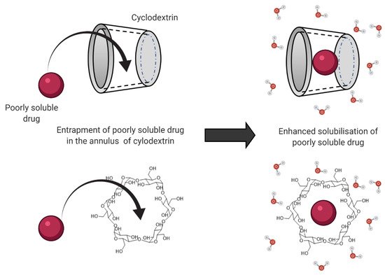
  7. Devasari, N.; Dora, C.P.; Singh, C.; Paidi, S.R.; Kumar, V.; Sobhia, M.E.; Suresh, S. Inclusion complex of erlotinib with sulfobutyl ether-β-cyclodextrin: Preparation, characterization, in silico, in vitro and in vivo evaluation. Carbohydr. Polym. 2015, 134, 547–556.
  8. Rubim, A.M.; Rubenick, J.B.; Maurer, M.; LaPorta, L.V.; Rolim, C.M.B. Inclusion complex of amiodarone hydrochloride with cyclodextrins: Preparation, characterization and dissolution rate evaluation. Braz. J. Pharm. Sci. 2017, 53.
  9. Rasheed, A. Cyclodextrins as Drug Carrier Molecule: A Review. Sci. Pharm. 2008, 76, 567–598.
  10. Wiedmann, T.S.; Kamel, L. Examination of the Solubilization of Drugs by Bile Salt Micelles. J. Pharm. Sci. 2002, 91, 1743–1764.
  11. Loftsson, T.; Moya-Ortega, M.D.; Alvarez-Lorenzo, C.; Concheiro, A. Pharmacokinetics of cyclodextrins and drugs after oral and parenteral administration of drug/cyclodextrin complexes. J. Pharm. Pharmacol. 2015, 68, 544–555.
  12. Pavlović, N.; Goločorbin-Kon, S.; Đanić, M.; Stanimirov, B.; Al-Salami, H.; Stankov, K.; Mikov, M. Bile Acids and Their Derivatives as Potential Modifiers of Drug Release and Pharmacokinetic Profiles. Front. Pharmacol. 2018, 9, 1283.
  13. Porter, C.; Trevaskis, N.; Charman, W. Lipids and lipid-based formulations: Optimizing the oral delivery of lipophilic drugs. Nat. Rev. Drug Discov. 2007, 6, 231–248.
  14. Zughaid, H.; Forbes, B.; Martin, G.P.; Patel, N. Bile salt composition is secondary to bile salt concentration in determining hydrocortisone and progesterone solubility in intestinal mimetic fluids. Int. J. Pharm. 2012, 422, 295–301.
  15. Shaikh Permeability Enhancement Techniques for Poorly Permeable Drugs: A Review. J. Appl. Pharm. Sci. 2012, 2, 34–39.
  16. Kotze, A.; Luessen, H.; Thanou, M.; Verhoef, J.; De Boer, A.; Junginger, H. Chitosan and Chitosan Derivatives as Absorption Enhancers for Peptide Drugs Across Mucosal Epithelia. Drugs Pharm. Sci. 1999, 98, 341–386.
  17. Borchard, G.; Lueβen, H.L.; de Boer, A.G.; Verhoef, J.; Lehr, C.-M.; Junginger, H.E. The potential of mucoadhesive polymers in enhancing intestinal peptide drug absorption. III: Effects of chitosan-glutamate and carbomer on epithelial tight junctions in vitro. J. Control. Release 1996, 39, 131–138.
  18. Fan, D.; Wu, X.; Dong, W.; Sun, W.; Li, J.; Tang, X. Enhancement by sodium caprate and sodium deoxycholate of the gastrointestinal absorption of berberine chloride in rats. Drug Dev. Ind. Pharm. 2012, 39, 1447–1456.
  19. Mathavan, S.; Chen-Tan, N.; Arfuso, F.; Al-Salami, H. A comprehensive study of novel microcapsules incorporating gliclazide and a permeation enhancing bile acid: Hypoglycemic effect in an animal model of Type-1 diabetes. Drug Deliv. 2015, 23, 2869–2880.
  20. Yu, Q.; Cao, Y.; Liu, L.-F.; Jiang, S.-X.; Yang, Q. Enhancement of sodium taurocholate to the absorption of cefquinome. Pak. J. Pharm. Sci. 2016, 29, 139–143.
  21. Handali, S.; Moghimipour, E.; Tabassi, S.A.S.; Ramezani, M.; Lobenberg, R. Brush border membrane vesicle and Caco-2 cell line: Two experimental models for evaluation of absorption enhancing effects of saponins, bile salts, and some synthetic surfactants. J. Adv. Pharm. Technol. Res. 2016, 7, 75–79.
  22. Gaowa, A.; Horibe, T.; Kohno, M.; Kawakami, K. Bile Acid as an Effective Absorption Enhancer for Oral Delivery of Epidermal Growth Factor Receptor–Targeted Hybrid Peptide. J. Pharm. Sci. 2018, 107, 1322–1329.
  23. Tajdini, F.; Amini, M.A.; Nafissi-Varcheh, N.; Faramarzi, M.A. Production, physiochemical and antimicrobial properties of fungal chitosan from Rhizomucor miehei and Mucor racemosus. Int. J. Biol. Macromol. 2010, 47, 180–183.
  24. He, Z.G.; Lian, H.; Sun, J.; Yu, Y.P.; Liu, Y.H.; Wang, Y.J. Supramolecular micellar nanoaggregates based on a novel chitosan/vitamin E succinate copolymer for paclitaxel selective delivery. Int. J. Nanomed. 2011, 6, 3323–3334.
  25. Nadai, M.; Tajiri, C.; Yoshizumi, H.; Suzuki, Y.; Zhao, Y.L.; Kimura, M.; Tsunekawa, Y.; Hasegawa, T. Effect of Chitosan on Gastrointestinal Absorption of Water-Insoluble Drugs Following Oral Administration in Rats. Biol. Pharm. Bull. 2006, 29, 1941–1946.
  26. Dodane, V. Effect of chitosan on epithelial permeability and structure. Int. J. Pharm. 1999, 182, 21–32.
  27. Liang, K.; Jia, Z.-Y.; Jin, X.; Zhang, S.-B.; Li, S.-M. Influence of chitosan nanoparticles as the absorption enhancers on salvianolic acid B In vitro and In vivo evaluation. Pharmacogn. Mag. 2016, 12, 57–63.
  28. Smith, J.; Wood, E.; Dornish, M. Effect of Chitosan on Epithelial Cell Tight Junctions. Pharm. Res. 2004, 21, 43–49.
  29. Schipper, N.G.M.; Vårum, K.M.; Artursson, P. Chitosans as Absorption Enhancers for Poorly Absorbable Drugs. 1: Influence of Molecular Weight and Degree of Acetylation on Drug Transport Across Human Intestinal Epithelial (Caco-2) Cells. Pharm. Res. 1996, 13, 1686–1692.
  30. Thanou, M.; Verhoef, J.; Junginger, H. Oral drug absorption enhancement by chitosan and its derivatives. Adv. Drug Deliv. Rev. 2001, 52, 117–126.
  31. Kubbinga, M.; Augustijns, P.; García, M.A.; Heinen, C.; Wortelboer, H.M.; Verwei, M.; Langguth, P. The effect of chitosan on the bioaccessibility and intestinal permeability of acyclovir. Eur. J. Pharm. Biopharm. 2019, 136, 147–155.
  32. Srinivas, N.R. The Interesting Case of Acyclovir Delivered Using Chitosan in Humans: Is it a Drug Issue or Formulation Issue? Pharm. Res. 2015, 33, 543–547.
  33. Valdes, S.A.; Alzhrani, R.F.; Lansakara-P, D.S.P.; Cui, Z. Effect of a Solid Lipid Nanoparticle Formulation on the Bioavailability of 4-(N)-Docosahexaenoyl 2′, 2′-Difluorodeoxycytidine After Oral Administration. AAPS PharmSciTech 2020, 21, 77.
  34. Chen, W.; Fan, D.; Meng, L.; Miao, Y.; Yang, S.; Weng, Y.; He, H.; Tang, X. Enhancing effects of chitosan and chitosan hydrochloride on intestinal absorption of berberine in rats. Drug Dev. Ind. Pharm. 2011, 38, 104–110.
  35. Kontogiannidou, E.; Meikopoulos, T.; Virgiliou, C.; Bouropoulos, N.; Gika, H.; Vizirianakis, I.; Müllertz, A.; Fatouros, D.G. Towards the development of Self-Nano-Emulsifying Drug Delivery Systems (SNEDDS) containing trimethyl chitosan for the oral delivery of amphotericin B: In vitro assessment and cytocompatibility studies. J. Drug Deliv. Sci. Technol. 2020, 56, 101524.
  36. Qian, S.; Zhang, Q.; Wang, Y.; Lee, B.; Betageri, G.V.; Chow, M.S.; Huang, M.; Zuo, Z. Bioavailability enhancement of glucosamine hydrochloride by chitosan. Int. J. Pharm. 2013, 455, 365–373.
  37. Babadi, D.; Dadashzadeh, S.; Osouli, M.; Daryabari, M.S.; Haeri, A. Nanoformulation strategies for improving intestinal permeability of drugs: A more precise look at permeability assessment methods and pharmacokinetic properties changes. J. Control. Release 2020, 321, 669–709.
  38. Teixeira, M.; Carbone, C.; Souto, E. Beyond liposomes: Recent advances on lipid based nanostructures for poorly soluble/poorly permeable drug delivery. Prog. Lipid Res. 2017, 68, 1–11.
  39. Baek, J.-S.; Cho, C.-W. Surface modification of solid lipid nanoparticles for oral delivery of curcumin: Improvement of bioavailability through enhanced cellular uptake, and lymphatic uptake. Eur. J. Pharm. Biopharm. 2017, 117, 132–140.
  40. Porter, C.; Charman, W.N. Uptake of drugs into the intestinal lymphatics after oral administration. Adv. Drug Deliv. Rev. 1997, 25, 71–89.
  41. Garg, A.; Bhalala, K.; Tomar, D.S.; Wahajuddin. In-situ single pass intestinal permeability and pharmacokinetic study of developed Lumefantrine loaded solid lipid nanoparticles. Int. J. Pharm. 2017, 516, 120–130.
  42. Ansari, M.J.; Anwer, K.; Jamil, S.; Al-Shdefat, R.; Ali, B.E.; Ahmad, M.M. Enhanced oral bioavailability of insulin-loaded solid lipid nanoparticles: Pharmacokinetic bioavailability of insulin-loaded solid lipid nanoparticles in diabetic rats. Drug Deliv. 2015, 23, 1972–1979.
  43. Palmerston Mendes, L.; Pan, J.; Torchilin, V.P. Dendrimers as Nanocarriers for Nucleic Acid and Drug Delivery in Cancer Therapy. Molecules 2017, 22, 1401.
  44. Beezer, A.; King, A.; Martin, I.; Mitchel, J.; Twyman, L.; Wain, C. Dendrimers as potential drug carriers; encapsulation of acidic hydrophobes within water soluble PAMAM derivatives. Tetrahedron 2003, 59, 3873–3880.
  45. Yan, C.; Gu, J.; Lv, Y.; Shi, W.; Jing, H. Improved intestinal absorption of water-soluble drugs by acetylation of G2 PAMAM dendrimer nanocomplexes in rat. Drug Deliv. Transl. Res. 2017, 11, 408–415.
  46. Sadekar, S.; Thiagarajan, G.; Bartlett, K.; Hubbard, D.; Ray, A.; McGill, L.; Ghandehari, H. Poly(amido amine) dendrimers as absorption enhancers for oral delivery of camptothecin. Int. J. Pharm. 2013, 456, 175–185.
  47. Qi, R.; Zhang, H.; Xu, L.; Shen, W.; Chen, C.; Wang, C.; Cao, Y.; Wang, Y.; van Dongen, M.A.; He, B.; et al. G5 PAMAM dendrimer versus liposome: A comparison study on the in vitro transepithelial transport and in vivo oral absorption of simvastatin. Nanomed. Nanotechnol. Biol. Med. 2015, 11, 1141–1151.
  48. Yiyun, C.; Tongwen, X. Dendrimers as Potential Drug Carriers. Part I. Solubilization of Non-Steroidal Anti-Inflammatory Drugs in the Presence of Polyamidoamine Dendrimers. Eur. J. Med. Chem. 2005, 40, 1188–1192.
  49. D’Emanuele, A.; Jevprasesphant, R.; Penny, J.; Attwood, D. The use of a dendrimer-propranolol prodrug to bypass efflux transporters and enhance oral bioavailability. J. Control. Release 2004, 95, 447–453.
  50. Choudhury, H.; Pandey, M.; Gorain, B.; Chatterjee, B.; Madheswaran, T.; Shadab; Mak, K.-K.; Tambuwala, M.; Chourasia, M.K.; Kesharwani, P. Nanoemulsions as Effective Carriers for the Treatment of Lung Cancer. In Nanotechnology-Based Targeted Drug Delivery Systems for Lung Cancer; Academic Press: Cambridge, MA, USA, 2019; pp. 217–247.
  51. Kawakami, K.; Yoshikawa, T.; Moroto, Y.; Kanaoka, E.; Takahashi, K.; Nishihara, Y.; Masuda, K. Microemulsion formulation for enhanced absorption of poorly soluble drugs: I. Prescription design. J. Control. Release 2002, 81, 65–74.
  52. Aboofazeli, R. Nanometric-Scaled Emulsions (Nanoemulsions). Iran. J. Pharm. Res. 2010, 9, 325–326.
  53. Isailović, T.M.; Todosijević, M.N.; Dordević, S.M.; Savić, S.D. Natural Surfactants-Based Micro/Nanoemulsion Systems for NSAIDs—Practical Formulation Approach, Physicochemical and Biopharmaceutical Characteristics/Performances. In Microsized and Nanosized Carriers for Nonsteroidal Anti-Inflammatory Drugs; Academic Press: Cambridge, MA, USA, 2017.
  54. Baral, K.; Song, J.-G.; Lee, S.; Bajracharya, R.; Sreenivasulu, G.; Kim, M.; Lee, K.; Han, H.-K. Enhanced Bioavailability of AC1497, a Novel Anticancer Drug Candidate, via a Self-Nanoemulsifying Drug Delivery System. Pharmaceutics 2021, 13, 1142.
  55. Kuncahyo, I.; Choiri, S.; Fudholi, A.; Martien, R.; Rohman, A. Development of pitavastatin-loaded super-saturable self-nano emulsion: A continues screening and optimization approach using statistical technique. J. Dispers. Sci. Technol. 2021, 1–10.
  56. Li, M.; Cui, J.; Ngadi, M.O.; Ma, Y. Absorption mechanism of whey-protein-delivered curcumin using Caco-2 cell monolayers. Food Chem. 2015, 180, 48–54.
  57. Anuar, N.; Sabri, A.H.; Effendi, T.J.B.; Hamid, K.A. Development and characterisation of ibuprofen-loaded nanoemulsion with enhanced oral bioavailability. Heliyon 2020, 6, e04570.
  58. Gao, F.; Zhang, Z.; Bu, H.; Huang, Y.; Gao, Z.; Shen, J.; Zhao, C.; Li, Y. Nanoemulsion improves the oral absorption of candesartan cilexetil in rats: Performance and mechanism. J. Control. Release 2011, 149, 168–174.
  59. Date, A.A.; Desai, N.; Dixit, R.; Nagarsenker, M. Self-nanoemulsifying drug delivery systems: Formulation insights, applications and advances. Nanomedicine 2010, 5, 1595–1616.
  60. Thakkar, H.P.; Khunt, A.; Dhande, R.D.; Patel, A.A. Formulation and evaluation of Itraconazole nanoemulsion for enhanced oral bioavailability. J. Microencapsul. 2015, 32, 559–569.
  61. Li, Y.-J.; Hu, X.-B.; Lu, X.-L.; Liao, D.-H.; Tang, T.-T.; Wu, J.-Y.; Xiang, D.-X. Nanoemulsion-based delivery system for enhanced oral bioavailability and Caco-2 cell monolayers permeability of berberine hydrochloride. Drug Deliv. 2017, 24, 1868–1873.
  62. Chen, S.; Zhang, J.; Wu, L.; Wu, H.; Dai, M. Paeonol nanoemulsion for enhanced oral bioavailability: Optimization and mechanism. Nanomedicine 2018, 13, 269–282.
More
Information
Contributors MDPI registered users' name will be linked to their SciProfiles pages. To register with us, please refer to https://encyclopedia.pub/register : , , , ,
View Times: 537
Revisions: 3 times (View History)
Update Date: 26 Aug 2022
1000/1000
Video Production Service