Submitted Successfully!
To reward your contribution, here is a gift for you: A free trial for our video production service.
Thank you for your contribution! You can also upload a video entry or images related to this topic.
Version Summary Created by Modification Content Size Created at Operation
1 -- 2537 2022-08-01 07:53:43 |
2 Reference format revised. Meta information modification 2537 2022-08-01 11:32:36 |

Video Upload Options

Do you have a full video?

Confirm

Are you sure to Delete?
Cite
If you have any further questions, please contact Encyclopedia Editorial Office.
Wang, Y.;  Lu, J. Chaperone-Mediated Autophagy in Neurodegenerative Diseases. Encyclopedia. Available online: https://encyclopedia.pub/entry/25715 (accessed on 28 March 2024).
Wang Y,  Lu J. Chaperone-Mediated Autophagy in Neurodegenerative Diseases. Encyclopedia. Available at: https://encyclopedia.pub/entry/25715. Accessed March 28, 2024.
Wang, Yi-Ting, Jia-Hong Lu. "Chaperone-Mediated Autophagy in Neurodegenerative Diseases" Encyclopedia, https://encyclopedia.pub/entry/25715 (accessed March 28, 2024).
Wang, Y., & Lu, J. (2022, August 01). Chaperone-Mediated Autophagy in Neurodegenerative Diseases. In Encyclopedia. https://encyclopedia.pub/entry/25715
Wang, Yi-Ting and Jia-Hong Lu. "Chaperone-Mediated Autophagy in Neurodegenerative Diseases." Encyclopedia. Web. 01 August, 2022.
Chaperone-Mediated Autophagy in Neurodegenerative Diseases
Edit

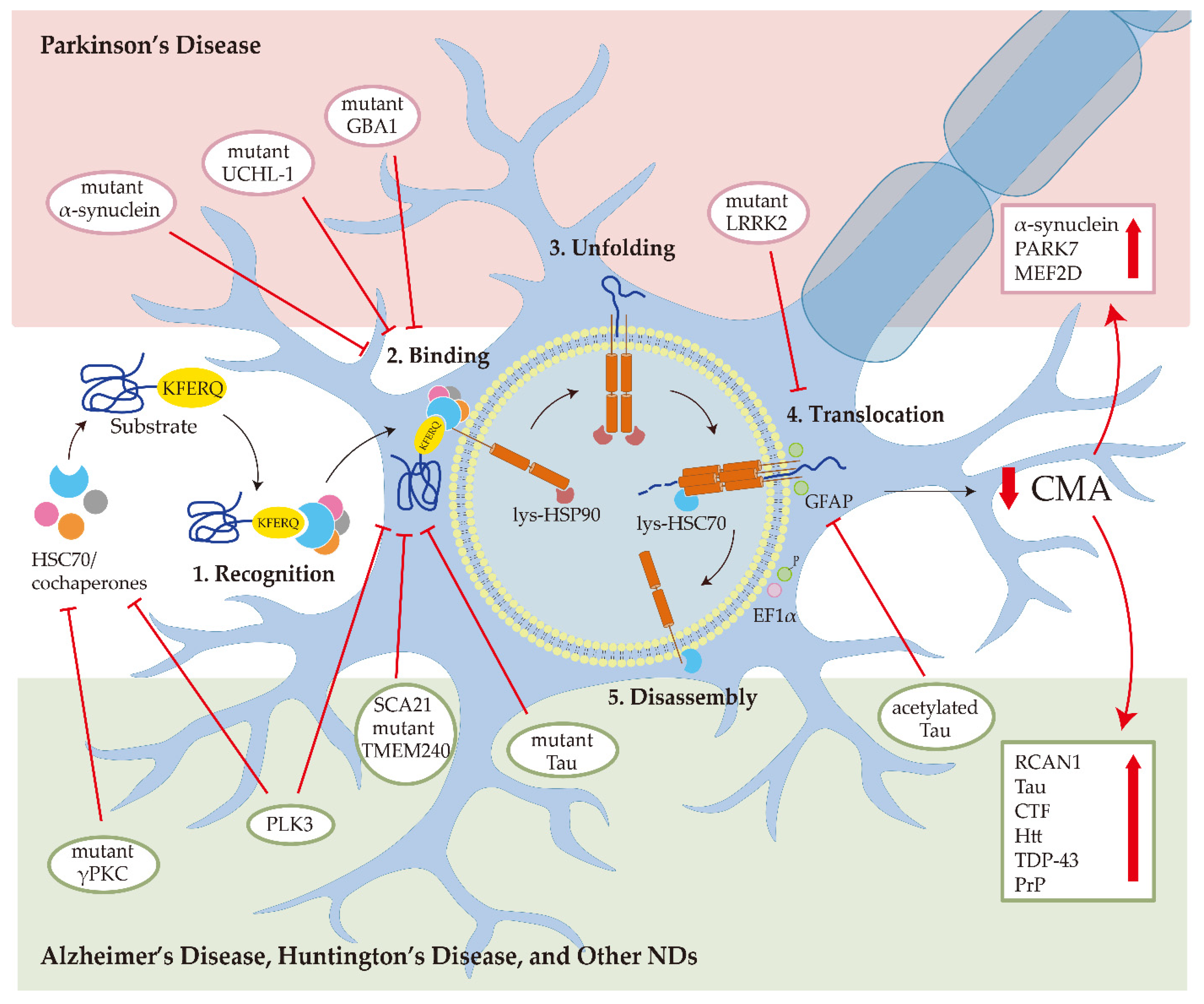
Chaperone-mediated autophagy (CMA) is a protein degradation mechanism through lysosomes. By targeting the KFERQ motif of the substrate, CMA is responsible for the degradation of about 30% of cytosolic proteins, including a series of proteins associated with neurodegenerative diseases (NDs). The fact that decreased activity of CMA is observed in NDs, and ND-associated mutant proteins, including alpha-synuclein and Tau, directly impair CMA activity reveals a possible vicious cycle of CMA impairment and pathogenic protein accumulation in ND development. Given the intrinsic connection between CMA dysfunction and ND, enhancement of CMA has been regarded as a strategy to counteract ND. Indeed, genetic and pharmacological approaches to modulate CMA have been shown to promote the degradation of ND-associated proteins and alleviate ND phenotypes in multiple ND models.

chaperone-mediated autophagy autophagy neurodegenerative disease Parkinson’s disease Alzheimer’s disease Huntington’s disease

1. Introduction

Neurodegenerative diseases (NDs), one of the major health threats to human, affect 50 million people worldwide [1]. As ND is an age-related disease, population aging also makes ND an urgent health issue. The most common NDs include Alzheimer’s disease (AD), Parkinson’s disease (PD), Huntington’s disease (HD), amyotrophic lateral sclerosis (ALS), frontotemporal lobar degeneration (FTLD), and the spinocerebellar ataxias (SCA) [2]. The common feature of ND is the intracytoplasmic deposition of aggregate-prone proteins in neurons. The removal of undesired, damaged, misfolded, and aggregated proteins is a therapeutic focus of ND.
Autophagy is a self-degradation pathway via lysosome. It plays a homeostatic function and is involved in the pathophysiological process of anti-aging, anti-microbial, anti-tumor, differentiation, development, and immunity [3].
Autophagy is classified as macroautophagy, microautophagy, and chaperone-mediated autophagy (CMA) based on how cargo is delivered to the lysosome. Macroautophagy is the best-studied type of autophagy. During macroautophagy, the cargo is sequestrated by double-membrane vesicles-autophagosomes and transported to the lysosome. In microautophagy, the lysosomes directly uptake the cytosolic compounds through membrane enwrapping. In CMA, heat shock cognate 71 kDa protein (HSC70) chaperones bind to damaged or defective proteins containing KFERQ-like sequences and transport them to the lysosomes via lysosome-associated membrane protein 2A (LAMP2A) [4]. With increasing age, the efficiency of CMA is lowered, which increases the risk of harmful proteins accumulating into insoluble clumps that damage cells. The common feature of AD and other NDs is the presence of toxic protein aggregates in the patient’s brain [5]. Moreover, the large number of defective proteins overwhelm CMA and eventually paralyze it.

2. CMA as a Therapeutic Target for Neurodegenerative Diseases

2.1. Molecular Mechanism of CMA

2.1.1. CMA Substrate Recognition

The CMA-targeting motif is a pentapeptide sequence related to KFERQ biochemically, which must contain up to two positively charged residues: arginine (R), lysine (K); up to two of the hydrophobic residues: isoleucine (I), phenylalanine (F), leucine (L), or valine (V); one single negative residue: glutamate (E) or aspartate (D); and one single glutamine (Q) flanked at either the N- or C-terminus of the pentapeptide [6]. This canonical motif can be found in about 40% of the mammalian proteome [7][8][9].

2.1.2. Transportation of CMA Substrate by HSC70

Once the KFERQ sequence in the CMA substrate binds to HSC70, the substrate is delivered to the lysosomal membrane. The HSC70 belongs to the heat shock protein family, one of the largest groups of chaperones. The HSC70 can be found in the cellular membrane, extracellular exosomes, the nucleus, and the cytosol [10]. Unlike the HSC70 in the cytosol, the lysosomal HSC70 (lys-HSC70) is involved in CMA substrate uptake [11]. When the pathway is triggered by starvation or oxidative stress, the lys-HSC70 amount and the number of lysosomes that contain lys-HSC70 are increased [12]. Cochaperones, including the carboxyl terminus of HSC70-interacting protein, heat shock protein 40 kDa (HSP40), HSP70-interacting protein, and HSP70-HSP90 organizing protein, are also involved in substrate unfolding and lysosomal translocation [4].

2.1.3. Translocation of CMA Substrate by LAMP2A

LAMP2A contains a cytosolic tail different from the other variants of the LAMP2 genes [13]. The binding between substrate and LAMP2A promotes LAMP2A multimerization to form a translocation complex during CMA. After the substrate translocates into the lysosome lumen, the LAMP2A multimer disassembles into monomers and returns to the cytosol for reuse [14]. The efficiency of the CMA pathway can be affected by the velocity of assembly and disassembly of the LAMP2A translocation complex. Glial fibrillary acidic protein (GFAP) and elongation factor 1 alpha (EF1α) are identified as the regulators of LAMP2A oligomerization. GFAP contributes to the stability of the translocation complex, whereas the EF1α-GTP binding promotes the self-association between GFAP molecules, further disrupting the translocation complex’s stability. Thus, GTP has been shown to be a CMA inhibitor [15]. Moreover, prolonged starvation, oxidative stress, or inhibition of the proteolytic pathway can raise the LAMP2A levels and LAMP2A-positive lysosomes and further increase CMA activity [16].

2.2. Physiological Function of CMA

As a recycling and protein quality control mechanism, CMA plays an important role in maintaining cellular homeostasis and exerting specific physiological functions in regulating the cell cycle, cell survival, cell stemness, transcriptional regulation, metabolic pathways, and immune responses [17].

2.2.1. Starvation

CMA can be activated during nutritional starvation to promote amino acid recycling for maintaining protein synthesis and gluconeogenesis [17]. After removal of serum from the medium for 8 to 10 h, CMA will be gradually activated and persists at a high level for 3 days while the activation of macroautophagy is shorter [18][19][20]. The starvation-activated CMA can degrade less critical proteins and keep protein synthesis by recycling amino acids [21]. However, the starvation-activated CMA is tissue and cell-type selective, which can be more effectively stimulated in the liver, spleen, kidney, and heart [22].

2.2.2. Protein Quality Control

The protein quality control is another most characterized function of CMA, which can be activated by oxidative stress and hypoxic stress [12][23], and during aging and neurodegenerative disease [24]. CMA can electively remove the damaged and misfolded proteins from the cytosol. The upregulation of LAMP2A transcription is involved in the oxidation-induced activation of CMA [25]. Issa et al.’s research on the Drosophila brain indicated that oxidative stress increased LAMP2A expression and CMA activation. The expression of LAMP2A further prevented the accumulation of Ref(2)P, the Drosophila melanogaster homolog of mammalian SQSTM1/p62, under acute oxidative stress. These results indicate that CMA increases autophagic flux in the Drosophila brain and has neuroprotective properties [26]. The hypoxic stress can also effectively activate the CMA process, which reduces damaged protein and promotes neuron survival [23].

2.2.3. Metabolic Regulation

Since CMA’s discovery, it has been associated with cellular energetics and activated by starvation. The alterations in carbohydrate and lipid metabolism caused by the selective degradation of the main enzymes by CMA are responsible for cellular energetics, which is also associated with brain aging and ND [27][28][29]. Most glycolytic enzymes contain KFERQ motifs. For example, glyceraldehyde 3-phosphate dehydrogenase (GAPDH) and pyruvate kinase were first identified as CMA substrates [30]. CMA modulation of carbohydrate metabolism has been established in a mouse model. CMA modulates hepatic glycolysis by regulating the levels of glycolytic enzymes and enzymes involved in the tricarboxylic acid cycle process [31]. The aging-related decline in CMA may raise rates of glycolysis and break the energy balance in old organisms [29][31].

2.2.4. Cell Cycle Control

CMA modulates several cellular mechanisms by regulating protein degradation and controlling the abundance of proteins. Some cell cycle modulators that carry the KFERQ-like motif are involved in the CMA-induced cell cycle process [32]. For example, the level of myocyte enhancer factor-2 (MEF2) protein can regulate neuronal proliferation and survival [33] while CMA mediates the MEF2D degradation. Both wild-type α-synuclein and a PD-associated mutant blocked MEF2D-HSC70 binding, leading to MEF2D accumulation and neuronal death and further increasing the risk of PD [34][35]. MEF2A, another isoform of MEF2, can be degraded under the oxidative stress-induced lysosome destabilization condition [36]. CMA impairment is related to cellular senescence by triggering the DNA damage response, SA-β-gal activity, upregulation of p21, and accumulation of p16 and lipofuscin. Evidence showed that the dysfunctional CMA led to physiological aging and neurodegeneration by accumulating senescent cells [37].

2.2.5. Immune Responses

Downregulating LAMP2A or HSC70 in the immune system can limit the distribution of cytoplasmic epitopes by class II molecules in antigen-presenting cells [38]. CMA has also exhibited the capacity to help regulate the CD4+ T cell response, as it selectively degrades the ubiquitin ligase Itch and the calcineurin inhibitor regulator of calcineurin 1 (RCAN1), which are two T cell receptor signaling negative regulators. The relationship between an age-dependent decrease in CMA activity in T cells and the impaired T cell function associated with aging has been verified [39]. Moreover, CMA plays an important role in the immunosuppressive function of mesenchymal stromal cells by IFN-γ plus TNF-α-induced activation of NF-κB and STAT1 [40].

2.3. Role of CMA in Neurodegenerative Diseases

In the central nervous system (CNS), the hallmarks of many NDs are the misfolding, aggregation, and accumulation of proteins, which result in cellular dysfunction, synaptic damage, and brain damage [41]. Some ND-related proteins are identified as CMA substrate proteins, such as α-synuclein (Figure 1). Several pieces of research showed that CMA activity was reduced in several NDs, suggesting that dysfunctional CMA is implicated in ND [4].
Figure 1. Chaperone-mediated autophagy (CMA) in neurodegenerative diseases (NDs). In Parkinson’s disease, the mutant α-synuclein interacts with LAMP2A with higher affinity and blocks CMA degradation of other substrates; the mutant UCHL-1 and GBA1 reduce CMA activity by aberrantly interacting with the HSC70 and LAMP2A; mutant LRRK2 impairs the formation of translocation complex; the downregulation of CMA can increase the α-synuclein, PARK7, and MEF2D accumulation. In Alzheimer’s disease, mutant Tau binds with LAMP2A and inhibits CMA activity; acetylated Tau prevents the translocation of substrates into the lysosome; the downregulation of CMA can increase the RCAN1, Tau, and CTF levels. In Huntington’s disease, CMA malfunction contributes to Htt aggregation. In prion disease, PLK3 affects the levels of HSC70 and LAMP2A to mediate PrP degradation through CMA. For amyotrophic lateral sclerosis and frontotemporal lobar degeneration, TDP-43 is degraded by CMA. For spinocerebellar ataxias, mutant γPKC interacts with HSC70 and SCA21 mutant TMEM240 blocks the LAMP2A transport.

2.3.1. Parkinson’s Disease

PD is the second most prevalent ND and is presently incurable. The clinical symptoms of PD include motor abnormalities such as the symptomatic triad of bradykinesia, resting tremors, and rigidity, and non-motor symptoms such as neurobehavioral disorders, autonomic dysfunctions, sensory impairments, and sleep disturbances [42]. The pathogenic feature of PD is the loss of nigrostriatal dopaminergic innervation. The main pathogenic molecular mechanisms include α-synuclein misfolding and aggregate formation, mitochondrial dysfunction, protein clearance impairment associated with the ubiquitin-proteasome system (UPS) and autophagy-lysosomal, neuroinflammation, and oxidative stress [43], most of which are related to autophagy. α-synuclein (SNCA), Parkin, ubiquitin C-terminal hydrolase L1 (UCHL-1), PTEN-induced kinase 1 (PINK1), PARK7 (DJ-1), leucine-rich repeat kinase 2 (LRRK2), ATPase cation transporting 13A2 (ATP13A2), glucocerebrosidase (GBA), vacuolar protein sorting ortholog 35 (VPS35), Eukaryotic Translation Initiation Factor 4 Gamma 1 (EIF4G1), and PARK16 are identified as the causative genes of PD [44]. Some of these genes have been found as CMA substrates and CMA regulators in PD [45].
Since α-synuclein was identified as a CMA substrate and wild-type α-synuclein was degraded through CMA, PD is the first ND associated with CMA [25]. In CMA, the α-synuclein directly interacts with the key protein player of CMA, LAMP2A [46]. For HSC70, miR-320a (HSC70 miRNA) inhibits CMA and promotes α-synuclein accumulation [47]. Reduced CMA in PD is caused by the loss of LAMP2A and HSC70 proteins, which occurs primarily in brain areas, and also caused by accumulating membrane-associated α-synuclein and other recognized CMA substrates [45]. CMA degrades α-synuclein, whereas pathogenic α-synuclein impairs CMA progress. A30P and A53T are two mutant variants of α-synuclein found in familial forms of PD. These two mutant types of α-synuclein have a higher affinity for the LAMP2A than other substrates. However, they block the degradation of themselves and other substrates through CMA [25].
Pathogenic mutations of the LRRK2 are the most common factor for familial PD. LRRK2 has been linked to several putative PD pathogenic mechanisms, including α-synuclein accumulation, Tau hyperphosphorylation, the inflammatory response, oxidative stress, mitochondrial dysfunction, synaptic dysfunction, and autophagy-lysosomal system impairment [48]. A recent study suggested that DNL201, an LRRK2 kinase inhibitor, rescues lysosomal dysfunction in PD patients [49]. CMA could induce LRRK2 degradation in lysosomes. However, the G2019S, a mutant form of LRRK2, can suppress CMA via interfering with the formation of translocation complex at the lysosomal membrane [50]. In neurons from LRRK2 G2019S mice, the mutant LRRK2 interferes with CMA activity, increases colocalization between LAMP2A and α-synuclein, and causes α-synuclein aggregation [51].
In addition to α-synuclein, LRRK2, and their mutants, the UCHL-1, PARK7, MEF2D, VPS35 mutant, and GBA1 mutant are PD-related proteins associated with CMA pathways as well. UCHL-1 and GBA1 mutants aberrantly interact with the HSC70 and LAMP2A, further inducing α-synuclein accumulation by blocking CMA activity [52][53]. PARK7 and MEF2D protein levels can be regulated by CMA activity. CMA was demonstrated to selectively degrade the oxidized and altered PARK7, protecting mitochondria from damaging and affecting cell survival [54]. The mislocalized and inactive MEF2D accumulation caused by CMA dysfunctions is also a potential reason for PD [34]. Pathogenic VPS35 affects LAMP2A retrieval from the endosome to the Golgi complex and increases LAMP2A degradation [55].

2.3.2. Alzheimer’s Disease

AD is the most prevalent form of dementia among senior citizens, and its clinical symptoms are progressive memory impairment and cognitive function loss [56]. The primary lesions of AD are the deposition of extracellular amyloid β (Aβ) and intraneuronal Tau neurofibrillary tangles in particular brain areas [57]. Autophagy has a significant impact on the metabolism of Aβ and Tau proteins. The accumulation of toxic proteins in the AD brain is thought to be caused by autophagy malfunction [58]. Like PD-related proteins, several AD-related proteins have also been identified as CMA substrates. The presence of two CMA-targeting motifs in Tau’s C-terminal region suggests that CMA can degrade the Tau aggregation [59]. Mutant Tau protein can lower CMA activity by interacting with LAMP2A and prevent translocation to the lysosome lumen [59].

2.3.3. Huntington’s Disease and Other NDs

HD, a dominantly inherited late-onset ND, is caused by accumulation and aggregation of mutant huntingtin protein (Htt). Htt is identified as a CMA substrate and interacts with HSC70 and LAMP2A. There is an expended N-terminal polyglutamine (polyQ) tract in Htt [60]. The polyQ-binding protein forces mutant Htt to CMA machinery, resulting in Htt degradation. Interestingly, CMA activity is upregulated in the early stages of HD due to compensatory adjustment of the decreased macroautophagy in HD. For the late stage of HD, the lower level of LAMP2A suggests that CMA is impaired [61].
Other NDs such as prion diseases, ALS, FTLD, and SCA are related to CMA. The prion protein (PrP) is the main content of the prion. Overexpression of polo-like kinase 3 (PLK3) induces the degradation of mutated PrP. In both in vivo and in vitro abnormal PrP models, the levels of HSC70 and LAMP2A are downregulated. In addition, the overexpression of PLK3 can upregulate HSC70 and LAMP2A levels. Taken together, PLK3 mediates the degradation of PrP through CMA [62][63]. Accumulation of transactivation response DNA-binding protein 43 kDa (TDP-43) is the hallmark of ALS and FLTD. CMA can maintain the physiological and pathology forms of TDP-43 as the KFERQ motif in TDP-43 can bind with HSC70 [64]. SCA is a heterogeneous group of progressive ND. SCA14 is caused by mutant γPKC, which is a CMA substrate. The interaction between mutant γPKC and HSC70 blocked CMA activity in neuronal cells [65].

References

  1. Gammon, K. Neurodegenerative disease: Brain windfall. Nature 2014, 515, 299–300.
  2. Lamptey, R.N.L.; Chaulagain, B.; Trivedi, R.; Gothwal, A.; Layek, B.; Singh, J. A Review of the Common Neurodegenerative Disorders: Current Therapeutic Approaches and the Potential Role of Nanotherapeutics. Int. J. Mol. Sci. 2022, 23, 1851.
  3. Levine, B.; Kroemer, G. Autophagy in the pathogenesis of disease. Cell 2008, 132, 27–42.
  4. Kaushik, S.; Cuervo, A.M. The coming of age of chaperone-mediated autophagy. Nat. Rev. Mol. Cell Biol. 2018, 19, 365–381.
  5. Auzmendi-Iriarte, J.; Matheu, A. Impact of Chaperone-Mediated Autophagy in Brain Aging: Neurodegenerative Diseases and Glioblastoma. Front. Aging Neurosci. 2021, 12, 630743.
  6. Fred Dice, J. Peptide sequences that target cytosolic proteins for lysosomal proteolysis. Trends Biochem. Sci. 1990, 15, 305–309.
  7. Kaushik, S.; Cuervo, A.M. AMPK-dependent phosphorylation of lipid droplet protein PLIN2 triggers its degradation by CMA. Autophagy 2016, 12, 432–438.
  8. Lv, L.; Li, D.; Zhao, D.; Lin, R.; Chu, Y.; Zhang, H.; Zha, Z.; Liu, Y.; Li, Z.; Xu, Y.; et al. Acetylation targets the M2 isoform of pyruvate kinase for degradation through chaperone-mediated autophagy and promotes tumor growth. Mol. Cell 2011, 42, 719–730.
  9. Ferreira, J.V.; Soares, A.R.; Ramalho, J.S.; Pereira, P.; Girao, H. K63 linked ubiquitin chain formation is a signal for HIF1A degradation by Chaperone-Mediated Autophagy. Sci. Rep. 2015, 5, 10210.
  10. Bonam, S.R.; Ruff, M.; Muller, S. HSPA8/HSC70 in Immune Disorders: A Molecular Rheostat that Adjusts Chaperone-Mediated Autophagy Substrates. Cells 2019, 8, 849.
  11. Cuervo, A.M.; Dice, J.F.; Knecht, E. A population of rat liver lysosomes responsible for the selective uptake and degradation of cytosolic proteins. J. Biol. Chem. 1997, 272, 5606–5615.
  12. Kiffin, R.; Christian, C.; Knecht, E.; Cuervo, A.M. Activation of Chaperone-mediated Autophagy during Oxidative Stress. Mol. Biol. Cell 2004, 15, 4829–4840.
  13. Cuervo, A.M.; Dice, J.F. Unique properties of lamp2a compared to other lamp2 isoforms. J. Cell Sci. 2000, 113, 4441–4450.
  14. Cuervo, A.M.; Wong, E. Chaperone-mediated autophagy: Roles in disease and aging. Cell Res. 2014, 24, 92–104.
  15. Bandyopadhyay, U.; Sridhar, S.; Kaushik, S.; Kiffin, R.; Cuervo, A.M. Identification of Regulators of Chaperone-Mediated Autophagy. Mol. Cell 2010, 39, 535–547.
  16. Catarino, S.; Pereira, P.; Girão, H. Molecular control of chaperone-mediated autophagy. Essays Biochem. 2017, 61, 663–674.
  17. Hosaka, Y.; Araya, J.; Fujita, Y.; Kuwano, K. Role of chaperone-mediated autophagy in the pathophysiology including pulmonary disorders. Inflamm. Regen. 2021, 41, 29.
  18. Mizushima, N.; Yamamoto, A.; Matsui, M.; Yoshimori, T.; Ohsumi, Y. In Vivo Analysis of Autophagy in Response to Nutrient Starvation Using Transgenic Mice Expressing a Fluorescent Autophagosome Marker. Mol. Biol. Cell 2004, 15, 1101–1111.
  19. Massey, A.C.; Kaushik, S.; Sovak, G.; Kiffin, R.; Cuervo, A.M. Consequences of the selective blockage of chaperone-mediated autophagy. Proc. Natl. Acad. Sci. USA 2006, 103, 5805–5810.
  20. Singh, R.; Kaushik, S.; Wang, Y.; Xiang, Y.; Novak, I.; Komatsu, M.; Tanaka, K.; Cuervo, A.M.; Czaja, M.J. Autophagy regulates lipid metabolism. Nature 2009, 458, 1131–1135.
  21. Cuervo, A.M.; Knecht, E.; Terlecky, S.R.; Dice, J.F. Activation of a selective pathway of lysosomal proteolysis in rat liver by prolonged starvation. Am. J. Physiol. Cell Physiol. 1995, 269, C1200–C1208.
  22. Orenstein, S.J.; Cuervo, A.M. Chaperone-mediated autophagy: Molecular mechanisms and physiological relevance. Semin. Cell Dev. Biol. 2010, 21, 719–726.
  23. Dohi, E.; Tanaka, S.; Seki, T.; Miyagi, T.; Hide, I.; Takahashi, T.; Matsumoto, M.; Sakai, N. Hypoxic stress activates chaperone-mediated autophagy and modulates neuronal cell survival. Neurochem. Int. 2012, 60, 431–442.
  24. López-Otín, C.; Blasco, M.A.; Partridge, L.; Serrano, M.; Kroemer, G. The Hallmarks of Aging. Cell 2013, 153, 1194–1217.
  25. Cuervo, A.M.; Stefanis, L.; Fredenburg, R.; Lansbury, P.T.; Sulzer, D. Impaired Degradation of Mutant α-Synuclein by Chaperone-Mediated Autophagy. Science 2004, 305, 129–295.
  26. Issa, A.R.; Sun, J.; Petitgas, C.; Mesquita, A.; Dulac, A.; Robin, M.; Mollereau, B.; Jenny, A.; Chérif-Zahar, B.; Birman, S. The lysosomal membrane protein LAMP2A promotes autophagic flux and prevents SNCA-induced Parkinson disease-like symptoms in the Drosophila brain. Autophagy 2018, 14, 1898–1910.
  27. Tasset, I.; Cuervo, A.M. Role of chaperone-mediated autophagy in metabolism. FEBS J 2016, 283, 2403–2413.
  28. Alfaro, I.E.; Albornoz, A.; Molina, A.; Moreno, J.; Cordero, K.; Criollo, A.; Budini, M. Chaperone Mediated Autophagy in the Crosstalk of Neurodegenerative Diseases and Metabolic Disorders. Front. Endocrinol. 2018, 9, 778.
  29. Hallett, P.J.; Huebecker, M.; Brekk, O.R.; Moloney, E.B.; Rocha, E.M.; Priestman, D.A.; Platt, F.M.; Isacson, O. Glycosphingolipid levels and glucocerebrosidase activity are altered in normal aging of the mouse brain. Neurobiol. Aging 2018, 67, 189–200.
  30. Aniento, F.; Roche, E.; Cuervo, A.M.; Knecht, E. Uptake and degradation of glyceraldehyde-3-phosphate dehydrogenase by rat liver lysosomes. J. Biol. Chem. 1993, 268, 10463–10470.
  31. Schneider, J.L.; Suh, Y.; Cuervo, A.M. Deficient chaperone-mediated autophagy in liver leads to metabolic dysregulation. Cell Metab. 2014, 20, 417–432.
  32. Andrade-Tomaz, M.; de Souza, I.; Rocha, C.R.R.; Gomes, L.R. The Role of Chaperone-Mediated Autophagy in Cell Cycle Control and Its Implications in Cancer. Cells 2020, 9, 2140.
  33. She, H.; Mao, Z. Regulation of myocyte enhancer factor-2 transcription factors by neurotoxins. Neurotoxicology 2011, 32, 563–566.
  34. Yang, Q.; She, H.; Gearing, M.; Colla, E.; Lee, M.; Shacka, J.J.; Mao, Z. Regulation of neuronal survival factor MEF2D by chaperone-mediated autophagy. Science 2009, 323, 124–127.
  35. Gan-Or, Z.; Dion, P.A.; Rouleau, G.A. Genetic perspective on the role of the autophagy-lysosome pathway in Parkinson disease. Autophagy 2015, 11, 1443–1457.
  36. Zhang, L.; Sun, Y.; Fei, M.; Tan, C.; Wu, J.; Zheng, J.; Tang, J.; Sun, W.; Lv, Z.; Bao, J.; et al. Disruption of chaperone-mediated autophagy-dependent degradation of MEF2A by oxidative stress-induced lysosome destabilization. Autophagy 2014, 10, 1015–1035.
  37. Moreno-Blas, D.; Gorostieta-Salas, E.; Castro-Obregón, S. Connecting chaperone-mediated autophagy dysfunction to cellular senescence. Ageing Res. Rev. 2018, 41, 34–41.
  38. Zhou, D.; Li, P.; Lin, Y.; Lott, J.M.; Hislop, A.D.; Canaday, D.H.; Brutkiewicz, R.R.; Blum, J.S. Lamp-2a facilitates MHC class II presentation of cytoplasmic antigens. Immunity 2005, 22, 571–581.
  39. Valdor, R.; Mocholi, E.; Botbol, Y.; Guerrero-Ros, I.; Chandra, D.; Koga, H.; Gravekamp, C.; Cuervo, A.M.; Macian, F. Chaperone-mediated autophagy regulates T cell responses through targeted degradation of negative regulators of T cell activation. Nat. Immunol. 2014, 15, 1046–1054.
  40. Zhang, J.; Huang, J.; Gu, Y.; Xue, M.; Qian, F.; Wang, B.; Yang, W.; Yu, H.; Wang, Q.; Guo, X.; et al. Inflammation-induced inhibition of chaperone-mediated autophagy maintains the immunosuppressive function of murine mesenchymal stromal cells. Cell. Mol. Immunol. 2021, 18, 1476–1488.
  41. Soto, C.; Pritzkow, S. Protein misfolding, aggregation, and conformational strains in neurodegenerative diseases. Nat. Neurosci. 2018, 21, 1332–1340.
  42. Gupta, S.; Shukla, S. Non-motor symptoms in Parkinson’s disease: Opening new avenues in treatment. Curr. Res. Behav. Sci. 2021, 2, 100049.
  43. Jankovic, J.; Tan, E.K. Parkinson’s disease: Etiopathogenesis and treatment. J. Neurol. Neurosurg. Psychiatry 2020, 91, 795–808.
  44. Klein, C.; Westenberger, A. Genetics of Parkinson’s disease. Cold Spring Harb. Perspect. Med. 2012, 2, a008888.
  45. Li, B.; Zhang, Y.; Yuan, Y.; Chen, N. A new perspective in Parkinson’s disease, chaperone-mediated autophagy. Parkinsonism Relat. Disord. 2011, 17, 231–235.
  46. Vogiatzi, T.; Xilouri, M.; Vekrellis, K.; Stefanis, L. Wild type α-synuclein is degraded by chaperone-mediated autophagy and macroautophagy in neuronal cells. J. Biol. Chem. 2008, 283, 23542–23556.
  47. Li, G.; Yang, H.; Zhu, D.; Huang, H.; Liu, G.; Lun, P. Targeted Suppression of Chaperone-Mediated Autophagy by miR-320a Promotes α-Synuclein Aggregation. Int. J. Mol. Sci. 2014, 15, 15845–15857.
  48. Li, J.-Q.; Tan, L.; Yu, J.-T. The role of the LRRK2 gene in Parkinsonism. Mol. Neurodegener. 2014, 9, 47.
  49. Jennings, D.; Huntwork-Rodriguez, S.; Henry, A.G.; Sasaki, J.C.; Meisner, R.; Diaz, D.; Solanoy, H.; Wang, X.; Negrou, E.; Bondar, V.V.; et al. Preclinical and clinical evaluation of the LRRK2 inhibitor DNL201 for Parkinson’s disease. Sci. Transl. Med. 2022, 14, eabj2658.
  50. Orenstein, S.J.; Kuo, S.-H.; Tasset, I.; Arias, E.; Koga, H.; Fernandez-Carasa, I.; Cortes, E.; Honig, L.S.; Dauer, W.; Consiglio, A.; et al. Interplay of LRRK2 with chaperone-mediated autophagy. Nat. Neurosci. 2013, 16, 394–406.
  51. Kuo, S.-H.; Orenstein, S.; Koga, H.; Tang, G.; Kanter, E.; Alcalay, R.; Cuervo, A.M.; Sulzer, D. LRRK2 G2019S Impairs Chaperone-Mediated Autophagy in Neurons (IN2-1.001). Neurology 2013, 80, IN2-1.001-IN002-001.001.
  52. Kabuta, T.; Furuta, A.; Aoki, S.; Furuta, K.; Wada, K. Aberrant interaction between Parkinson disease-associated mutant UCH-L1 and the lysosomal receptor for chaperone-mediated autophagy. J. Biol. Chem. 2008, 283, 23731–23738.
  53. Kuo, S.-H.; Tasset, I.; Cheng, M.M.; Diaz, A.; Pan, M.-K.; Lieberman, O.J.; Hutten, S.J.; Alcalay, R.N.; Kim, S.; Ximénez-Embún, P.; et al. Mutant glucocerebrosidase impairs α-synuclein degradation by blockade of chaperone-mediated autophagy. Sci. Adv. 2022, 8, eabm6393.
  54. Wang, B.; Cai, Z.; Tao, K.; Zeng, W.; Lu, F.; Yang, R.; Feng, D.; Gao, G.; Yang, Q. Essential control of mitochondrial morphology and function by chaperone-mediated autophagy through degradation of PARK7. Autophagy 2016, 12, 1215–1228.
  55. Tang, F.-L.; Erion, J.R.; Tian, Y.; Liu, W.; Yin, D.-M.; Ye, J.; Tang, B.; Mei, L.; Xiong, W.-C. VPS35 in Dopamine Neurons Is Required for Endosome-to-Golgi Retrieval of Lamp2a, a Receptor of Chaperone-Mediated Autophagy That Is Critical for α-Synuclein Degradation and Prevention of Pathogenesis of Parkinson’s Disease. J. Neurosci. 2015, 35, 10613–10628.
  56. Masters, C.L.; Bateman, R.; Blennow, K.; Rowe, C.C.; Sperling, R.A.; Cummings, J.L. Alzheimer’s disease. Nat. Rev. Dis. Primers 2015, 1, 15056.
  57. Block, M.L. NADPH oxidase as a therapeutic target in Alzheimer’s disease. BMC Neurosci. 2008, 9 (Suppl. S2), S8.
  58. Yu, W.H.; Cuervo, A.M.; Kumar, A.; Peterhoff, C.M.; Schmidt, S.D.; Lee, J.-H.; Mohan, P.S.; Mercken, M.; Farmery, M.R.; Tjernberg, L.O.; et al. Macroautophagy—A novel β-amyloid peptide-generating pathway activated in Alzheimer’s disease. J. Cell Biol. 2005, 171, 87–98.
  59. Wang, Y.; Martinez-Vicente, M.; Krüger, U.; Kaushik, S.; Wong, E.; Mandelkow, E.-M.; Cuervo, A.M.; Mandelkow, E. Tau fragmentation, aggregation and clearance: The dual role of lysosomal processing. Hum. Mol. Genet. 2009, 18, 4153–4170.
  60. Bauer, P.O.; Goswami, A.; Wong, H.K.; Okuno, M.; Kurosawa, M.; Yamada, M.; Miyazaki, H.; Matsumoto, G.; Kino, Y.; Nagai, Y.; et al. Harnessing chaperone-mediated autophagy for the selective degradation of mutant huntingtin protein. Nat. Biotechnol. 2010, 28, 256–263.
  61. Koga, H.; Martinez-Vicente, M.; Arias, E.; Kaushik, S.; Sulzer, D.; Cuervo, A.M. Constitutive upregulation of chaperone-mediated autophagy in Huntington’s disease. J. Neurosci. 2011, 31, 18492–18505.
  62. Wang, H.; Tian, C.; Sun, J.; Chen, L.-N.; Lv, Y.; Yang, X.-D.; Xiao, K.; Wang, J.; Chen, C.; Shi, Q.; et al. Overexpression of PLK3 Mediates the Degradation of Abnormal Prion Proteins Dependent on Chaperone-Mediated Autophagy. Mol. Neurobiol. 2017, 54, 4401–4413.
  63. Soto, C.; Satani, N. The intricate mechanisms of neurodegeneration in prion diseases. Trends Mol. Med. 2011, 17, 14–24.
  64. Oshikawa, M.; Okada, K.; Tabata, H.; Nagata, K.I.; Ajioka, I. Dnmt1-dependent Chk1 pathway suppression is protective against neuron division. Development 2017, 144, 3303–3314.
  65. Seki, T.; Yoshino, K.I.; Tanaka, S.; Dohi, E.; Onji, T.; Yamamoto, K.; Hide, I.; Paulson, H.L.; Saito, N.; Sakai, N. Establishment of a novel fluorescence-based method to evaluate chaperone-mediated autophagy in a single neuron. PLoS ONE 2012, 7, e31232.
More
Information
Subjects: Cell Biology
Contributors MDPI registered users' name will be linked to their SciProfiles pages. To register with us, please refer to https://encyclopedia.pub/register : ,
View Times: 275
Revisions: 2 times (View History)
Update Date: 01 Aug 2022
1000/1000