Submitted Successfully!
To reward your contribution, here is a gift for you: A free trial for our video production service.
Thank you for your contribution! You can also upload a video entry or images related to this topic.
Version Summary Created by Modification Content Size Created at Operation
1 -- 2521 2022-05-11 12:28:13 |
2 format change Meta information modification 2521 2022-05-11 12:33:08 |

Video Upload Options

Do you have a full video?

Confirm

Are you sure to Delete?
Cite
If you have any further questions, please contact Encyclopedia Editorial Office.
Alauddin, M.; Alauddin, M.S.; , .; Sabarudin, M.A.; Mat Baharin, N.H. Barrier Membrane in Regenerative Therapy. Encyclopedia. Available online: https://encyclopedia.pub/entry/22811 (accessed on 29 March 2024).
Alauddin M, Alauddin MS,  , Sabarudin MA, Mat Baharin NH. Barrier Membrane in Regenerative Therapy. Encyclopedia. Available at: https://encyclopedia.pub/entry/22811. Accessed March 29, 2024.
Alauddin, Muhammad, Muhammad Syafiq Alauddin,  , Muhammad Annurdin Sabarudin, Nor Haliza Mat Baharin. "Barrier Membrane in Regenerative Therapy" Encyclopedia, https://encyclopedia.pub/entry/22811 (accessed March 29, 2024).
Alauddin, M., Alauddin, M.S., , ., Sabarudin, M.A., & Mat Baharin, N.H. (2022, May 11). Barrier Membrane in Regenerative Therapy. In Encyclopedia. https://encyclopedia.pub/entry/22811
Alauddin, Muhammad, et al. "Barrier Membrane in Regenerative Therapy." Encyclopedia. Web. 11 May, 2022.
Barrier Membrane in Regenerative Therapy
Edit

Guided bone and tissue regeneration remains an integral treatment modality to regenerate bone surrounding teeth and dental implants. Barrier membranes have been developed and produced commercially to allow space for bone regeneration and prevent the migration of unwanted cells. Ideal membrane properties, including biocompatibility, sufficient structural integrity and suitable shelf life with easy clinical application, are important to ensure good clinical regenerative outcomes. Membranes have various types, and their clinical application depends on the origin, material, structure and properties. 

bone regeneration tissue scaffolds guided tissue regeneration periodontal dental implants

1. Introduction

Various barrier membranes have been used generally in dentistry to complement bone augmentation in implant therapy and periodontal regenerative dentistry. Guided bone regeneration (GBR) and guided tissue regeneration (GTR) have been explored extensively and accepted clinically as a core procedure to regenerate the loss of periodontal tissue [1][2]. Conventionally, a barrier membrane is implanted on a regenerative area which has lost its volumetric tissue to prevent the migration of undesired cells from the gingival epithelium and connective tissue [3][4]. Ideally, the implanted barrier membrane provides a shielding effect for up to 6 weeks and approximately 24 weeks for periodontal tissue regeneration and bone augmentation therapy, respectively. Thus, this membrane provides the desired space for tissue regeneration and ultimately pre-selectively guides periodontal ligament cells and bone regeneration [5][6][7].
Ideal membrane properties are important to ensure good clinical regenerative outcomes. Such properties include biocompatibility and non-toxicity to the surrounding tissue and the body of the organism; high tissue tolerance to ensure progressive and complete integration with the periodontal fibers; adequate structural integrity and good dimensional stability (i.e., ability of the membrane to maintain its shape and position until degradation or removal); tolerable clinical handling, along with suitable storage time; simple application and modification with a tack pin or sutured through; selective permeability to prevent the invasion of epithelial cells, while promoting the proliferation of osteogenic cells; space maintenance for regenerative cells; and adequate blood-clot formation to enhance angiogenesis and vascularity for regeneration. The criteria for ideal regenerative procedure are described as “PASS”, which consists of primary, non-tension wound closure that enables healing by primary intention, angiogenesis to promote blood supply to the regenerative area, stability of clot to allow development and proliferation of osteogenic cell and space maintenance for undifferentiated mesenchymal cells platform [8][9][10].

2. Resorbable Membranes

Several resorbable membranes have been developed and proved clinically effective in the management of periodontal and peri-implant defects. The nature of these membranes being resorbable prevents the need for second surgery. Thus, they are preferred by patients over non-resorbable membranes. For the past 10 years, commercially available resorbable membranes have been used to treat periodontal and peri-implant defects through GTR, based on epithelial exclusion principle [11].

2.1. Collagen Membranes

Collagen membranes are natural, resorbable membranes made from human, porcine or bovine sources, such as pericardium, dermis and Achilles’ tendon. Type I collagen is abundant in the periodontal connective tissue and, thus, has been widely used to develop commercial collagen membranes. The attractive properties of collagen membranes include biocompatibility, hemostatic and chemotactic support and wound-healing enhancement through clot stabilization. The capability of collagen membranes to prevent epithelial downgrowth and weak immunogenicity has made them suitable for periodontal regeneration.

2.2. Clinical Evidence

The performance of collagen membranes in the management of intrabony periodontal defects through regeneration has been recognized since the late 1980s. A probing pocket depth reduction as high as 4 mm has been reported following GTR procedures using collagen membranes [12]. Cortellini et al. compared the clinical attachment level gain after GTR and access flap surgery in intrabony defects [12]. Greater attachment level gain was observed after GTR, using resorbable membranes, compared with access flap alone. The results may be attributed to several factors, such as the crosslinking technique, width of intrabony defect, initial probing depth and measurement technique.

2.3. Fibrin

Wound healing involves clearing bacterial infection through leukocytes and tissue formation through the attraction of fibroblasts. The process is concurrent with angiogenesis, which accelerates healing by increasing the supply of leukocytes and growth factors. The understanding of wound healing has led to the use of platelet concentrates to enhance perfect wound healing and increase the degree of regeneration. Platelet-rich fibrin mimics natural wound healing and amplifies it when the blood supply is deemed insufficient. Autologous platelet concentrates (APCs) are further discussed in a subsection below.

2.4. Placenta

Other natural biomaterials that have recently gained attention are chorion membrane (CM) and amniotic membrane (AM) derived from human placenta. When used in oral soft tissue management, these membranes secrete anti-inflammatory cytokines, growth factors and chemokines and exert antimicrobial effects. They also have low immunogenicity and improve epithelization. Gulameabasse et al. recently published a systematic review of 21 studies conducted on 375 human patients on the use of CM and amnion/chorion membrane (ACM). They found that CM and ACM are effective alternatives to current techniques in treating various oral soft-tissue defects, including gingival recession, intrabony and furcation defects, alveolar ridge preservation, keratinized tissue width augmentation around dental implants, maxillary sinus repair and large bone reconstruction [13]. However, further studies are necessary to investigate their role in bone regeneration.

2.5. Chitosan

Chitosan is a deacylated chitin derivative which is biocompatible, self-resorbed and has antimicrobial properties. It exerts osteoinducing effect, acts as a hydrating agent and enhances tissue healing. A laboratory test on human periodontal ligament cells showed that composite membranes composed of chitosan and bioactive glass promote cell metabolic activity and mineralization. Chitosan is a potential candidate for GTR [14]. However, the applications of chitosan are limited by its low biodegradation, poor mechanical properties and ineffective hemostasis maintenance. Electrospinning and lyophilization improve the properties of membranes as effective scaffolds [15][16]. Chitosan-infused membranes prepared by using electrospinning produce an aligned and random fiber morphology with a surface conducive to cellular attachment. The fibers support matrix deposition, and the surface layer prevents junctional epithelium, thereby maintaining space for periodontal regeneration [16]. Zhang et al. generated a unique multifunctional scaffold by combining chitosan, polycaprolactone and gelatin through electrospinning and lyophilization and then implanted the membrane subcutaneously; the results reveal that the membrane has low immunogenicity, its degradation rate resembles tissue regeneration and it prevents external cell invasion [15]. Nevertheless, comprehensive clinical studies are needed prior to the recommendation of this membrane for clinical use. Chitosan is a potential candidate for affordable and low-cost GTR biomaterials in the future.

2.6. Current Development of Resorbable Membranes

The porcine-derived collagen bioactive membrane CelGroTM (Orthocell Ltd., Murdoch, Australia) was developed for GBR in dental and orthopedic applications [17]. CelGroTM promotes vascularization [18], induces cellular recruitment [19] and upregulates pro-osteogenic factors at the implant site [20]. Compared to with the commercially available collagen membrane Bio-Gide®, CelGroTM shows much better cortical alignment and lower porosity at the defect interface. CelgroTM can restore bone defects without complications or adverse events. Cone-beam computed tomography (CBCT) images show significantly increased bone formation horizontally and vertically, which provides sufficient support to the implants within 4 months [21].
Collagen membranes can modulate the osteoimmune response of macrophages. Chen et al. modified a collagen membrane by coating it with a nanometer bioactive glass (hardysonite) through pulsed laser deposition for GBR and evaluated its ability to enhance osteogenesis through osteoimmunomodulation [22]. They found that the modified collagen membrane can enhance the osteogenic differentiation of bone-marrow-derived mesenchymal stem cells, suggesting that collagen membranes with nanometer-sized hardysonite coating are promising for GBR applications. In addition, Annen et al. developed a collagen membrane with prolonged resorption time to overcome early resorption limitation. However, the results showed significantly higher membrane exposure in the new collagen membrane than in the native collagen membrane [23].

3. Non-Resorbable Membranes
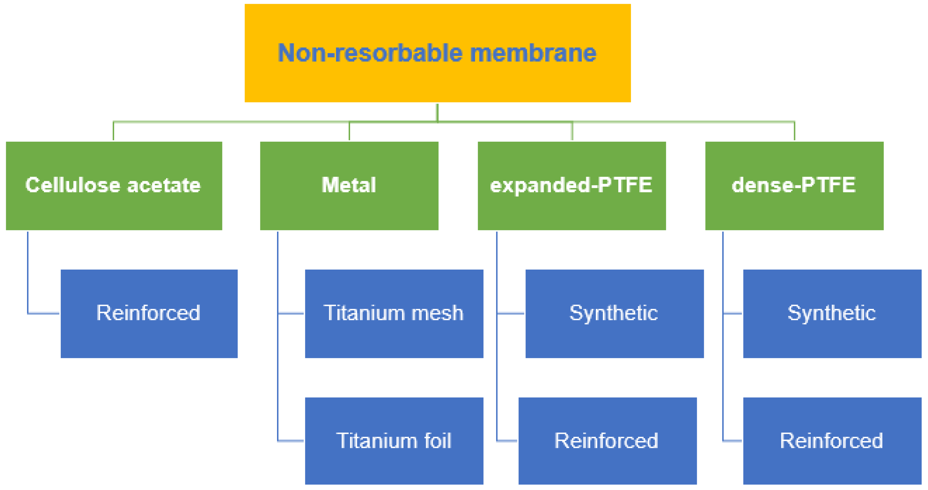
Cellulose acetate (CA) was the earliest material used in non-resorbable membranes, which are intended to keep the gingival connective tissue away from the root surface and allow periodontal regeneration [24]. CA has been used because of its outstanding properties, including neutrality, biocompatibility, low cost and renewability [25]. Non-resorbable membranes, which can be further classified into metal, ePTFE and dense PTFE (dPTFE) with or without titanium reinforcement, are widely used in periodontal regenerative approaches, such as GTR/GBR, are collectively depicted in Figure 1. GTR/GBR requires a membrane that works as a physical barrier that can prevent the competitive invasion of highly proliferative cells of the surrounding tissue, mainly fibroblasts and epithelial cells. Meanwhile, the native cell proliferation properties of the natural regeneration region should be promoted [26][27].
Figure 1. Various types of non-resorbable membranes.
Overall, ePTFE has shown positive results in regenerative procedures, provided it has primary closure [28]. An ePTFE membrane features a chemically stable, biocompatible, inert polymer, but its structure is porous and flexible. These properties enable this membrane to resist degradation produced by microbiological or enzymatic reactions [28][29]. These membranes consist of two distinct parts: an open microstructure (100–300 µm porosity) and an occlusive structure (<8 µm porosity). The porous microstructure stimulates the ingrowth of collagen fibrils, thereby improving membrane stability and facilitating nutrient transport through its pores, which, together, stimulate new bone formation during the first healing period [30]. By contrast, the occlusive component is generally impervious to fluids and prevents soft tissue cells from migrating into the area of bone development [31]. As a result, barrier materials should have a porous fraction to generate optimal regenerative therapy results [30][31][32]. However, premature exposure of ePTFE membranes is relatively common and is reportedly approximately 30–40%, and this may lead to infection and lack of new bone formation as a result of fibrous tissue ingrowth [33]. Therefore, primary closure is deemed necessary over ePTFE membranes, but this can be challenging in larger defects [34]. The need for additional surgery to remove the membrane increases the risk of exposing newly regenerated bone to bacteria. The timing of membrane removal is also important, because early removal can lead to resorption of regenerated bone, whereas late removal can increase the risks of bacterial contamination and infection [35]. The conventional product available worldwide with the types of material are briefly describe in Table 1.
Table 1. Commercially available non-resorbable barrier membrane.

Product (Company)

Material

Ti- Micromesh (ACE)

Titanium mesh

Tocksystem (MeshTM)

Titanium mesh

Millipore

Cellulose acetate

Gore-Tex®

ePTFE

Cytoplast™

dPTFE

Ti-Reinforced Gore-Tex®

Titanium-reinforced ePTFE

Cytoplast™ Ti-Reinforced 250

Titanium-reinforced dPTFE

4. Synthetic Membranes

Synthetic biodegradable membranes are made of polymers. Natural polymers, such as collagen, are readily biocompatible, and the protein may enhance cell adhesion and proliferation. However, the mechanical and physical properties of natural polymers are inferior to those of synthetic polymers. For instance, natural polymers have less tensile strength and a higher degradation rate than synthetic polymers [36]. Synthetic polymers are biocompatible, making them suitable as biomaterials. In addition, synthetic polymers have tailorable physiochemical properties according to the desired outcomes and manufacturing reproducibility [37][38][39]. Some synthetic polymers are degradable, and they have gained much attention for membrane development in tissue engineering, because secondary surgery is not needed to remove the membrane.
Biodegradable synthetic polymers to be used as biomaterials for tissue engineering should have several advantageous properties. For instance, they should be biocompatible and not induce inflammatory changes around the tissue. They should also be degraded when the surrounding tissue is ready to function. Excellent physiochemical properties, according to the intended outcome, are also required. Therefore, the current research in biodegradable synthetic polymers is attempting to synthesize polymers and copolymers that can match the ideal properties of the desired function of the biomaterial.
Synthetic biodegradable membranes for biomedical and tissue engineering have several types, including polylactic acid (PLA), polyglycolic acid (PGA), polycaprolactone (PCL), poly(glycolide-co-lactide) copolymer and other copolymers. The investigation of other types of biodegradable synthetic polymers and copolymers is still underway.

5. Autologous Platelet Concentrate (APC)

5.1. Types of Autologous Platelet Concentrate

The challenge in GTR involves the replacement and reconstruction of massive tissue defects, especially in the presence of local and systemic contributing factors, such as habitual smoking, diabetes mellitus and multi-walled defects. Utilizing grafting materials, membrane barriers and additional therapy of biologic agents will be beneficial to regenerate the desirable amount of defect quality and quantity. In challenging cases, additional biologic agents will help promote healing induction and conduction in the local surgical area, making the healing process predictable and faster to produce true periodontal regeneration [40][41]. APC has various types, including pure platelet-rich fibrin (PRP), leukocyte platelet-rich fibrin, advanced platelet-rich fibrin (A-PRF), injectable platelet-rich fibrin, titanium platelet-rich fibrin, prepared platelet-rich lysate and concentrated growth factor (CGF). PRP was initially developed for medical purposes, such as the management of severe thrombopenia, and further elaborated into multiple applications in the medical field, such as orthopedics, dermatology, sports medicine and others, because of its capacity to retain growth factors and, thus, improve healing response at the application site.
Second-generation plasma concentrates consist of PRF and CGF. PRF was initially developed by Chakroun et al., using simple centrifugation for the application in surgical fields of dentistry, without using any additive materials, such as anticoagulants and thrombin [42][43]. The variation of these plasma-derived blood products is determined by two key predeterminants, which are the leukocyte volume and fibrin mass. The original derivatives, such as pure PRP, contain an immature, minute fibrin and fibrillae diameter, thus forming a less dense fibrin tissue adhesive. In general, these derivatives produce an unstable network and a high rate of tissue dissolution.
PRF consists of a stable, mature fibrin network, due to accomplished tissue polymerization accompanied by platelets and leukocytes forming a biomaterial with enhanced biomechanical tissue with structural integrity compared with the original PRP [43]. Nevertheless, CGF developed by Sacco produces an autologous membrane that is thicker, denser and more durable than the conventional PRF [44]. Ultimately, the demand and quest for soft- and hard-tissue healing response with optimal bone and tissue regeneration and remodeling are paramount. The plasma concentrates are still controversial with regard to their efficiency and effectiveness in integrating the bone–graft–implant–tissue complex, especially in the long term. However, the theoretical concepts of local application of growth factors will eventually enhance and support local healing and regeneration.

6. High-Performance Polymer

Polyetheretherketone (PEEK) is a semicrystallized thermosoftening polymer derived from the polyaryletherketone group. It is widely used in the medical field as an excellent alternative to titanium in orthopedics [45]. The research and application of PEEK in dentistry are extensive; specifically, it has been used as a dental implant, provisional abutment, obturator, denture base, clasp for dentures and others because of its good biological, mechanical, aesthetic and handling properties [46][47][48][49][50][51]. Given its excellent mechanical properties and structural integrity, PEEK has been suggested by Papia et al. to be used as a barrier membrane in complex three-dimensional surgery, because of its satisfactory mechanical properties under tensile and flexural strength with the thickness range of 0.5–1.0 mm, making it a desirable material for regeneration therapy [52]. In addition, it possesses the required general stiffness, strength and hardness, while maintaining ductility and light compared with other materials, such as polymer and ceramic [53][54]. The versatility of this material in manufacturing method, either milling under substractive computer-aided design and manufacturing (CAD-CAM) or rapid prototyping by additive manufacturing, makes it a favorable material to be utilized as a barrier membrane [55]. A study showed that the 3D bone augmentation utilizing a customized virtually designed PEEK sheet has satisfactory vertical and horizontal bone gain, with mean values of 3.47 and 3.42 mm, respectively [56]. A continuation study comparing the customized PEEK sheet and pre-bent titanium mesh achieved satisfactory outcomes in bone gain for both groups under CBCT assessment [57]. Additional clinical studies must be conducted to investigate the applicability of high-performance polymer PEEK or polyetherimide as a barrier membrane [58].

References

  1. Elgali, I.; Omar, O.; Dahlin, C.; Thomsen, P. Guided bone regeneration: Materials and biological mechanisms revisited. Eur. J. Oral Sci. 2017, 125, 315–337.
  2. Khojasteh, A.; Kheiri, L.; Motamedian, S.R.; Khoshkam, V. Guided bone regeneration for the reconstruction of alveolar bone defects. Ann. Maxillofac. Surg. 2017, 7, 263–277.
  3. Masquelet, A.C.; Begue, T. The Concept of Induced Membrane for Reconstruction of Long Bone Defects. Orthop. Clin. N. Am. 2010, 41, 27–37.
  4. Liu, J.; Kerns, D.G. Mechanisms of Guided Bone Regeneration: A Review. Open Dent. J. 2014, 8, 56–65.
  5. Hoornaert, A.; D’Arros, C.; Heymann, M.-F.; Layrolle, P. Biocompatibility, resorption and biofunctionality of a new synthetic biodegradable membrane for guided bone regeneration. Biomed. Mater. 2016, 11, 045012.
  6. Caballé-Serrano, J.; Sawada, K.; Miron, R.J.; Bosshardt, D.D.; Buser, D.; Gruber, R. Collagen barrier membranes adsorb growth factors liberated from autogenous bone chips. Clin. Oral Implants Res. 2016, 28, 236–241.
  7. Huang, H.-L.; Ma, Y.-H.; Tu, C.-C.; Chang, P.-C. Radiographic Evaluation of Regeneration Strategies for the Treatment of Advanced Mandibular Furcation Defects: A Retrospective Study. Membranes 2022, 12, 219.
  8. Sasaki, J.-I.; Abe, G.L.; Li, A.; Thongthai, P.; Tsuboi, R.; Kohno, T.; Imazato, S. Barrier membranes for tissue regeneration in dentistry. Biomater. Investig. Dent. 2021, 8, 54–63.
  9. Sbricoli, L.; Guazzo, R.; Annunziata, M.; Gobbato, L.; Bressan, E.; Nastri, L. Selection of collagen membranes for bone regenera-tion: A literature review. Materials 2020, 13, 786.
  10. Wang, H.L.; Boyapati, L. “PASS” principles for predictable bone regeneration. Implant Dent. 2006, 15, 8–17.
  11. Gottlow, J.; Nyman, S.; Karring, T.; Lindhe, J. New attachment formation as the result of controlled tissue regeneration. J. Clin. Periodontol. 1984, 11, 494–503.
  12. Cortellini, P.; Carnevale, G.; Sanz, M.; Tonetti, M.S. Treatment of deep and shallow intrabony defects A multicenter randomized controlled clinical trial. J. Clin. Periodontol. 1998, 25, 981–987.
  13. Gulameabasse, S.; Gindraux, F.; Catros, S.; Fricain, J.C.; Fenelon, M. Chorion and amnion/chorion membranes in oral and per-iodontal surgery: A systematic review. J. Biomed. Mater. Res. Part B Appl. Biomater. 2021, 109, 1216–1229.
  14. Qasim, S.S.B.; Baig, M.R.; Matinlinna, J.P.; Daood, U.; Al-Asfour, A. Highly Segregated Biocomposite Membrane as a Functionally Graded Template for Periodontal Tissue Regeneration. Membranes 2021, 11, 667.
  15. Zhang, L.; Dong, Y.; Zhang, N.; Shi, J.; Zhang, X.; Qi, C.; Midgley, A.C.; Wang, S. Potentials of sandwich-like chitosan/polycaprolactone/gelatin scaffolds for guided tissue regeneration membrane. Mater. Sci. Eng. C 2020, 109, 110618.
  16. Qasim, S.B.; Najeeb, S.; Delaine-Smith, R.M.; Rawlinson, A.; Rehman, I.U. Potential of electrospun chitosan fibers as a surface layer in functionally graded GTR membrane for periodontal regeneration. Dent. Mater. 2017, 33, 71–83.
  17. Hassan, M.; Prakasam, S.; Bain, C.; Ghoneima, A.; Liu, S.S.-Y. A randomized split-mouth clinical trial on effectiveness of am-nion-chorion membranes in alveolar ridge preservation: A clinical, radiologic, and morphometric study. Int. J. Oral Maxillofac. Implants 2017, 32, 1389–1398.
  18. Chan, E.C.; Kuo, S.-M.; Kong, A.M.; Morrison, W.A.; Dusting, G.J.; Mitchell, G.M.; Lim, S.Y.; Liu, G.-S. Three Dimensional Collagen Scaffold Promotes Intrinsic Vascularisation for Tissue Engineering Applications. PLoS ONE 2016, 11, e0149799.
  19. Turri, A.; Elgali, I.; Vazirisani, F.; Johansson, A.; Emanuelsson, L.; Dahlin, C.; Thomsen, P.; Omar, O. Guided bone regeneration is promoted by the molecular events in the membrane compartment. Biomaterials 2016, 84, 167–183.
  20. Taguchi, Y.; Amizuka, N.; Nakadate, M.; Ohnishi, H.; Fujii, N.; Oda, K.; Nomura, S.; Maeda, T. A histological evaluation for guided bone regeneration induced by a collagenous membrane. Biomaterials 2005, 26, 6158.
  21. Allan, B.; Ruan, R.; Landao-Bassonga, E.; Gillman, N.; Wang, T.; Gao, J.; Ruan, Y.; Xu, Y.; Lee, C.; Goonewardene, M.; et al. Collagen Membrane for Guided Bone Regeneration in Dental and Orthopedic Applications. Tissue Eng. Part A 2021, 27, 372–381.
  22. Chen, Z.; Chen, L.; Liu, R.; Lin, Y.; Chen, S.; Lu, S.; Lin, Z.; Chen, Z.; Wu, C.; Xiao, Y. The osteoimmunomodulatory property of a barrier collagen membrane and its manipulation via coating nanometer-sized bioactive glass to improve guided bone re-generation. Biomater. Sci. 2018, 6, 1007–1019.
  23. Annen, B.M.; Ramel, C.F.; Hämmerle, C.H.; Jung, R.E. Use of a new cross-linked collagen membrane for the treatment of pe-ri-implant dehiscence defects: A randomised controlled double-blinded clinical trial. Eur. J. Oral Implantol. 2011, 4, 87–100.
  24. Ronda, M.; Rebaudi, A.; Torelli, L.; Stacchi, C. Expanded vs. dense polytetrafluoroethylene membranes in vertical ridge augmentation around dental implants: A prospective randomized controlled clinical trial. Clin. Oral Implants Res. 2014, 25, 859–866.
  25. Ruggiero, R.; de Almeida Carvalho, V.; da Silva, L.G.; de Magalhães, D.; Ferreira, J.A.; de Menezes, H.H.M.; de Melo, G.P.; Naves, M.M. Study of in vitro degradation of cellulose acetate membranes modified and incorporated with tetracycline for use as an adjuvant in periodontal reconstitution. Ind. Crop. Prod. 2015, 72, 2–6.
  26. Nyman, S.; Lindhe, J.; Karring, T.; Rylander, H. New attachment following surgical treatment of human periodontal disease. J. Clin. Periodontol. 1982, 9, 290–296.
  27. Liang, Y.; Luan, X.; Liu, X. Recent advances in periodontal regeneration: A biomaterial perspective. Bioact. Mater. 2020, 5, 297–308.
  28. Soldatos, N.K.; Stylianou, P.; Angelov, N.; Koidou, P.; Yukna, R.; Romanos, G.E. Limitations and options using resorbable versus nonresorbable membranes for successful guided bone regeneration. Quintessence Int. 2017, 48, 131–147.
  29. Retzepi, M.; Donos, N. Guided Bone Regeneration: Biological principle and therapeutic applications. Clin. Oral Implants Res. 2010, 21, 567–576.
  30. Gutta, R.; Baker, R.A.; Bartolucci, A.A.; Louis, P.J. Barrier Membranes Used for Ridge Augmentation: Is There an Optimal Pore Size? J. Oral Maxillofac. Surg. 2009, 67, 1218–1225.
  31. Lee, J.-Y.; Kim, Y.-K.; Yun, P.-Y.; Oh, J.-S.; Kim, S.-G. Guided bone regeneration using two types of non-resorbable barrier mem-branes. J. Korean Assoc. Oral Maxillofac. Surg. 2010, 36, 275–279.
  32. Rakhmatia, Y.D.; Ayukawa, Y.; Furuhashi, A.; Koyano, K. Current barrier membranes: Titanium mesh and other membranes for guided bone regeneration in dental applications. J. Prosthodont. Res. 2013, 57, 3–14.
  33. Vroom, M.; Gründemann, L. Nietresorbeerbare membranen. Tandartspraktijk 2014, 35, 8–13.
  34. Barber, H.D.; Lignelli, J.; Smith, B.M.; Bartee, B.K. Using a Dense PTFE Membrane Without Primary Closure to Achieve Bone and Tissue Regeneration. J. Oral Maxillofac. Surg. 2007, 65, 748–752.
  35. Rodriguez, I.A.; Selders, G.S.; Fetz, A.E.; Gehrmann, C.J.; Stein, S.H.; Evensky, J.A.; Green, M.S.; Bowlin, G.L. Barrier membranes for dental applications: A review and sweet advancement in membrane developments. Mouth Teeth 2018, 2, 1–9.
  36. Castro, A.G.; Diba, M.; Kersten, M.; Jansen, J.A.; Beucken, J.J.V.D.; Yang, F. Development of a PCL-silica nanoparticles composite membrane for Guided Bone Regeneration. Mater. Sci. Eng. C 2018, 85, 154–161.
  37. Wang, J.; Wang, L.; Zhou, Z.; Lai, H.; Xu, P.; Liao, L.; Wei, J. Biodegradable Polymer Membranes Applied in Guided Bone/Tissue Regeneration: A Review. Polymers 2016, 8, 115.
  38. Ulery, B.D.; Nair, L.S.; Laurencin, C.T. Biomedical applications of biodegradable polymers. J. Polym. Sci. Part B Polym. Phys. 2011, 49, 832–864.
  39. Iqbal, N.; Khan, A.S.; Asif, A.; Yar, M.; Haycock, J.W.; Rehman, I.U. Recent concepts in biodegradable polymers for tissue engi-neering paradigms: A critical review. Int. Mater. Rev. 2019, 64, 91–126.
  40. Alauddin, M.S.; Ramli, H. Management of Membrane Exposure Utilizing Concentrated Growth Factor (CFG) in Guided Bone Regeneration: A Clinical Report. Open Dent. J. 2020, 14, 763–768.
  41. Alauddin, M.S.; Yusof, N.M.; Adnan, A.S.; Said, Z. Preliminary Novel Analysis on Antimicrobial Properties of Concentrated Growth Factor against Bacteria-Induced Oral Diseases. Eur. J. Dent. 2022, 1–239.
  42. Dohan, D.M.; Choukroun, J.; Diss, A.; Dohan, S.L.; Dohan, A.; Mouhyi, J.; Gogly, B. Platelet-rich fibrin (PRF): A second-generation platelet concentrate. Part I: Technological concepts and evolution. Oral Surg. Oral Med. Oral Pathol. Oral Radiol. Endodontol. 2006, 101, e37–e44.
  43. Dohan Ehrenfest, D.M.; Rasmusson, L.; Albrektsson, T. Classification of platelet concentrates: From pure platelet-rich plasma (P-PRP) to leucocyte- and platelet-rich fibrin (L-PRF). Trends Biotechnol. 2009, 27, 158–167.
  44. Rodella, L.F.; Favero, G.; Boninsegna, R.; Buffoli, B.; Labanca, M.; Scarì, G.; Sacco, L.; Batani, T.; Rezzani, R. Growth factors, CD34 positive cells, and fibrin network analysis in concentrated growth factors fraction. Microsc. Res. Tech. 2011, 74, 772–777.
  45. Kurtz, S.M.; Devine, J.N. PEEK biomaterials in trauma, orthopedic, and spinal implants. Biomaterials 2007, 28, 4845–4869.
  46. Schwitalla, A.; Müller, W.-D. PEEK Dental Implants: A Review of the Literature. J. Oral Implant. 2013, 39, 743–749.
  47. Toth, J.M.; Wang, M.; Estes, B.T.; Scifert, J.L.; Seim, H.B., III; Turner, A.S. Polyetheretherketone as a biomaterial for spinal applica-tions. Biomaterials 2006, 27, 324–334.
  48. Santing, H.J.; Meijer, H.J.; Raghoebar, G.M.; Özcan, M. Fracture strength and failure mode of maxillary implant-supported provisional single crowns: A comparison of composite resin crowns fabricated directly over PEEK abutments and solid titanium abutments. Clin. Implant Dent. Relat. Res. 2012, 14, 882–889.
  49. Tannous, F.; Steiner, M.; Shahin, R.; Kern, M. Retentive forces and fatigue resistance of thermoplastic resin clasps. Dent. Mater. 2012, 28, 273–278.
  50. Costa-Palau, S.; Torrents-Nicolas, J.; Barberà, M.B.-D.; Cabratosa-Termes, J. Use of polyetheretherketone in the fabrication of a maxillary obturator prosthesis: A clinical report. J. Prosthet. Dent. 2014, 112, 680–682.
  51. Aldhuwayhi, S.; Alauddin, M.S.; Martin, N. The Structural Integrity and Fracture Behaviour of Teeth Restored with PEEK and Lithium-Disilicate Glass Ceramic Crowns. Polymers 2022, 14, 1001.
  52. Papia, E.; Brodde, S.A.; Becktor, J.P. Deformation of polyetheretherketone, PEEK, with different thicknesses. J. Mech. Behav. Biomed. Mater. 2022, 125, 104928.
  53. Alauddin, M.S. A Review of Polymer Crown Materials: Biomechanical and Material Science. J. Clin. Diagn. Res. 2019, 13, ZE01–ZE05.
  54. Najeeb, S.; Zafar, M.S.; Khurshid, Z.; Siddiqui, F. Applications of polyetheretherketone (PEEK) in oral implantology and prosthodontics. J. Prosthodont. Res. 2016, 60, 12–19.
  55. Alauddin, M.S.; Baharuddin, A.S.; Mohd Ghazali, M.I. The modern and digital transformation of oral health care: A mini review. Healthcare 2021, 9, 118.
  56. El Morsy, O.A.; Barakat, A.; Mekhemer, S.; Mounir, M. Assessment of 3-dimensional bone augmentation of severely atrophied maxillary alveolar ridges using patient-specific poly ether-ether ketone (PEEK) sheets. Clin. Implant Dent. Relat. Res. 2020, 22, 148–155.
  57. Mounir, M.; Shalash, M.; Mounir, S.; Nassar, Y.; El Khatib, O. Assessment of three dimensional bone augmentation of severely atrophied maxillary alveolar ridges using prebent titanium mesh vs customized poly-ether-ether-ketone (PEEK) mesh: A randomized clinical trial. Clin. Implant Dent. Relat. Res. 2019, 21, 960–967.
  58. Alqurashi, H.; Khurshid, Z.; Syed, A.U.; Habib, S.R.; Rokaya, D.; Zafar, M.S. Polyetherketoneketone (PEKK): An emerging bio-material for oral implants and dental prostheses. J. Adv. Res. 2021, 28, 87–95.
More
Information
Contributors MDPI registered users' name will be linked to their SciProfiles pages. To register with us, please refer to https://encyclopedia.pub/register : , , , ,
View Times: 506
Revisions: 2 times (View History)
Update Date: 11 May 2022
1000/1000