 +1 credit
| Version | Summary | Created by | Modification | Content Size | Created at | Operation |
|---|---|---|---|---|---|---|
| 1 | Katya Rubia | + 14293 word(s) | 14293 | 2022-03-25 09:54:14 | | | |
| 2 | Vivi Li | -6603 word(s) | 7690 | 2022-03-28 03:39:36 | | |
Video Upload Options
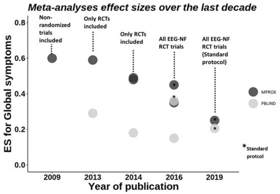
Attention-deficit/hyperactivity disorder (ADHD) is characterised by persisting and impairing symptoms of age-inappropriate inattention and/or hyperactivity/impulsivity (DSM-5). EEG-neurofeedback has been tested for about 45 years, with the latest meta-analyses of randomised controlled trials (RCT) showing small/medium effects compared to non-active controls only. Three small studies piloted neurofeedback of frontal activations in ADHD using functional magnetic resonance imaging or near-infrared spectroscopy, finding no superior effects over control conditions. Brain stimulation has been applied to ADHD using mostly repetitive transcranial magnetic and direct current stimulation (rTMS/tDCS). rTMS has shown mostly negative findings on improving cognition or symptoms. Meta-analyses of tDCS studies targeting mostly the dorsolateral prefrontal cortex show small effects on cognitive improvements with only two out of three studies showing clinical improvements. Trigeminal nerve stimulation has been shown to improve ADHD symptoms with medium effect in one RCT. Modern neurotherapeutics are attractive due to their relative safety and potential neuroplastic effects. However, they need to be thoroughly tested for clinical and cognitive efficacy across settings and beyond core symptoms and for their potential for individualised treatment.
1. Introduction
2. Neurotherapeutics in ADHD
2.1. Neurofeedback
2.1.1. EEG-NF
Meta-Analyses of EEG-NF
2.2. Brain Stimulation
2.2.1. Repetitive Transcranial Magnetic Stimulation (rTMS)
| Stimulation Protocol | Outcome Measures (Bold/Underlined = Improvement) | ||||||||
|---|---|---|---|---|---|---|---|---|---|
| Study | Design | N | Age | Target | Sessions | Frequency | Duration | Clinical | Cognitive |
| Children | |||||||||
| Cao et al., 2020 [77] |
Single-blind, randomised, parallel (2 active controls: ATX, ATX-rTMS; no sham) | 64 (~20 each) | 6–13 | R DLPFC a | 20 | 18 Hz (100% MT) | 2000 pulses (4 s on, 26 s off) | SNAP-IV | CPT; WISC; IGT |
| Gomez et al., 2014 [76] |
Open label | 10 | 7–12 | L DLPFC | 5 | 1 Hz (90% MT) | 1500 pulses (on, off n/r) | DSM-IV ADHD symptom checklist (hyperactivity/imp., inattention) | n/t |
| Adults | |||||||||
| Bloch et al., 2010 [72] |
Single-blind, sham-controlled, randomised, crossover | 13 | NR (adults) | R DLPFC a | 1 | 20 Hz (100% MT) | 1680 pulses (2 s on, 30 s off) | PANAS (inattention, total score; mood, anxiety, hyperactivity); VAS (inattention, mood) b | n/t |
| Paz et al., 2018 [74] |
Double-blind, sham-controlled, randomised, parallel | A: 13 S: 9 | A: 32 S: 30 |
L DLPFC c | 20 | 18 Hz (120% MT) | 1980 pulses (2 s on, 20 s off) | CAARS | TOVA |
| Weaver et al., 2012 [73] |
Single-blind, sham-controlled, randomised, crossover | 9 | 18 | R DLPFC a | 10 | 10 Hz (100% MT) | 2000 pulses (4 s on, 26 s off) | CGI-I scale; ADHD-IV scale | WAIS/WISC-IV; Connors CPT; DKEFS; Buschke Selective Reminding Test; Symbol Digit Coding test; Finger Oscillation tasks |
| Alyagon et al., 2020 [75] |
Double-semi-blind, randomised, active and sham-controlled | 52 (15, 14, 14) | 21–46 | R IFC & DLPFC | 15 | 18 Hz (120% MT) | 1440 pulses (2 s on, 20 s off) | CAARS (global ADHD symptoms; hyperactivity/impulsiveness) (BAARS-IV (hyperactivity/impulsiveness), BRIEF-A, BDI) | STROOP; STOP |
2.2.2. Transcranial Direct Current Stimulation (tDCS)
| Stimulation Protocol | Outcome Measures (Bold/Underlined = Improvement; Cursive = Impairment) |
|||||||||
|---|---|---|---|---|---|---|---|---|---|---|
| Study | Design | n | Mean Age | Anode/Cathode | mA | Sessions | Timing a | Duration (mins) | Clinical | Cognitive |
| Children | ||||||||||
| † Bandeira et al., 2016 [98] |
Open label | 9 | 11 | L DLPFC/R SOA | 2 | 5 | Online | 28 | Patient Global Impression of Improvement | Visual Attention Test (OM); NEPSY-II-inhibition (Switch errors); Digit Span; Corsi Cubes |
| Breitling et al., 2016 [99] |
Single-blind, sham-controlled, randomised, crossover | 21 | 14 | R IFC/L Cheek | 1 | 1 | Online | 20 | n/t | Flanker (Incongruent trials: COM c,d & RTV c) e |
| L Cheek/R IFC | 1 | 1 | Online | 20 | n/t | Flanker | ||||
| Munz et al., 2015 [97] |
Double-blind, sham-controlled, randomised, crossover | 14 | 12 | L DLPFC/R Cheek; R DLPFC/L Cheek |
0.25 | 1 | Offline | 25 (5 on, 1 off) | n/t | Go/No-Go (Go RT & RTV); Motor memory; Alertness |
| Nejati et al., 2020, Exp 1 [101] |
Double-blind, sham-controlled, randomised, crossover | 15 | 10 | L DLPFC/R DLPFC | 1 | 1 | Offline | 15 | n/t | Go/No-Go; N-back (Acc, RT); Stroop (Incongruent trials: COM & RT); WCST (Completion time) |
| Nejati et al., 2020, Exp 2 [101] |
Double-blind, sham-controlled, randomised, crossover | 10 | 9 | L DLPFC/R SOA | 1 | 1 | Offline | 15 | n/t | Go/No-Go; N-back (Acc c, RT) d; WCST (Total categories completed, total & pers errors) d |
| R SOA/L DLPFC | 1 | 1 | Offline | 15 | n/t | Go/No-Go (No--Go acc) d; N-back; WCST (Total categories completed, total & pers errors c) d | ||||
| Prehn-Kristensen et al., 2014 [96] |
Double-blind, sham-controlled, randomised, parallel | 12 | 12 | L DLPFC/R Cheek; R DLPFC/L Cheek | 0.25 | 1 | Offline | 25 (5 on, 1 off) | n/t | Declarative Memory (Acc); Alertness; Digit Span |
| Soff et al., 2017 [94] |
Double-blind, sham-controlled, randomised, crossover | 15 | 14 | L DLPFC/Vertex | 1 | 5 | Online | 20 | FBB-ADHD(Inattention f) g,h | QbTest (Inattention f; hyperactivity i) g,h |
| Soltaninejad et al., 2019 [91] | Single-blind, sham-controlled, randomised, crossover | 20 | 16 | L DLPFC/R SOA | 1.5 | 1 | Online | 15 | n/t | Go/No-Go (Go Acc) c,d; Stroop |
| R SOA/L DLPFC | 1.5 | 1 | Online | 15 | n/t | Go/No-Go (NoGo Acc) c,j; Stroop | ||||
| ‡ Soltaninejad et al., 2015 [91] |
Single-blind, sham-controlled, randomised, crossover | 20 | 16 | rIFC/L SOA | 1 | 1 | Online | 15 | n/t | Go/No-Go (Go Acc); Stroop |
| Sotnikova et al., 2017 [95] |
Double-blind, sham-controlled, randomised, crossover | 13 | 14 | L DLPFC/Vertex | 1 | 1 | Online | 20 | n/t | QbTest (RT, RTV k, OMs, Acc) l |
| Breitling et al., 2020 [99] |
Double-blind, sham- and HD-tDCS controlled, randomised, crossover | ADHD: 15HC: 15 | 13 (10–16) |
R IFC/L SOA | 1 | 3 with CT | Online | 20 | n/t | WM task; ERPs N200; P300 |
| Salehinejad et al., 2020 [100] |
Single-blind, sham-controlled, randomised, cross-over | 19 | 9 (8–12) |
1 | 2 | Online | 23 | n/t | ANT (orienting); GNG; SAT; Stroop | |
| † Westwood et al., 2021 [92] |
Double-blind, sham-controlled, randomised, parallel | 50 | 14 | R IFC/L SOA | 1 | 15 | Online | 20 | ADHD-RS; Conners 3P | GNG; Stop; Simon; WCST; CPT; MCT; time estimation; NIH WM; Verbal Fluency |
| Nejati et al., 2020 [101] |
Double-blind, sham-controlled, randomised, cross-over | 20 | 9 | L DLPFC/R vmPFC R DLPFC/L vmPFC Sham |
1 | 1 | Online | 20 | n/t | BART; CDDT (k20, k10) |
| † Berger et al., 2021 [104] |
Double-blind, active controlled, randomised, cross-over | 19 | 7–12 | L DLPFC (tDCS)/R SOA L DLPFC/R IFC (tRNS) |
0.75 | 5 | Online | 5 | n/t | ADHD-RS; Working & short-term memory, Moxo-CPT (all improved with tRNS vs. tDCS) |
| Adults | ||||||||||
| † Allenby et al., 2018 [107] |
Double-blind, sham-controlled, randomised, crossover | 37 | 32 | L DLPFC/R SOA | 2 | 3 | Online | 20 | n/t | Conners CPT (COM m); Stop Task |
| Cachoeira et al., 2017 [108] |
Double-blind, sham-controlled, randomised, parallel | A: 9 S: 8 |
A: 31 S: 34 |
R DLPFC/L DLPFC | 2 | 5 | Offline | 20 | ADHD Checklist (Inattention, Total) n; SDS (after tDCS); ADHD total score 2 weeks | None |
| Cosmo et al., 2015 [105] |
Double-blind, sham-controlled, randomised, parallel | A: 30 S: 30 |
A: 32 S: 33 |
LDLPFC/R DLPFC | 1 | 1 | Offline | 20 | n/t | Go/No-Go |
| Jacoby et al., 2018 [106] |
Single-blind, sham-controlled, randomised, crossover | 20 | 23 | L&R DLPFC/Cerebellum | 1.8 | 1 | Offline | 20 | n/t | CPT (multi-button presses) |
| Dubreuil-Vall et al., 2020 [109] |
Double-blind, sham-controlled, randomised, crossover | 37 | 18–67 | L DLPFC/R SOA R DLPFC/R SOA |
2 | 1 | Offline | 30 | n/t | Flanker (incongruent RT) n = 18; L P300; L N200. Stop (go RTs); L P200. n = 19 Flanker; Stop |
References
- American Psychiatric Association. Diagnostic and Statistical Manual of Mental Disorders, 5th ed.; American Psychiatric Publishing, Inc.: Washington, DC, USA, 2013.
- Thomas, R.; Sanders, S.; Doust, J.; Beller, E.; Glasziou, P. Prevalence of Attention-Deficit/Hyperactivity Disorder: A Systematic Review and Meta-analysis. Pediatrics 2015, 135, e994–e1001.
- Rubia, K. Functional brain imaging across development: A review. Eur. Child Adolesc. Psychiatry 2013, 22, 719–731.
- Rubia, K. “Cool” inferior fronto-striatal dysfunction in Attention Deficit Hyperactivity Disorder (ADHD) versus “hot” ventromedial orbitofronto-limbic dysfunction in conduct disorder: A review. Biol. Psychiatry 2011, 69, e69–e87.
- Willcutt, E.G.; Sonuga-Barke, E.J.S.; Nigg, J.T.; Sergeant, G.A. Recent Developments in Neuropsychological Models of Childhood Psychiatric Disorders. In Biological Child Psychiatry; Banaschewski, T., Rohde, L.A., Eds.; Karger: Basel, Switzerland, 2008; Volume 24, pp. 195–226.
- Pievsky, M.A.; McGrath, R.E. The Neurocognitive Profile of Attention-Deficit/Hyperactivity Disorder: A Review of Meta-Analyses. Arch. Clin. Neuropsychol. 2018, 33, 143–157.
- Rubia, K.; Halari, R.; Christakou, A.; Taylor, E. Impulsiveness as a timing disturbance: Neurocognitive abnormalities in attention-deficit hyperactivity disorder during temporal processes and normalization with methylphenidate. Philos. Trans. R. Soc. B Biol. Sci. 2009, 364, 1919–1931.
- Noreika, V.; Falter, C.M.; Rubia, K. Timing deficits in attention-deficit/hyperactivity disorder (ADHD): Evidence from neurocognitive and neuroimaging studies. Neuropsychologia 2013, 51, 235–266.
- Plichta, M.M.; Scheres, A. Ventral–striatal responsiveness during reward anticipation in ADHD and its relation to trait impulsivity in the healthy population: A meta-analytic review of the fMRI literature. Neurosci. Biobehav. Rev. 2014, 38, 125–134.
- Groen, Y.; Gaastra, G.F.; Lewis-Evans, B.; Tucha, O. Risky Behavior in Gambling Tasks in Individuals with ADHD—A Systematic Literature Review. PLoS ONE 2013, 8, e74909.
- Nigg, J.T.; Stavro, G.; Ettenhofer, M.; Hambrick, D.Z.; Miller, T.; Henderson, J.M. Executive functions and adhd in adults: Evidence for selective effects on ADHD symptom domains. J. Abnorm. Psychol. 2005, 114, 706–717.
- Roberts, B.A.; Martel, M.M.; Nigg, J.T. Are There Executive Dysfunction Subtypes Within ADHD? J. Atten. Disord. 2017, 21, 284–293.
- Cortese, S.; Adamo, N.; Del Giovane, C.; Mohr-Jensen, C.; Hayes, A.J.; Carucci, S.; Atkinson, L.; Tessari, L.; Banaschewski, T.; Coghill, D.; et al. Comparative efficacy and tolerability of medications for attention-deficit hyperactivity disorder in children, adolescents, and adults: A systematic review and network meta-analysis. Lancet Psychiatry 2018, 5, 727–738.
- Rubia, K.; Alzamora, A.; Cubillo, A.; Smith, A.B.; Radua, J.; Brammer, M.J. Effects of stimulants on brain function in ADHD: A systematic review and meta-analysis. Biol. Psychiatry 2014, 76, 616–628.
- Coghill, D.R.; Seth, S.; Pedroso, S.; Usala, T.; Currie, J.; Gagliano, A. Effects of Methylphenidate on Cognitive Functions in Children and Adolescents with Attention-Deficit/Hyperactivity Disorder: Evidence from a Systematic Review and a Meta-Analysis. Biol. Psychiatry 2014, 76, 603–615.
- Pievsky, M.A.; McGrath, R.E. Neurocognitive effects of methylphenidate in adults with attention-deficit/hyperactivity disorder: A meta-analysis. Neurosci. Biobehav. Rev. 2018, 90, 447–455.
- Swanson, J.M. Debate: Are Stimulant Medications for Attention-Deficit/Hyperactivity Disorder Effective in the Long Term? (Against). J. Am. Acad. Child Adolesc. Psychiatry 2019, 58, 936–938.
- Coghill, D. Debate: Are Stimulant Medications for Attention-Deficit/Hyperactivity Disorder Effective in the Long Term? (For). J. Am. Acad. Child Adolesc. Psychiatry 2019, 58, 938–939.
- Connolly, J.J.; Glessner, J.T.; Elia, J.; Hakonarson, H. ADHD & Pharmacotherapy: Past, Present and Future: A Review of the Changing Landscape of Drug Therapy for Attention Deficit Hyperactivity Disorder. Ther. Innov. Regul. Sci. 2015, 49, 632–642.
- Lubar, J.F.; Shouse, M.N. EEG and behavioral changes in a hyperkinetic child concurrent with training of the sensorimotor rhythm (SMR). Appl. Psychophysiol. Biofeedback 1976, 1, 293–306.
- Satterfield, J.H. EEG issues in children with minimal brain dysfunction. Semin. Psychiatry 1973, 5, 35–46.
- Satterfield, J.H.; Lesser, L.I.; Saul, R.E.; Cantwell, D.P. EEG aspects in the diagnosis and treatment of minimal brain dysfunction. Ann. N. Y. Acad. Sci. 1973, 205, 274–282.
- Jäncke, L. The plastic human brain. Restor. Neurol. Neurosci. 2009, 27, 521–538.
- Rapoport, J.L.; Gogtay, N. Brain Neuroplasticity in Healthy, Hyperactive and Psychotic Children: Insights from Neuroimaging. Neuropsychopharmacology 2008, 33, 181–197.
- Draganski, B.; Gaser, C.; Busch, V.; Schuierer, G.; Bogdahn, U.; May, A. Neuroplasticity: Changes in grey matter induced by training—Newly honed juggling skills show up as a transient feature on a brain-imaging scan. Nature 2004, 427, 311–312.
- Draganski, B.; May, A. Training-induced structural changes in the adult human brain. Behav. Brain Res. 2008, 192, 137–142.
- Draganski, B.; Gaser, C.; Kempermann, G.; Kuhn, H.-G.; Winkler, J.; Büchel, C.; May, A. Temporal and Spatial Dynamics of Brain Structure Changes during Extensive Learning. J. Neurosci. 2006, 26, 6314–6317.
- Dodich, A.; Zollo, M.; Crespi, C.; Cappa, S.; Martinez, D.L.; Falini, A.; Canessa, N. Short-term Sahaja Yoga meditation training modulates brain structure and spontaneous activity in the executive control network. Brain Behav. 2019, 9, e01159.
- Rubia, K. Cognitive Neuroscience of Attention Deficit Hyperactivity Disorder (ADHD) and Its Clinical Translation. Front. Hum. Neurosci. 2018, 12, 100.
- Ashkan, K.; Shotbolt, P.; David, A.; Samuel, M. Deep brain stimulation: A return journey from psychiatry to neurology. Postgrad. Med. J. 2013, 89, 323–328.
- Anderson, V.; Spencer-Smith, M.; Wood, A. Do children really recover better? Neurobehavioural plasticity after early brain insult. Brain 2011, 134, 2197–2221.
- Brunoni, A.R.; Nitsche, M.A.; Bolognini, N.; Bikson, M.; Wagner, T.; Merabet, L.; Edwards, D.; Valero-Cabré, A.; Rotenberg, A.; Pascual-Leone, A.; et al. Clinical research with transcranial direct current stimulation (tDCS): Challenges and future directions. Brain Stimul. 2012, 5, 175–195.
- Schachar, R.J.; Tannock, R.; Logan, G. Inhibitory Control, Impulsiveness, and Attention-Deficit Hyperactivity Disorder. Clin. Psychol. Rev. 1993, 13, 721–739.
- Arns, M.; Heinrich, H.; Strehl, U. Evaluation of neurofeedback in ADHD: The long and winding road. Biol. Psychol. 2014, 95, 108–115.
- Ros, T.; Enriquez-Geppert, S.; Zotev, V.; Young, K.D.; Wood, G.; Whitfield-Gabrieli, S.; Wan, F.; Vuilleumier, P.; Vialatte, F.; Van De Ville, D.; et al. Consensus on the reporting and experimental design of clinical and cognitive-behavioural neurofeedback studies (CRED-nf checklist). Brain 2020, 143, 1674–1685.
- Arns, M.; de Ridder, S.; Strehl, U.; Breteler, M.; Coenen, A. Efficacy of Neurofeedback Treatment in ADHD: The Effects on Inattention, Impulsivity and Hyperactivity: A Meta-Analysis. Clin. EEG Neurosci. 2009, 40, 180–189.
- Arns, M.; Clark, C.R.; Trullinger, M.; Debeus, R.; Mack, M.; Aniftos, M. Neurofeedback and Attention-Deficit/Hyperactivity-Disorder (ADHD) in Children: Rating the Evidence and Proposed Guidelines. Appl. Psychophysiol. Biofeedback 2020, 45, 39–48.
- Cortese, S.; Ferrin, M.; Brandeis, D.; Holtmann, M.; Aggensteiner, P.; Daley, D.; Santosh, P.; Simonoff, E.; Stevenson, J.; Stringaris, A.; et al. Neurofeedback for Attention-Deficit/Hyperactivity Disorder: Meta-Analysis of Clinical and Neuropsychological Outcomes from Randomized Controlled Trials. J. Am. Acad. Child Adolesc. Psychiatry 2016, 55, 444–455.
- Van Doren, J.; Arns, M.; Heinrich, H.; Vollebregt, M.A.; Strehl, U.; Loo, S.K. Sustained effects of neurofeedback in ADHD: A systematic review and meta-analysis. Eur. Child Adolesc. Psychiatry 2018, 28, 293–305.
- Micoulaud-Franchi, J.-A.; Geoffroy, P.A.; Fond, G.; Lopez, R.; Bioulac, S.; Philip, P. EEG neurofeedback treatments in children with ADHD: An updated meta-analysis of randomized controlled trials. Front. Hum. Neurosci. 2014, 8, 906.
- Riesco-Matías, P.; Yela-Bernabé, J.R.; Crego, A.; Sánchez-Zaballos, E. What Do Meta-Analyses Have to Say About the Efficacy of Neurofeedback Applied to Children With ADHD? Review of Previous Meta-Analyses and a New Meta-Analysis. J. Atten. Disord. 2021, 25, 473–485.
- Sonuga-Barke, E.; Brandeis, D.; Cortese, S.; Daley, D.; Danckaerts, M.; Dopfner, M.; Ferrin, M.; Holtmann, M.; Van der Oord, S. Evidence for Efficacy of Neurofeedback in ADHD? Response. Am. J. Psychiatry 2013, 170, 800–802.
- Yan, L.; Wang, S.; Yuan, Y.; Zhang, J. Effects of neurofeedback versus methylphenidate for the treatment of ADHD: Systematic review and meta-analysis of head-to-head trials. Evidence-Based Ment. Health 2019, 22, 111–117.
- Lambez, B.; Harwood-Gross, A.; Golumbic, E.Z.; Rassovsky, Y. Non-pharmacological interventions for cognitive difficulties in ADHD: A systematic review and meta-analysis. J. Psychiatr. Res. 2020, 120, 40–55.
- Bussalb, A.; Congedo, M.; Barthélemy, Q.; Ojeda, D.; Acquaviva, E.; Delorme, R.; Mayaud, L. Clinical and Experimental Factors Influencing the Efficacy of Neurofeedback in ADHD: A Meta-Analysis. Front. Psychiatry 2019, 10, 35.
- Hodgson, K.; Hutchinson, A.; Denson, L. Nonpharmacological treatments for ADHD: A meta-analytic review. J. Atten. Disord. 2014, 18, 275–282.
- Norris, S.L. Challenges in Using Nonrandomized Studies in Systematic Reviews of Treatment Interventions. Ann. Intern. Med. 2005, 142, 1112–1119.
- Sonuga-Barke, E.J.; Brandeis, D.; Cortese, S.; Daley, D.; Ferrin, M.; Holtmann, M.; Stevenson, J.; Danckaerts, M.; Van Der Oord, S.; Döpfner, M.; et al. Nonpharmacological Interventions for ADHD: Systematic Review and Meta-Analyses of Randomized Controlled Trials of Dietary and Psychological Treatments. Am. J. Psychiatry 2013, 170, 275–289.
- Arnold, L.E.; Arns, M.; Barterian, J.; Bergman, R.; Black, S.; Conners, C.K.; Connor, S.; Dasgupta, S.; Debeus, R.; Higgins, T.; et al. Double-Blind Placebo-Controlled Randomized Clinical Trial of Neurofeedback for Attention-Deficit/Hyperactivity Disorder With 13-Month Follow-up. J. Am. Acad. Child Adolesc. Psychiatry 2020, 60, 841–855.
- Strehl, U.; Aggensteiner, P.; Wachtlin, D.; Brandeis, D.; Albrecht, B.; Arana, M.; Bach, C.; Banaschewski, T.; Bogen, T.; Flaig-Röhr, A.; et al. Neurofeedback of Slow Cortical Potentials in Children with Attention-Deficit/Hyperactivity Disorder: A Multicenter Randomized Trial Controlling for Unspecific Effects. Front. Hum. Neurosci. 2017, 11, 135.
- Bussalb, A.; Collin, S.; Barthélemy, Q.; Ojeda, D.; Bioulac, S.; Blasco-Fontecilla, H.; Brandeis, D.; Ouakil, D.P.; Ros, T.; Mayaud, L. Is there a cluster of high theta-beta ratio patients in attention deficit hyperactivity disorder? Clin. Neurophysiol. 2019, 130, 1387–1396.
- Faraone, S.V.; Banaschewski, T.; Coghill, D.; Zheng, Y.; Biederman, J.; Bellgrove, M.A.; Newcorn, J.H.; Gignac, M.; Al Saud, N.M.; Manor, I.; et al. The World Federation of ADHD International Consensus Statement: 208 Evidence-based conclusions about the disorder. Neurosci. Biobehav. Rev. 2021, 128, 789–818.
- Demirtas-Tatlidede, A.; Vahabzadeh-Hagh, A.M.; Pascual-Leone, A. Can noninvasive brain stimulation enhance cognition in neuropsychiatric disorders? Neuropharmacology 2013, 64, 566–578.
- Ruf, S.P.; Fallgatter, A.J.; Plewnia, C. Augmentation of working memory training by transcranial direct current stimulation (tDCS). Sci. Rep. 2017, 7, 876.
- Katz, B.; Au, J.; Buschkuehl, M.; Abagis, T.; Zabel, C.; Jaeggi, S.M.; Jonides, J. Individual Differences and Long-term Consequences of tDCS-augmented Cognitive Training. J. Cogn. Neurosci. 2017, 29, 1498–1508.
- Fonteneau, C.; Redoute, J.; Haesebaert, F.; Le Bars, D.; Costes, N.; Suaud-Chagny, M.-F.; Brunelin, J. Frontal Transcranial Direct Current Stimulation Induces Dopamine Release in the Ventral Striatum in Human. Cereb. Cortex 2018, 28, 2636–2646.
- Meyer, B.; Mann, C.; Götz, M.; Gerlicher, A.; Saase, V.; Yuen, K.S.; Aedo-Jury, F.; Gonzalez-Escamilla, G.; Stroh, A.; Kalisch, R. Increased Neural Activity in Mesostriatal Regions after Prefrontal Transcranial Direct Current Stimulation and l-DOPA Administration. J. Neurosci. 2019, 39, 5326–5335.
- Borwick, C.; Lal, R.; Lim, L.W.; Stagg, C.J.; Aquili, L. Dopamine depletion effects on cognitive flexibility as modulated by tDCS of the dlPFC. Brain Stimul. 2020, 13, 105–108.
- Fukai, M.; Bunai, T.; Hirosawa, T.; Kikuchi, M.; Ito, S.; Minabe, Y.; Ouchi, Y. Endogenous dopamine release under transcranial direct-current stimulation governs enhanced attention: A study with positron emission tomography. Transl. Psychiatry 2019, 9, 115.
- Adelhöfer, N.; Mückschel, M.; Teufert, B.; Ziemssen, T.; Beste, C. Anodal tDCS affects neuromodulatory effects of the norepinephrine system on superior frontal theta activity during response inhibition. Brain Struct. Funct. 2019, 224, 1291–1300.
- Mishima, T.; Nagai, T.; Yahagi, K.; Akther, S.; Oe, Y.; Monai, H.; Kohsaka, S.; Hirase, H. Transcranial Direct Current Stimulation (tDCS) Induces Adrenergic Receptor-Dependent Microglial Morphological Changes in Mice. eNeuro 2019, 6.
- Moretti, J.; Poh, E.Z.; Rodger, J. rTMS-Induced Changes in Glutamatergic and Dopaminergic Systems: Relevance to Cocaine and Methamphetamine Use Disorders. Front. Neurosci. 2020, 14, 137.
- Poh, E.Z.; Hahne, D.; Moretti, J.; Harvey, A.R.; Clarke, M.W.; Rodger, J. Simultaneous quantification of dopamine, serotonin, their metabolites and amino acids by LC-MS/MS in mouse brain following repetitive transcranial magnetic stimulation. Neurochem. Int. 2019, 131, 104546.
- Kuo, M.-F.; Nitsche, M.A. Effects of Transcranial Electrical Stimulation on Cognition. Clin. EEG Neurosci. 2012, 43, 192–199.
- Ziemann, U.; Siebner, H.R. Modifying motor learning through gating and homeostatic metaplasticity. Brain Stimul. 2008, 1, 60–66.
- Cramer, S.C.; Sur, M.; Dobkin, B.H.; O’Brien, C.; Sanger, T.D.; Trojanowski, J.Q.; Rumsey, J.M.; Hicks, R.; Cameron, J.; Chen, D.; et al. Harnessing neuroplasticity for clinical applications. Brain 2011, 134, 1591–1609.
- Lefaucheur, J.-P.; André-Obadia, N.; Antal, A.; Ayache, S.S.; Baeken, C.; Benninger, D.; Cantello, R.M.; Cincotta, M.; De Carvalho, M.; De Ridder, D.; et al. Evidence-based guidelines on the therapeutic use of repetitive transcranial magnetic stimulation (rTMS). Clin. Neurophysiol. 2014, 125, 2150–2206.
- Janicak, P.G.; Dokucu, M.E. Transcranial magnetic stimulation for the treatment of major depression. Neuropsychiatry Dis. Treat. 2015, 11, 1549–1560.
- Mehta, U.M.; Naik, S.S.; Thanki, M.V.; Thirthalli, J. Investigational and Therapeutic Applications of Transcranial Magnetic Stimulation in Schizophrenia. Curr. Psychiatry Rep. 2019, 21, 89.
- Parkin, B.L.; Ekhtiari, H.; Walsh, V.F. Non-invasive Human Brain Stimulation in Cognitive Neuroscience: A Primer. Neuron 2015, 87, 932–945.
- Rossi, S.; Hallett, M.; Rossini, P.M.; Pascual-Leone, A. Safety, ethical considerations, and application guidelines for the use of transcranial magnetic stimulation in clinical practice and research. Clin. Neurophysiol. 2009, 120, 2008–2039.
- Bloch, Y. Transcranial magnetic stimulation (TMS) as a treatment in ADHD. Isr. J. Psychiatry Relat. Sci. 2012, 49, 18.
- Weaver, L.; Rostain, A.L.; Mace, W.; Akhtar, U.; Moss, E.; O’Reardon, J.P. Transcranial Magnetic Stimulation (TMS) in the Treatment of Attention-Deficit/Hyperactivity Disorder in Adolescents and Young Adults. J. ECT 2012, 28, 98–103.
- Paz, Y.; Friedwald, K.; Levkovitz, Y.; Zangen, A.; Alyagon, U.; Nitzan, U.; Segev, A.; Maoz, H.; Koubi, M.; Bloch, Y. Randomised sham-controlled study of high-frequency bilateral deep transcranial magnetic stimulation (dTMS) to treat adult attention hyperactive disorder (ADHD): Negative results. World J. Biol. Psychiatry 2017, 19, 561–566.
- Alyagon, U.; Shahar, H.; Hadar, A.; Barnea-Ygael, N.; Lazarovits, A.; Shalev, H.; Zangen, A. Alleviation of ADHD symptoms by non-invasive right prefrontal stimulation is correlated with EEG activity. NeuroImage Clin. 2020, 26, 102206.
- Gómez, L.; Vidal, B.; Morales, L.; Báez, M.; Maragoto, C.; Galvizu, R.; Vera, H.; Cabrera, I.; Zaldívar, M.; Sánchez, A. Low Frequency Repetitive Transcranial Magnetic Stimulation in Children With Attention Deficit/Hyperactivity Disorder. Preliminary Results. Brain Stimul. 2014, 7, 760–762.
- Cao, P.; Xing, J.; Cao, Y.; Cheng, Q.; Sun, X.; Kang, Q.; Dai, L.; Zhou, X.; Song, Z. Clinical effects of repetitive transcranial magnetic stimulation combined with atomoxetine in the treatment of attention-deficit hyperactivity disorder. Neuropsychiatry Dis. Treat. 2018, 14, 3231–3240.
- Krishnan, C.; Santos, L.; Peterson, M.; Ehinger, M. Safety of Noninvasive Brain Stimulation in Children and Adolescents. Brain Stimul. 2015, 8, 76–87.
- Zewdie, E.; Ciechanski, P.; Kuo, H.; Giuffre, A.; Kahl, C.; King, R.; Cole, L.; Godfrey, H.; Seeger, T.; Swansburg, R.; et al. Safety and tolerability of transcranial magnetic and direct current stimulation in children: Prospective single center evidence from 3.5 million stimulations. Brain Stimul. 2020, 13, 565–575.
- Boggio, P.S.; Ferrucci, R.; Mameli, F.; Martins, D.; Martins, O.; Vergari, M.; Tadini, L.; Scarpini, E.; Fregni, F.; Priori, A. Prolonged visual memory enhancement after direct current stimulation in Alzheimer’s disease. Brain Stimul. 2012, 5, 223–230.
- Kuo, M.-F.; Paulus, W.; Nitsche, M.A. Therapeutic effects of non-invasive brain stimulation with direct currents (tDCS) in neuropsychiatric diseases. NeuroImage 2014, 85, 948–960.
- Polanía, R.; Nitsche, M.A.; Paulus, W. Modulating functional connectivity patterns and topological functional organization of the human brain with transcranial direct current stimulation. Hum. Brain Mapp. 2011, 32, 1236–1249.
- Pogarell, O.; Koch, W.; Pöpperl, G.; Tatsch, K.; Jakob, F.; Mulert, C.; Grossheinrich, N.; Rupprecht, R.; Möller, H.-J.; Hegerl, U.; et al. Acute prefrontal rTMS increases striatal dopamine to a similar degree as d-amphetamine. Psychiatry Res. Neuroimaging 2007, 156, 251–255.
- Kuo, M.-F.; Chen, P.-S.; Nitsche, M.A. The application of tDCS for the treatment of psychiatric diseases. Int. Rev. Psychiatry 2017, 29, 146–167.
- Nejati, V.; Salehinejad, M.A.; Nitsche, M.A.; Najian, A.; Javadi, A.-H. Transcranial Direct Current Stimulation Improves Executive Dysfunctions in ADHD: Implications for Inhibitory Control, Interference Control, Working Memory, and Cognitive Flexibility. J. Atten. Disord. 2020, 24, 1928–1943.
- Rubia, K.; Lim, L.; Ecker, C.; Halari, R.; Giampietro, V.; Simmons, A.; Brammer, M.; Smith, A.B. Effects of age and gender on neural networks of motor response inhibition: From adolescence to mid-adulthood. NeuroImage 2013, 83, 690–703.
- Rubia, K.; Smith, A.B.; Brammer, M.J.; Taylor, E. Right inferior prefrontal cortex mediates response inhibition while mesial prefrontal cortex is responsible for error detection. NeuroImage 2003, 20, 351–358.
- Rubia, K.; Smith, A.B.; Taylor, E.; Brammer, M. Linear age-correlated functional development of right inferior fron-to-striato-cerebellar networks during response inhibition and anterior Cingulate during error-related processes. Hum. Brain Mapp. 2007, 28, 1163–1177.
- Breitling, C.; Zaehle, T.; Dannhauer, M.; Bonath, B.; Tegelbeckers, J.; Flechtner, H.-H.; Krauel, K. Improving Interference Control in ADHD Patients with Transcranial Direct Current Stimulation (tDCS). Front. Cell. Neurosci. 2016, 10, 72.
- Soltaninejad, Z.; Nejati, V.; Ekhtiari, H. Effect of Anodal and Cathodal Transcranial Direct Current Stimulation on DLPFC on Modulation of Inhibitory Control in ADHD. J. Atten. Disord. 2019, 23, 325–332.
- Soltaninejad, Z.; Nejati, V.; Ekhtiari, H. Effect of transcranial Direct Current Stimulation on Remediation of Inhibitory Con-trol on right Inferior Frontal Gyrus in Attention Deficit and Hyperactivity Symptoms. Rehabil. Med. 2015, 3, 1–9.
- Westwood, S.J.; Criaud, M.; Lam, S.-L.; Lukito, S.; Wallace-Hanlon, S.; Kowalczyk, O.S.; Kostara, A.; Mathew, J.; Agbedjro, D.; Wexler, B.E.; et al. Transcranial direct current stimulation (tDCS) combined with cognitive training in adolescent boys with ADHD: A double-blind, randomised, sham-controlled trial. Psychol. Med. 2021, 6, 1–16.
- Westwood, S.J.; Bozhilova, N.; Criaud, M.; Lam, S.L.; Lukito, S.; Wallace-Hanlon, S.; Kowalczyk, O.S.; Kostara, A.; Mathew, J.; Agbedjro, D.; et al. The effect of transcranial direct current stimulation (tDCS) combined with cognitive training on EEG spectral power in adolescent boys with ADHD: A double-blind, randomised, sham-controlled trial. medRxiv 2020.
- Soff, C.; Sotnikova, A.; Christiansen, H.; Becker, K.; Siniatchkin, M. Transcranial direct current stimulation improves clinical symptoms in adolescents with attention deficit hyperactivity disorder. J. Neural Transm. 2017, 124, 133–144.
- Sotnikova, A.; Soff, C.; Tagliazucchi, E.; Becker, K.; Siniatchkin, M. Transcranial Direct Current Stimulation Modulates Neuronal Networks in Attention Deficit Hyperactivity Disorder. Brain Topogr. 2017, 30, 656–672.
- Prehn-Kristensen, A.; Munz, M.; Göder, R.; Wilhelm, I.; Korr, K.; Vahl, W.; Wiesner, C.; Baving, L. Transcranial Oscillatory Direct Current Stimulation during Sleep Improves Declarative Memory Consolidation in Children With Attention-deficit/hyperactivity Disorder to a Level Comparable to Healthy Controls. Brain Stimul. 2014, 7, 793–799.
- Munz, M.T.; Prehn-Kristensen, A.; Thielking, F.; Mölle, M.; Göder, R.; Baving, L. Slow oscillating transcranial direct current stimulation during non-rapid eye movement sleep improves behavioral inhibition in attention-deficit/hyperactivity disorder. Front. Cell. Neurosci. 2015, 9, 307.
- Bandeira, I.D.; Guimarães, R.S.Q.; Jagersbacher, J.G.; Barretto, T.L.; De Jesus-Silva, J.R.; Santos, S.N.; Argollo, N.; Lucena, R. Transcranial Direct Current Stimulation in Children and Adolescents With Attention-Deficit/Hyperactivity Disorder (ADHD): A Pilot Study. J. Child Neurol. 2016, 31, 918–924.
- Breitling, C.; Zaehle, T.; Dannhauer, M.; Tegelbeckers, J.; Flechtner, H.-H.; Krauel, K. Comparison between conventional and HD-tDCS of the right inferior frontal gyrus in children and adolescents with ADHD. Clin. Neurophysiol. 2020, 131, 1146–1154.
- Salehinejad, M.A.; Ghayerin, E.; Nejati, V.; Yavari, F.; Nitsche, M.A. Domain-specific Involvement of the Right Posterior Parietal Cortex in Attention Network and Attentional Control of ADHD: A Randomized, Cross-over, Sham-controlled tDCS Study. Neuroscience 2020, 444, 149–159.
- Nejati, V.; Khorrami, A.S.; Nitsche, M.A. Transcranial Direct Current Stimulation Improves Reward Processing in Children With ADHD. J. Atten. Disord. 2020, 25, 1623–1631.
- Fertonani, A.; Miniussi, C. Transcranial Electrical Stimulation: What We Know and Do Not Know about Mechanisms. Neuroscientist 2017, 23, 109–123.
- McDonnell, M.D.; Ward, L.M. The benefits of noise in neural systems: Bridging theory and experiment. Nat. Rev. Neurosci. 2011, 12, 415–425.
- Berger, I.; Dakwar-Kawar, O.; Grossman, E.S.; Nahum, M.; Kadosh, R.C. Scaffolding the attention-deficit/hyperactivity disorder brain using transcranial direct current and random noise stimulation: A randomized controlled trial. Clin. Neurophysiol. 2021, 132, 699–707.
- Cosmo, C.; Baptista, A.F.; de Araújo, A.; Rosário, R.S.D.; Miranda, J.G.V.; Montoya, P.; De Sena, E. A Randomized, Double-Blind, Sham-Controlled Trial of Transcranial Direct Current Stimulation in Attention-Deficit/Hyperactivity Disorder. PLoS ONE 2015, 10, e0135371.
- Jacoby, N.; Lavidor, M.; Jacoby, N.; Lavidor, M. Null tDCS Effects in a Sustained Attention Task: The Modulating Role of Learning. Front. Psychol. 2018, 9, 476.
- Allenby, C.; Falcone, M.; Bernardo, L.; Wileyto, E.P.; Rostain, A.; Ramsay, J.; Lerman, C.; Loughead, J. Transcranial direct current brain stimulation decreases impulsivity in ADHD. Brain Stimul. 2018, 11, 974–981.
- Cachoeira, C.T.; Leffa, D.T.; Mittelstadt, S.D.; Mendes, L.S.T.; Brunoni, A.R.; Pinto, J.V.; Blazius, V.; Machado, V.; Bau, C.; Rohde, L.A.; et al. Positive effects of transcranial direct current stimulation in adult patients with attention-deficit/hyperactivity disorder A pilot randomized controlled study. Psychiatry Res. 2017, 247, 28–32.
- Dubreuil-Vall, L.; Gomez-Bernal, F.; Villegas, A.C.; Cirillo, P.; Surman, C.; Ruffini, G.; Widge, A.S.; Camprodon, J.A. Transcranial Direct Current Stimulation to the Left Dorsolateral Prefrontal Cortex Improves Cognitive Control in Patients With Attention-Deficit/Hyperactivity Disorder: A Randomized Behavioral and Neurophysiological Study. Biol. Psychiatry Cogn. Neurosci. Neuroimaging 2020, 6, 439–448.
- Salehinejad, M.A.; Wischnewski, M.; Nejati, V.; Vicario, C.M.; Nitsche, M.A. Transcranial direct current stimulation in attention-deficit hyperactivity disorder: A meta-analysis of neuropsychological deficits. PLoS ONE 2019, 14, e0215095.
- Westwood, S.J.; Radua, J.; Rubia, K. Non-Invasive Brain Stimulation in Attention Deficit Hyperactivity Disorder: A Systematic Review and Meta-Analysis. Psychol. Med. 2021, 6, 1–16, in press.
- Moliadze, V.; Schmanke, T.; Andreas, S.; Lyzhko, E.; Freitag, C.M.; Siniatchkin, M. Stimulation intensities of transcranial direct current stimulation have to be adjusted in children and adolescents. Clin. Neurophysiol. 2015, 126, 1392–1399.
- Lefaucheur, J.P.; Antal, A.; Ayache, S.S.; Benninger, D.H.; Brunelin, J.; Cogiamanian, F.; Cotelli, M.; De Ridder, D.; Ferrucci, R.; Langguth, B.; et al. Evidence-based guidelines on the therapeutic use of transcranial direct current stimulation (tDCS). Clin. Neurophysiol. 2014, 125, 2150–2206.
- Knudsen, E.I. Sensitive Periods in the Development of the Brain and Behavior. J. Cogn. Neurosci. 2004, 16, 1412–1425.
- Kekic, M.; Boysen, E.; Campbell, I.C.; Schmidt, U. A systematic review of the clinical efficacy of transcranial direct current stimulation (tDCS) in psychiatric disorders. J. Psychiatr. Res. 2016, 74, 70–86.
- Moffa, A.H.; Brunoni, A.R.; Nikolin, S.; Loo, C.K. Transcranial Direct Current Stimulation in Psychiatric Disorders: A Comprehensive Review. Psychiatr. Clin. N. Am. 2018, 41, 447–463.
- Nitsche, M.A.; Cohen, L.G.; Wassermann, E.M.; Priori, A.; Lang, N.; Antal, A.; Paulus, W.; Hummel, F.; Boggio, P.; Fregni, F.; et al. Transcranial direct current stimulation: State of the art 2008. Brain Stimul. 2008, 1, 206–223.
- Kim, S.; Stephenson, M.; Morris, P.G.; Jackson, S.R. tDCS-induced alterations in GABA concentration within primary motor cortex predict motor learning and motor memory: A 7T magnetic resonance spectroscopy study. NeuroImage 2014, 99, 237–243.
- Silvanto, J.; Muggleton, N.; Walsh, V. State-dependency in brain stimulation studies of perception and cognition. Trends Cogn. Sci. 2008, 12, 447–454.
- Krause, B.; Márquez-Ruiz, J.; Kadosh, R.C. The effect of transcranial direct current stimulation: A role for cortical excitation/inhibition balance? Front. Hum. Neurosci. 2013, 7, 602.
- Kadosh, R.C.; Levy, N.; O’Shea, J.; Shea, N.; Savulescu, J. The neuroethics of non-invasive brain stimulation. Curr. Biol. 2012, 22, R108–R111.
- Sarkar, A.; Dowker, A.; Kadosh, R.C. Cognitive Enhancement or Cognitive Cost: Trait-Specific Outcomes of Brain Stimulation in the Case of Mathematics Anxiety. J. Neurosci. 2014, 34, 16605–16610.
- Iuculano, T.; Kadosh, R.C. The Mental Cost of Cognitive Enhancement. J. Neurosci. 2013, 33, 4482–4486.

