Submitted Successfully!
To reward your contribution, here is a gift for you: A free trial for our video production service.
Thank you for your contribution! You can also upload a video entry or images related to this topic.
Version Summary Created by Modification Content Size Created at Operation
1 + 2447 word(s) 2447 2022-03-08 07:53:58 |
2 format is correct Meta information modification 2447 2022-03-25 04:22:36 |

Video Upload Options

Do you have a full video?

Confirm

Are you sure to Delete?
Cite
If you have any further questions, please contact Encyclopedia Editorial Office.
Nickoloff, J. Nucleases and Co-Factors in DNA Replication Stress Responses. Encyclopedia. Available online: https://encyclopedia.pub/entry/21011 (accessed on 29 March 2024).
Nickoloff J. Nucleases and Co-Factors in DNA Replication Stress Responses. Encyclopedia. Available at: https://encyclopedia.pub/entry/21011. Accessed March 29, 2024.
Nickoloff, Jac. "Nucleases and Co-Factors in DNA Replication Stress Responses" Encyclopedia, https://encyclopedia.pub/entry/21011 (accessed March 29, 2024).
Nickoloff, J. (2022, March 24). Nucleases and Co-Factors in DNA Replication Stress Responses. In Encyclopedia. https://encyclopedia.pub/entry/21011
Nickoloff, Jac. "Nucleases and Co-Factors in DNA Replication Stress Responses." Encyclopedia. Web. 24 March, 2022.
Nucleases and Co-Factors in DNA Replication Stress Responses
Edit

DNA replication stress is a constant threat that cells must manage to proliferate and maintain genome integrity. DNA replication stress responses, a subset of the broader DNA damage response (DDR), operate when the DNA replication machinery (replisome) is blocked or replication forks collapse during S phase. There are many sources of replication stress, such as DNA lesions caused by endogenous and exogenous agents including commonly used cancer therapeutics, and difficult-to-replicate DNA sequences comprising fragile sites, G-quadraplex DNA, hairpins at trinucleotide repeats, and telomeres. Replication stress is also a consequence of conflicts between opposing transcription and replication, and oncogenic stress which dysregulates replication origin firing and fork progression. Cells initially respond to replication stress by protecting blocked replisomes, but if the offending problem (e.g., DNA damage) is not bypassed or resolved in a timely manner, forks may be cleaved by nucleases, inducing a DNA double-strand break (DSB) and providing a means to accurately restart stalled forks via homologous recombination. However, DSBs pose their own risks to genome stability if left unrepaired or misrepaired.  

DNA damage replication stress structure-specific nucleases DNA damage response cancer biology cancer therapy

1. Introduction

Accurate DNA replication and proper chromosome segregation to daughter cells are critical to maintaining genome integrity and preventing cancer. Replication of the 6.3 billion bp of the diploid human genome during a typical eight-hour S phase requires >30,000 active origins, ~5000 of which are active at a time [1][2]. Replication forks travel in a highly processive manner, synthesizing ~3000 bp per min, yet forks frequently encounter obstacles that stall replisomes, causing replication stress and triggering stress responses including the intra-S checkpoint [3], fork protection to prevent replisome dissociation or fork collapse, and repair mechanisms that restart damaged forks. Replication stress is caused by a wide variety of endogenous and exogenous factors. Spontaneous DNA damage is caused by endogenous reactive oxygen species formed during cellular metabolism [4][5], misincorporation of ribonucleotides, and DNA lability [5]. DNA damage is also caused by exogenous genotoxic chemicals, and by ionizing and non-ionizing radiation. The vast majority of DNA lesions block replicative polymerases, necessitating lesion repair by an appropriate repair pathway, lesion bypass (damage tolerance) by translesion DNA synthesis (TLS) polymerases, repriming, homologous recombination (HR) mediated template switching, or passive rescue from an adjacent fork [6][7]. DNA lesions that block replicative polymerases include nucleotides with broken rings, oxidized bases, and chemical adducts, as well as single- and double-strand breaks (DSBs). DNA polymerase inhibitors and depletion of nucleotide pools with hydroxyurea are exogenous sources that cause global replication stress, slowing or stopping most or all replication forks [8].
Additional endogenous sources of replication stress are difficult to replicate DNA sequences and certain chromatin environments (e.g., G-quadraplex DNA, common fragile sites, telomeric DNA) [9][10][11][12][13][14][15][16][17]. Replication stress is also caused by stable R-loops which form by hybridization of RNA transcripts to DNA templates, especially in G-rich sequences [18][19][20], and by collisions between opposing transcription and replication machinery, particularly in highly transcribed ribosomal RNA gene arrays, fragile sites, and telomeres [21][22][23][24][25][26][27]. Topoisomerases avert replication stress by preventing DNA overwinding in front of replication forks, a type of intrinsic, topological replication stress. A recent yeast study showed that cohesin, a highly conserved protein with essential roles in sister chromatid cohesion required for proper chromosome segregation, increases replication stress in centromeric and ribosomal DNA by trapping topological stress [28]. Although cells suffer replication stress at random sites throughout the genome due to spontaneous (or induced) DNA damage, the stress associated with difficult to replicate sequences and challenging chromatin environments must be managed at those sites in every S phase.
When replication forks are blocked, the initial response has two aims: (1) protect the replication fork by stabilizing the replisome machinery, and (2) protect the fork from nucleolytic attack [29][30]. If a blocked fork is not restarted in a timely manner, it may be cleaved by structure-specific nucleases yielding a single-ended DSB (seDSB) that is processed by resection nucleases to suppress misrepair by canonical non-homologous end-joining (cNHEJ) and promote accurate fork restart by HR. This is important because cNHEJ is the dominant DSB repair pathway in mammalian cells [31][32] and cNHEJ of seDSBs can cause deletions and translocations that produce acentric and dicentric chromosomes that segregate improperly in mitosis or induce breakage-bridge-fusion cycles that further threaten genome integrity [33].

2. DDR Signaling in Response to Replication Stress

DNA repair and DNA damage checkpoint systems minimize replication fork encounters with blocking lesions [34][35][36], but with a steady state of ~10,000 DNA lesions per cell in unstressed cells [5] and ~5000 simultaneously active replisomes during S phase [2], fork encounters with blocking lesions are unavoidable. Acute or chronic exposures to genotoxic chemicals and radiation greatly increase replication stress, as does dysregulated replication associated with oncogenic stress [37][38][39]. Under normal circumstances, the leading and lagging strand replication machines are coupled, traveling together with the MCM (minichromosome maintenance) replicative helicase. If the leading strand polymerase is blocked, MCM helicase may decouple and unwind DNA ahead of the fork, exposing hundreds of bases of single-stranded DNA (ssDNA) [40][41]. As with ssDNA exposed by 5′–3′ resection of broken ends at DSBs by resection nucleases EXO1 and DNA2 (with its cofactor BLM) [42][43][44], the ssDNA exposed by decoupled MCM helicase is rapidly bound by the abundant, heterotrimeric replication protein A (RPA) (Figure 1A). RPA-bound ssDNA is recognized by the ATR (ataxia telangiectasia and Rad3-related) cofactor ATRIP (ATR-interacting protein), leading to activation of ATR (Figure 1B), the central signaling kinase of the intra-S checkpoint response [45]. In addition to ssDNA-RPA and ATRIP, ATR activation requires several other factors including TopBP1, RAD17-RFC, and the 9-1-1 complex. Additional, distinct ATR activation mechanisms have been described involving NBS1, a component of the MRE11-RAD50-NBS1 (MRN) complex, and the RPA-binding factor ETAA1 [45][46][47].
Dna 02 00006 g001
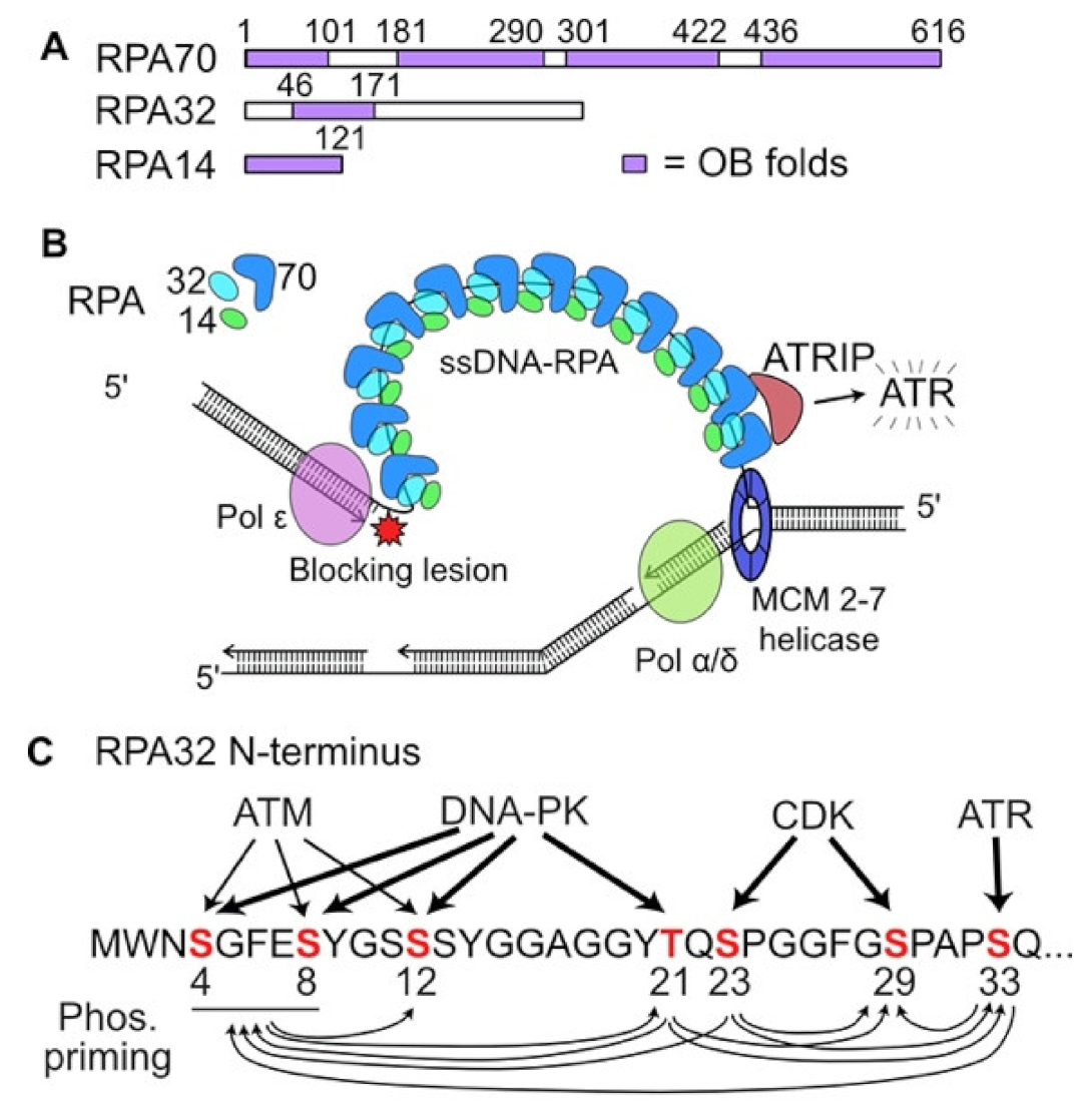
ATR is a member of the phosphatidyl inositol 3′ kinase-related kinase (PIKK) family, which also includes ATM and the catalytic subunit of DNA-dependent protein kinase (DNA-PKcs). PIKKs play central roles in DNA damage responses including DSB repair, checkpoint activation, apoptosis, suppression of transcription, and responses to telomere dysfunction and viral infection [48][49]. Activation of each PIKK involves a specific co-factor. The ATR interacting protein ATRIP recruits ATR to RPA-bound ssDNA, initiating ATR activation [50]. The MRN complex and the Ku70/Ku80 heterodimer bind to frank DSB ends, the NBS1 component of MRN recruits and activates ATM, and Ku70/Ku80 recruits and activates DNA-PKcs [48]. Activated PIKKs are autophosphorylated, and they phosphorylate each other and many other targets, showing various degrees of signaling pathway crosstalk [48].

3. Protecting and Rescuing Blocked Replication Forks

DNA replication initiates at origins in a complex, highly regulated process involving assembly of pre-replication complexes and licensing factors that ensure DNA is replicated only once per cell cycle [51]. For this reason, there is a premium on protecting replisomes at stalled replication forks to prevent replisome dissociation and fork collapse. Stalled forks are protected by a plethora of repair and replication factors, including RIF1, which inhibits end resection, the MRN-interacting protein MRNIP, the TLS suppressor USP1 which regulates PCNA via de-ubiquitination, HR proteins (RAD51, BRCA1, BRCA2, FANCD2), and RADX which regulates RAD51 [30][52][53][54][55][56][57]. Cells with defects in any of these fork protection factors are hypersensitive to replication stress.
Maintaining replisomes to protect stalled forks often involves fork regression to a ‘chicken foot’ structure that resembles four-way branched Holliday junctions of HR reactions (Figure 2A) [29]. Chicken foot structures have a one-ended DSB that at least initially includes ssDNA to which the HR factors RAD51, BRCA1, BRCA2, and the RAD51 paralogs (RAD51B/C/D and XRCC2/3) are recruited [58], although HR factors appear to play distinct roles in HR and fork protection [59].
Dna 02 00006 g002

4. MUS81: An Ancient Structure-Specific Nuclease Involved in HR and Restart of Stressed Replication Forks

MUS81 is a structure-specific 3′ endonuclease in the XPF 3′ endonuclease family that cleaves a variety of branched DNA structures including 3′ flaps and Holliday junctions. Yeast Mus81 was first discovered in 2000 in a two-hybrid screen for proteins that interacted with the RAD54 HR protein and was named for the sensitivity of Mus81-defective cells to methyl methanesulfonate and UV light [60]. Mus81-defective yeast also have a severe meiotic HR defect that together with its interaction with RAD54 suggested an important role in HR [60]. Indeed, yeast Mus81 and its Eme1 cofactor resolve Holliday junctions and human MUS81 cleaves four-way (Holliday) junctions and 3′ flap structures [61][62] (Figure 2B). In human cells, MUS81 with its EME1 cofactor resolves Holliday junctions in HR intermediates [63][64][65][66], and reversed forks that resemble Holliday junctions [67]. In contrast, MUS81 with its EME2 cofactor cleaves blocked replication forks, causing fork collapse to a seDSB [68][69][70] (Figure 2B). The seDSB is apparently resected to allow formation of a RAD51-ssDNA nucleoprotein filament that catalyzes fork restart by a mechanism that resembles break-induced replication (BIR) [71], although the resection nuclease(s) involved in processing MUS81-cleaved forks are not known (Figure 2C). Yeast Mus81 also mediates resolution of structures in G2/M that arise when blocked forks are rescued by converging forks to complete DNA replication [72].

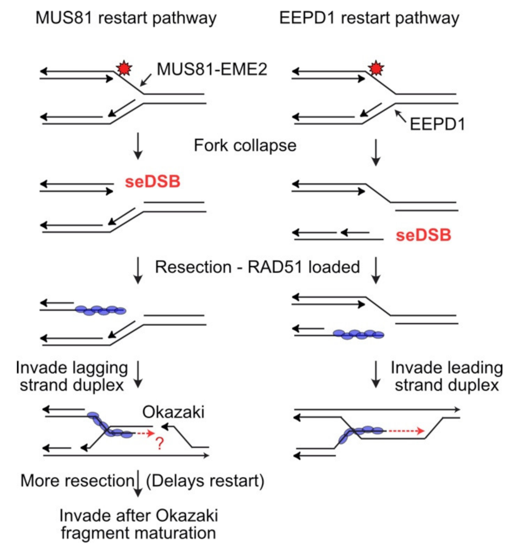
5. EEPD1: A 5′ Structure-Specific Endonuclease That Complements the 3′ MUS81 Nuclease

EEPD1 (endo- exonuclease phosphatase domain protein 1) has a DNase I-like nuclease domain and a DNA binding domain with two helix–hairpin–helix motifs similar to those in prokaryotic RuvA2. As with MUS81, cells with defective EEPD1 are hypersensitive to a variety of genotoxic chemicals and radiation, and replication stress induces chromosome aberrations and mitotic catastrophe [73][74]. iPOND (isolation of proteins on nascent DNA) is a technique that reveals proteins associated with replication forks, including replisome components and proteins recruited to stressed forks [75][76]. iPOND analysis demonstrated that EEPD1 is recruited to stalled replication forks, and similar to MUS81, EEPD1 cleaves fork structures in vitro, and stalled replication forks in vivo (Figure 3) [73]
Dna 02 00006 g003

6. Metnase: A Recently Evolved Nuclease-Protein Methyl Transferase That Promotes Replication Fork Restart

Metnase evolved ~50 million years ago when a Mariner transposon integrated downstream of a SET protein methylase, and subsequent genetic changes fused the SET and nuclease domains [77]. Metnase is a structure-specific nuclease with numerous genome stabilization functions including promotion of cNHEJ, chromosome decatenation, and restart of stressed replication forks [78][79][80]. Although defects in the Metnase nuclease delay replication fork restart [81] and Metnase cleaves replication fork structures in vitro [82], Metnase does not cleave stalled forks in vivo like MUS81 and EEPD1 [74]. These findings suggest that Metnase nuclease functions in a later step in replication fork restart, such as trimming flaps in HR-mediated fork repair intermediates [74]. The Metnase protein methylase targets histone H3 K36 to promote recruitment of cNHEJ factors Ku and NBS1 [83], Metnase automethylation regulates its chromosome decatenation function [84], and its methylase also plays an as yet undefined role in promoting restart of stressed replication forks [85]. Metnase is phosphorylated by Chk1, and this modification promotes cNHEJ, but suppresses replication fork restart [86]. Indeed, Metnase regulates Chk1 stability, suggesting a feedback loop between Metnase and Chk1 that coordinates DNA repair and checkpoint processes [87].

7. Conclusions

Despite the major advances in molecular characterization of tumors that inform targeted cancer therapies, the majority of cancer patients still receive non-targeted, genotoxic chemo- or radiotherapy, and these genotoxins universally cause replication stress. This has stimulated drug development efforts to augment chemo- and radiotherapy with agents that block general DDR signaling, such as inhibitors of ATM and ATR [88][89][90], as well as HR factors and replication stress nucleases (Table 1). Characterizing expression levels of MUS81, EEPD1, and Metnase may help illuminate tumor resistance to traditional therapeutics and inform personalized treatments, such as higher genotoxin doses to counteract the enhanced replication stress resistance associated with overexpression of these nucleases. Because of the central nature of DNA replication in cell division, replication stress responses also provide a rich environment for the development of targeted, synthetic lethal treatment strategies [34]
Table 1. Functions and inhibitors of key replication stress nucleases and co-factors.
Protein Biochemical Activities Biological Functions Inhibitor References
RPA Binds ssDNA, ATRIP, and itself DNA replication and repair; activates ATR through ATRIP binding to RPA-bound ssDNA; replaced by RAD51 on ssDNA during HR [91][92][93][94][95]
MRE11 DSB end binding, 3′-5′ exonuclease, endonuclease Early DSB sensor, ATM activation, promotes cNHEJ, initiates resection for HR [96][97][98]
CtIP Endonuclease Promotes limited resection by MRE11 [99][100]
EXO1 5′-3′ exonuclease Extensive end resection [101] *
DNA2 5′-3′ exonuclease Extensive end resection [102][103]
BLM 3′-5′ helicase Unwinds DNA structures during HR, promotes resection by DNA2 [104][105][106][107]
RAD51 Strand invasion (recombinase) Binds dsDNA, ssDNA and itself, catalyzes HR [108][109][110][111][112][113]
MUS81-EME2 3′ structure specific endonuclease Cleaves stalled forks, promotes fork restart [114]
EEPD1 5′ structure specific endonuclease Cleaves stalled forks, promotes fork restart and fork resection by EXO1 None 
Metnase 5′ structure specific endonuclease, protein methylase Cleaves stalled forks, promotes fork restart and fork resection by EXO1 [115]
SLX1-SLX4 5′ structure specific endonuclease Cleaves branched structures, promotes HR, crosslink repair, and telomere maintenance None 
XPF-ERCC1 5′ structure specific endonuclease Nucleotide excision repair, inter-strand crosslink repair, HR (replication stress?) [116][117]
* EXO1 activity inhibited indirectly by diallyl disulfide through reduced protein levels.  These proteins have been inhibited by using siRNA knockdown.

References

  1. Löb, D.; Lengert, N.; Chagin, V.O.; Reinhart, M.; Casas-Delucchi, C.S.; Cardoso, M.C.; Drossel, B. 3D replicon distributions arise from stochastic initiation and domino-like DNA replication progression. Nat. Commun. 2016, 7, 11207.
  2. Chagin, V.O.; Casas-Delucchi, C.S.; Reinhart, M.; Schermelleh, L.; Markaki, Y.; Maiser, A.; Bolius, J.J.; Bensimon, A.; Fillies, M.; Domaing, P.; et al. 4D Visualization of replication foci in mammalian cells corresponding to individual replicons. Nat. Commun. 2016, 7, 11231.
  3. IIyer, D.R.; Rhind, N. The Intra-S Checkpoint Responses to DNA Damage. Genes 2017, 8, 74.
  4. Sarmiento-Salinas, F.L.; Perez-Gonzalez, A.; Acosta-Casique, A.; Ix-Ballote, A.; Diaz, A.; Trevino, S.; Rosas-Murrieta, N.H.; Millán-Perez-Peña, L.; Maycotte., P. Reactive Oxygen Species: Role in Carcinogenesis, Cancer Cell Signaling and Tumor Progression. Life Sci. 2021, 284, 119942.
  5. Tubbs, A.; Nussenzweig, A. Endogenous DNA Damage as a Source of Genomic Instability in Cancer. Cell 2017, 168, 644–656.
  6. Nickoloff, J.A.; Sharma, N.; Taylor, L.; Allen, S.J.; Hromas, R. The Safe Path at the Fork: Ensuring Replication-Associated DNA Double-Strand Breaks are Repaired by Homologous Recombination. Front. Genet. 2021, 12.
  7. Conti, B.A.; Smogorzewska, A. Mechanisms of direct replication restart at stressed replisomes. DNA Repair 2020, 95, 102947.
  8. Lyu, X.; Chastain, M.; Chai, W. Genome-Wide Mapping and Profiling of Gammah2ax Binding Hotspots in Response to Different Replication Stress Inducers. BMC Genom. 2019, 20, 579.
  9. Poggi, L.; Richard, G.F. Alternative DNA Structures in Vivo: Molecular Evidence and Remaining Questions. Microbiol. Mol. Biol. Rev. 2020, 85, e00110–e00120.
  10. Spiegel, J.; Adhikari, S.; Balasubramanian, S. The Structure and Function of DNA G-Quadruplexes. Trends Chem. 2019, 2, 123–136.
  11. Barlow, J.H.; Faryabi, R.B.; Callen, E.; Wong, N.; Malhowski, A.; Chen, H.T.; Gutierrez-Cruz, G.; Sun, H.W.; McKinnon, P.; Wright, G.; et al. Identification of Early Replicating Fragile Sites That Contribute to Genome Instability. Cell 2013, 152, 620–632.
  12. Kaushal, S.; Freudenreich, C.H. The role of fork stalling and DNA structures in causing chromosome fragility. Genes Chromosom. Cancer 2018, 58, 270–283.
  13. Bochman, M.L.; Paeschke, K.; Zakian, V.A. DNA secondary structures: Stability and function of G-quadruplex structures. Nat. Rev. Genet. 2012, 13, 770–780.
  14. Gadaleta, M.C.; Noguchi, E. Regulation of DNA Replication through Natural Impediments in the Eukaryotic Genome. Genes 2017, 8, 98.
  15. Zeman, M.K.; Cimprich, K.A. Causes and consequences of replication stress. Nat. Cell Biol. 2013, 16, 2–9.
  16. Cicconi, A.; Chang, S. Shelterin and the replisome: At the intersection of telomere repair and replication. Curr. Opin. Genet. Dev. 2020, 60, 77–84.
  17. Gaillard, H.; Garcia-Muse, T.; Aguilera, A. Replication stress and cancer. Nat. Cancer 2015, 15, 276–289.
  18. Santos-Pereira, J.M.; Aguilera, A. R loops: New modulators of genome dynamics and function. Nat. Rev. Genet. 2015, 16, 583–597.
  19. Hamperl, S.; Bocek, M.J.; Saldivar, J.C.; Swigut, T.; Cimprich, K.A. Transcription-Replication Conflict Orientation Modulates R-Loop Levels and Activates Distinct DNA Damage Responses. Cell 2017, 170, 774–786.
  20. Freudenreich, C.H. R-loops: Targets for nuclease cleavage and repeat instability. Curr. Genet. 2018, 64, 789–794.
  21. Ivessa, A.; Lenzmeier, B.A.; Bessler, J.B.; Goudsouzian, L.K.; Schnakenberg, S.L.; Zakian, V.A. The Saccharomyces cerevisiae Helicase Rrm3p Facilitates Replication Past Nonhistone Protein-DNA Complexes. Mol. Cell 2003, 12, 1525–1536.
  22. Billard, P.; A Poncet, D. Replication Stress at Telomeric and Mitochondrial DNA: Common Origins and Consequences on Ageing. Int. J. Mol. Sci. 2019, 20, 4959.
  23. Kotsantis, P.; Silva, L.M.; Irmscher, S.; Jones, R.; Folkes, L.; Gromak, N.; Petermann, E. Increased global transcription activity as a mechanism of replication stress in cancer. Nat. Commun. 2016, 7, 13087.
  24. Bermejo, R.; Lai, M.S.; Foiani, M. Preventing Replication Stress to Maintain Genome Stability: Resolving Conflicts between Replication and Transcription. Mol. Cell 2012, 45, 710–718.
  25. Gómez-González, B.; Aguilera, A. Transcription-mediated replication hindrance: A major driver of genome instability. Genes Dev. 2019, 33, 1008–1026.
  26. Hamperl, S.; Cimprich, K.A. Conflict Resolution in the Genome: How Transcription and Replication Make It Work. Cell 2016, 167, 1455–1467.
  27. Garcia-Muse, T.; Aguilera, A. Transcription–replication conflicts: How they occur and how they are resolved. Nat. Rev. Mol. Cell Biol. 2016, 17, 553–563.
  28. Minchell, N.E.; Keszthelyi, A.; Baxter, J. Cohesin Causes Replicative DNA Damage by Trapping DNA Topological Stress. Mol. Cell 2020, 78, 739–751.e8.
  29. Qiu, S.; Jiang, G.; Cao, L.; Huang, J. Replication Fork Reversal and Protection. Front. Cell Dev. Biol. 2021, 9, 670392.
  30. Rickman, K.; Smogorzewska, A. Advances in understanding DNA processing and protection at stalled replication forks. J. Cell Biol. 2019, 218, 1096–1107.
  31. Chang, H.H.Y.; Pannunzio, N.R.; Adachi, N.; Lieber, M.R. Non-homologous DNA end joining and alternative pathways to double-strand break repair. Nat. Rev. Mol. Cell Biol. 2017, 18, 495–506.
  32. Scully, R.; Panday, A.; Elango, R.; Willis, N.A. DNA double-strand break repair-pathway choice in somatic mammalian cells. Nat. Rev. Mol. Cell Biol. 2019, 20, 698–714.
  33. Toledo, F. Mechanisms Generating Cancer Genome Complexity: Back to the Future. Cancers 2020, 12, 3783.
  34. Nickoloff, J.A.; Jones, D.; Lee, S.-H.; Williamson, E.A.; Hromas, R. Drugging the Cancers Addicted to DNA Repair. JNCI J. Natl. Cancer Inst. 2017, 109, djx059.
  35. Chatterjee, N.; Walker, G.C. Mechanisms of DNA Damage, Repair, and Mutagenesis. Environ. Mol. Mutagen. 2017, 58, 235–263.
  36. Jossen, R.; Bermejo, R. The DNA damage checkpoint response to replication stress: A Game of Forks. Front. Genet. 2013, 4, 26.
  37. Hills, S.A.; Diffley, J.F. DNA Replication and Oncogene-Induced Replicative Stress. Curr. Biol. 2014, 24, R435–R444.
  38. Primo, L.M.F.; Teixeira, L.K. DNA replication stress: Oncogenes in the spotlight. Genet. Mol. Biol. 2020, 43, e20190138.
  39. Kotsantis, P.; Petermann, E.; Boulton, S.J. Mechanisms of Oncogene-Induced Replication Stress: Jigsaw Falling into Place. Cancer Discov. 2018, 8, 537–555.
  40. Cortez, D. Unwind and slow down: Checkpoint activation by helicase and polymerase uncoupling: Figure 1. Genes Dev. 2005, 19, 1007–1012.
  41. Berti, M.; Cortez, D.; Lopes, M. The plasticity of DNA replication forks in response to clinically relevant genotoxic stress. Nat. Rev. Mol. Cell Biol. 2020, 21, 633–651.
  42. Daley, J.M.; Niu, H.; Miller, A.S.; Sung, P. Biochemical mechanism of DSB end resection and its regulation. DNA Repair 2015, 32, 66–74.
  43. Symington, L.S. Mechanism and regulation of DNA end resection in eukaryotes. Crit. Rev. Biochem. Mol. Biol. 2016, 51, 195–212.
  44. Zhao, F.; Kim, W.; Kloeber, J.A.; Lou, Z. DNA end resection and its role in DNA replication and DSB repair choice in mammalian cells. Exp. Mol. Med. 2020, 52, 1705–1714.
  45. Yazinski, S.A.; Zou, L. Functions, Regulation, and Therapeutic Implications of the ATR Checkpoint Pathway. Annu. Rev. Genet. 2016, 50, 155–173.
  46. Shiotani, B.; Nguyen, H.D.; Håkansson, P.; Maréchal, A.; Tse, A.; Tahara, H.; Zou, L. Two Distinct Modes of ATR Activation Orchestrated by Rad17 and Nbs1. Cell Rep. 2013, 3, 1651–1662.
  47. Haahr, P.; Hoffmann, S.; Tollenaere, M.; Ho, T.; Toledo, L.; Mann, M.; Bekker-Jensen, S.; Räschle, M.; Mailand, N. Activation of the ATR kinase by the RPA-binding protein ETAA1. Nat. Cell Biol. 2016, 18, 1196–1207.
  48. Blackford, A.N.; Jackson, S.P. ATM, ATR, and DNA-PK: The Trinity at the Heart of the DNA Damage Response. Mol. Cell 2017, 66, 801–817.
  49. Shibata, A.; Jeggo, P.A. ATM’s Role in the Repair of DNA Double-Strand Breaks. Genes 2021, 12, 1370.
  50. Saldivar, J.C.; Cortez, D.; Cimprich, K.A. The Essential Kinase ATR: Ensuring Faithful Duplication of a Challenging Genome. Nat. Rev. Mol. Cell Biol. 2017, 18, 622–636.
  51. Parker, M.W.; Botchan, M.R.; Berger, J.M. Mechanisms and regulation of DNA replication initiation in eukaryotes. Crit. Rev. Biochem. Mol. Biol. 2017, 52, 107–144.
  52. Mukherjee, C.; Tripathi, V.; Manolika, E.M.; Heijink, A.M.; Ricci, G.; Merzouk, S.; De Boer, H.R.; Demmers, J.; Van Vugt, M.A.T.M.; Chaudhuri, A.R. RIF1 promotes replication fork protection and efficient restart to maintain genome stability. Nat. Commun. 2019, 10, 3287.
  53. Bennett, L.G.; Wilkie, A.M.; Antonopoulou, E.; Ceppi, I.; Sanchez, A.; Vernon, E.G.; Gamble, A.; Myers, K.N.; Collis, S.J.; Cejka, P.; et al. MRNIP is a replication fork protection factor. Sci. Adv. 2020, 6, eaba5974.
  54. Lim, K.S.; Li, H.; Roberts, E.; Gaudiano, E.F.; Clairmont, C.; Sambel, L.A.; Ponnienselvan, K.; Liu, J.C.; Yang, C.; Kozono, D.; et al. USP1 Is Required for Replication Fork Protection in BRCA1-Deficient Tumors. Mol. Cell 2018, 72, 925–941.e4.
  55. Rickman, K.A.; Noonan, R.J.; Lach, F.; Sridhar, S.; Wang, A.; Abhyankar, A.; Huang, A.; Kelly, M.; Auerbach, A.D.; Smogorzewska, A. Distinct roles of BRCA2 in replication fork protection in response to hydroxyurea and DNA interstrand cross-links. Genes Dev. 2020, 34, 832–846.
  56. Schlacher, K.; Wu, H.; Jasin, M. A Distinct Replication Fork Protection Pathway Connects Fanconi Anemia Tumor Suppressors to RAD51-BRCA1/2. Cancer Cell 2012, 22, 106–116.
  57. Bhat, K.; Krishnamoorthy, A.; Dungrawala, H.; Garcin, E.B.; Modesti, M.; Cortez, D. RADX Modulates RAD51 Activity to Control Replication Fork Protection. Cell Rep. 2018, 24, 538–545.
  58. Berti, M.; Teloni, F.; Mijic, S.; Ursich, S.; Fuchs, J.; Palumbieri, M.D.; Krietsch, J.; Schmid, J.; Garcin, E.B.; Gon, S.; et al. Sequential role of RAD51 paralog complexes in replication fork remodeling and restart. Nat. Commun. 2020, 11, 1–12.
  59. Tye, S.; Ronson, G.E.; Morris, J.R. A fork in the road: Where homologous recombination and stalled replication fork protection part ways. Semin. Cell Dev. Biol. 2020, 113, 14–26.
  60. Interthal, H.; Heyer, W.-D. MUS81 encodes a novel Helix-hairpin-Helix protein involved in the response to UV- and methylation-induced DNA damage in Saccharomyces cerevisiae. J. Cell Biol. 2000, 263, 812–827.
  61. Boddy, M.N.; Gaillard, P.H.L.; McDonald, W.H.; Shanahan, P.; Yates, J.R., 3rd; Russell., P. Mus81-Eme1 Are Essential Components of a Holliday Junction Resolvase. Cell 2001, 107, 537–548.
  62. Chen, X.-B.; Melchionna, R.; Aliouat-Denis, C.-M.; Gaillard, P.-H.; Blasina, A.; Van de Weyer, I.; Boddy, M.N.; Russell, P.; Vialard, J.; McGowan, C.H. Human Mus81-Associated Endonuclease Cleaves Holliday Junctions In Vitro. Mol. Cell 2001, 8, 1117–1127.
  63. Dehé, P.M.; Coulon, S.; Scaglione, S.; Shanahan, P.; Takedachi, A.; Wohlschlegel, J.A.; Yates, J.R., 3rd; Llorente, B.; Russell, P.; Gaillard, P.H. Regulation of Mus81-Eme1 Holliday Junction Resolvase in Response to DNA Damage. Nat. Struct. Mol. Biol. 2013, 20, 598–603.
  64. Naim, V.; Wilhelm, T.; Debatisse, M.; Rosselli, F. Ercc1 and Mus81-Eme1 Promote Sister Chromatid Separation by Processing Late Replication Intermediates at Common Fragile Sites During Mitosis. Nat. Cell Biol. 2013, 15, 1008–1015.
  65. Wyatt, H.D.; Sarbajna, S.; Matos, J.; West, S.C. Coordinated Actions of SLX1-SLX4 and MUS81-EME1 for Holliday Junction Resolution in Human Cells. Mol. Cell 2013, 52, 234–247.
  66. Sarbajna, S.; Davies, D.; West, S.C. Roles of SLX1–SLX4, MUS81–EME1, and GEN1 in avoiding genome instability and mitotic catastrophe. Genes Dev. 2014, 28, 1124–1136.
  67. Amangyeld, T.; Shin, Y.-K.; Lee, M.; Kwon, B.; Seo, Y.-S. Human MUS81-EME2 can cleave a variety of DNA structures including intact Holliday junction and nicked duplex. Nucleic Acids Res. 2014, 42, 5846–5862.
  68. Pepe, A.; West, S.C. MUS81-EME2 Promotes Replication Fork Restart. Cell Rep. 2014, 7, 1048–1055.
  69. Pepe, A.; West, S.C. Substrate Specificity of the Mus81-Eme2 Structure Selective Endonuclease. Nucleic Acids Res. 2014, 42, 3833–3845.
  70. Gao, H.; Chen, X.-B.; McGowan, C.H. Mus81 Endonuclease Localizes to Nucleoli and to Regions of DNA Damage in Human S-phase Cells. Mol. Biol. Cell 2003, 14, 4826–4834.
  71. Kramara, J.; Osia, B.; Malkova, A. Break-Induced Replication: The Where, the Why, and the How. Trends Genet. 2018, 34, 518–531.
  72. Pardo, B.; Moriel-Carretero, M.; Vicat, T.; Aguilera, A.; Pasero, P. Homologous recombination and Mus81 promote replication completion in response to replication fork blockage. EMBO Rep. 2020, 21, e49367.
  73. Wu, Y.; Lee, S.H.; Williamson, E.A.; Reinert, B.L.; Cho, J.H.; Xia, F.; Jaiswal, A.S.; Srinivasan, G.; Patel, B.; Brantley, A.; et al. Eepd1 Rescues Stressed Replication Forks and Maintains Genome Stability by Promoting End Resection and Homologous Recombination Repair. PLoS Genet. 2015, 11, e1005675.
  74. Sharma, N.; Speed, M.C.; Allen, C.P.; Maranon, D.G.; Williamson, E.; Singh, S.; Hromas, R.; Nickoloff, J.A. Distinct roles of structure-specific endonucleases EEPD1 and Metnase in replication stress responses. NAR Cancer 2020, 2, zcaa008.
  75. Cortez, D. Proteomic Analyses of the Eukaryotic Replication Machinery. Methods Enzymol. 2017, 591, 33–53.
  76. Sirbu, B.M.; Couch, F.B.; Feigerle, J.T.; Bhaskara, S.; Hiebert, S.W.; Cortez., D. Analysis of Protein Dynamics at Active, Stalled, and Collapsed Replication Forks. Genes. Dev. 2011, 25, 1320–1327.
  77. Cordaux, R.; Udit, S.; Batzer, M.A.; Feschotte, C. Birth of a Chimeric Primate Gene by Capture of the Transposase Gene from a Mobile Element. Proc. Natl. Acad. Sci. USA 2006, 103, 8101–8106.
  78. De Haro, L.P.; Wray, J.; Williamson, E.A.; Durant, S.T.; Corwin, L.; Gentry, A.C.; Osheroff, N.; Lee, S.-H.; Hromas, R.; Nickoloff, J.A. Metnase promotes restart and repair of stalled and collapsed replication forks. Nucleic Acids Res. 2010, 38, 5681–5691.
  79. Lee, S.-H.; Oshige, M.; Durant, S.T.; Rasila, K.K.; Williamson, E.A.; Ramsey, H.; Kwan, L.; Nickoloff, J.A.; Hromas, R. The SET domain protein Metnase mediates foreign DNA integration and links integration to nonhomologous end-joining repair. Proc. Natl. Acad. Sci. USA 2005, 102, 18075–18080.
  80. Nickoloff, J.A.; Sharma, N.; Taylor, L.; Allen, S.J.; Lee, S.-H.; Hromas, R. Metnase and EEPD1: DNA Repair Functions and Potential Targets in Cancer Therapy. Front. Oncol. 2022, 12.
  81. Kim, H.-S.; Chen, Q.; Kim, S.-K.; Nickoloff, J.A.; Hromas, R.; Georgiadis, M.M.; Lee, S.-K. The Ddn Catalytic Motif Is Required for Metnase Functions in Nhej Repair and Replication Restart. J. Biol. Chem. 2014, 289, 10930–10938.
  82. Kim, H.-S.; Williamson, E.A.; Nickoloff, J.A.; Hromas, R.A.; Lee, S.-H. Metnase Mediates Loading of Exonuclease 1 onto Single Strand Overhang DNA for End Resection at Stalled Replication Forks. J. Biol. Chem. 2017, 292, 1414–1425.
  83. Fnu, S.; Williamson, E.A.; De Haro, L.P.; Brenneman, M.; Wray, J.; Shaheen, M.; Radhakrishnan, K.; Lee, S.-H.; Nickoloff, J.A.; Hromas, R. Methylation of histone H3 lysine 36 enhances DNA repair by nonhomologous end-joining. Proc. Natl. Acad. Sci. USA 2010, 108, 540–545.
  84. Williamson, E.A.; Rasila, K.K.; Corwin, L.K.; Wray, J.; Beck, B.D.; Severns, V.; Mobarak, C.; Lee, S.-H.; Nickoloff, J.A.; Hromas, R. The SET and transposase domain protein Metnase enhances chromosome decatenation: Regulation by automethylation. Nucleic Acids Res. 2008, 36, 5822–5831.
  85. Kim, H.-S.; Kim, S.-K.; Hromas, R.; Lee, S.-H. The Set Domain Is Essential for Metnase Functions in Replication Restart and the 5′ End of Ss-Overhang Cleavage. PLoS ONE 2015, 10, e0139418.
  86. Hromas, R.; Williamson, E.A.; Fnu, S.; Lee, Y.-J.; Park, S.-J.; Beck, B.D.; You, J.-S.; Laitao, A.; Nickoloff, J.A.; Lee, S.-H. Chk1 Phosphorylation of Metnase Enhances DNA Repair but Inhibits Replication Fork Restart. Oncogene 2012, 31, 4245–4254.
  87. Williamson, E.A.; Wu, Y.; Singh, S.; Byrne, M.; Wray, J.; Lee, S.-H.; A Nickoloff, J.; Hromas, R. The DNA repair component Metnase regulates Chk1 stability. Cell Div. 2014, 9, 1.
  88. Nickoloff, J.A.; Taylor, L.; Sharma, N.; Kato, T.A. Exploiting DNA Repair Pathways for Tumor Sensitization, Mitigation of Resistance, and Normal Tissue Protection in Radiotherapy. Cancer Drug Resist. 2014, 4, 244–263.
  89. Carrassa, L.; Damia, G. DNA damage response inhibitors: Mechanisms and potential applications in cancer therapy. Cancer Treat. Rev. 2017, 60, 139–151.
  90. Jackson, S.P.; Helleday, T. DNA REPAIR. Drugging DNA Repair. Science 2016, 352, 1178–1179.
  91. Mishra, A.K.; Dormi, S.S.; Turchi, A.M.; Woods, D.S.; Turchi, J.J. Chemical inhibitor targeting the replication protein A–DNA interaction increases the efficacy of Pt-based chemotherapy in lung and ovarian cancer. Biochem. Pharmacol. 2014, 93, 25–33.
  92. Glanzer, J.G.; Liu, S.; Wang, L.; Mosel, A.; Peng, A.; Oakley, G.G. RPA Inhibition Increases Replication Stress and Suppresses Tumor Growth. Cancer Res. 2014, 74, 5165–5172.
  93. Gavande, N.S.; VanderVere-Carozza, P.S.; Pawelczak, K.S.; Vernon, T.L.; Jordan, M.R.; Turchi, J.J. Structure-Guided Optimization of Replication Protein a (Rpa)-DNA Interaction Inhibitors. ACS Med. Chem. Lett. 2020, 11, 1118–1124.
  94. Patrone, J.D.; Waterson, A.G.; Fesik, S.W. Recent advancements in the discovery of protein–protein interaction inhibitors of replication protein A. MedChemComm 2016, 8, 259–267.
  95. Neher, T.M.; Bodenmiller, D.; Fitch, R.; Jalal, S.I.; Turchi, J.J. Novel Irreversible Small Molecule Inhibitors of Replication Protein A Display Single-Agent Activity and Synergize with Cisplatin. Mol. Cancer Ther. 2011, 10, 1796–1806.
  96. Dupre, A.; Boyer-Chatenet, L.; Sattler, R.M.; Modi, A.P.; Lee, J.H.; Nicolette, M.L.; Kopelovich, L.; Jasin, M.; Baer, R.; Paull, T.T.; et al. A Forward Chemical Genetic Screen Reveals an Inhibitor of the Mre11-Rad50-Nbs1 Complex. Nat. Chem. Biol. 2008, 4, 119–125.
  97. Chow, T.Y.-K.; A Alaoui-Jamali, M.; Yeh, C.; Yuen, L.; Griller, D. The DNA Double-Stranded Break Repair Protein Endo-Exonuclease as a Therapeutic Target for Cancer. Mol. Cancer Ther. 2004, 3, 911–919.
  98. Wang, Y.-Y.; Hung, A.C.; Lo, S.; Hsieh, Y.-C.; Yuan, S.-S.F. MRE11 as a molecular signature and therapeutic target for cancer treatment with radiotherapy. Cancer Lett. 2021, 514, 1–11.
  99. Lin, Z.P.; Ratner, E.S.; Whicker, M.E.; Lee, Y.; Sartorelli, A.C. Triapine Disrupts Ctip-Mediated Homologous Recombination Repair and Sensitizes Ovarian Cancer Cells to Parp and Topoisomerase Inhibitors. Mol. Cancer Res. 2014, 12, 381–393.
  100. Kuster, A.; Mozaffari, N.L.; Wilkinson, O.J.; Wojtaszek, J.L.; Zurfluh, C.; Przetocka, S.; Zyla, D.; von Aesch, C.; Dillingham, M.S.; Williams, R.S.; et al. A stapled peptide mimetic of the CtIP tetramerization motif interferes with double-strand break repair and replication fork protection. Sci. Adv. 2021, 7, eabc6381.
  101. Kuo, C.-H.; Leu, Y.-L.; Wang, T.-H.; Tseng, W.-C.; Feng, C.-H.; Wang, S.-H.; Chen, C.-C. A novel DNA repair inhibitor, diallyl disulfide (DADS), impairs DNA resection during DNA double-strand break repair by reducing Sae2 and Exo1 levels. DNA Repair 2019, 82, 102690.
  102. Liu, W.; Zhou, M.; Li, Z.; Li, H.; Polaczek, P.; Dai, H.; Wu, Q.; Liu, C.; Karanja, K.K.; Popuri, V.; et al. A Selective Small, Olecule Dna2 Inhibitor for Sensitization of Human Cancer Cells to Chemotherapy. EBioMedicine 2016, 6, 73–86.
  103. Kumar, S.; Peng, X.; Daley, J.; Yang, L.; Shen, J.; Nguyen, N.; Bae, G.; Niu, H.; Peng, Y.; Hsieh, H.-J.; et al. Inhibition of DNA2 nuclease as a therapeutic strategy targeting replication stress in cancer cells. Oncogenesis 2017, 6, e319.
  104. Chen, X.; Ali, I.Y.; EL Fisher, C.; Arribas-Bosacoma, R.; Rajasekaran, M.B.; Williams, G.; Walker, S.; Booth, J.R.; Hudson, J.J.; Roe, S.M.; et al. Uncovering an allosteric mode of action for a selective inhibitor of human Bloom syndrome protein. eLife 2021, 10.
  105. Nguyen, G.H.; Dexheimer, T.S.; Rosenthal, A.S.; Chu, W.K.; Singh, D.K.; Mosedale, G.; Bachrati, C.Z.; Schultz, L.; Sakurai, M.; Savitsky, P.; et al. A Small Molecule Inhibitor of the BLM Helicase Modulates Chromosome Stability in Human Cells. Chem. Biol. 2013, 20, 55–62.
  106. Yin, Q.-K.; Wang, C.-X.; Wang, Y.-Q.; Guo, Q.-L.; Zhang, Z.-L.; Ou, T.-M.; Huang, S.-L.; Li, D.; Wang, H.-G.; Tan, J.-H.; et al. Huang. Discovery of Isaindigotone Derivatives as Novel Bloom’s Syndrome Protein (BLM) Helicase Inhibitors That Disrupt the BLM/DNA Interactions and Regulate the Homologous Recombination Repair. J. Med. Chem. 2019, 62, 3147–3162.
  107. Datta, A.; Brosh, R.M., Jr. New Insights Into DNA Helicases as Druggable Targets for Cancer Therapy. Front. Mol. Biosci. 2018, 5, 59.
  108. Budke, B.; Lv, W.; Kozikowski, A.P.; Connell, P.P. Recent Developments Using Small Molecules to Target RAD51: How to Best Modulate RAD51 for Anticancer Therapy? ChemMedChem 2016, 11, 2468–2473.
  109. King, H.O.; Brend, T.; Payne, H.L.; Wright, A.; Ward, T.; Patel, K.; Egnuni, T.; Stead, L.F.; Patel, A.; Wurdak, H.; et al. RAD51 Is a Selective DNA Repair Target to Radiosensitize Glioma Stem Cells. Stem Cell Rep. 2017, 8, 125–139.
  110. Balbous, A.; Cortes, U.; Guilloteau, K.; Rivet, P.; Pinel, B.; Duchesne, M.; Godet, J.; Boissonnade, O.; Wager, M.; Bensadoun, R.J.; et al. A radiosensitizing effect of RAD51 inhibition in glioblastoma stem-like cells. BMC Cancer 2016, 16, 1–13.
  111. Pastushok, L.; Fu, Y.; Lin, L.; Luo, Y.; DeCoteau, J.F.; Lee, K.; Geyer, C.R. A Novel Cell-Penetrating Antibody Fragment Inhibits the DNA Repair Protein RAD51. Sci. Rep. 2019, 9, 11227.
  112. Turchick, A.; Hegan, D.C.; Jensen, R.B.; Glazer, P.M. A cell-penetrating antibody inhibits human RAD51 via direct binding. Nucleic Acids Res. 2017, 45, 11782–11799.
  113. Turchick, A.; Liu, Y.; Zhao, W.; Cohen, I.; Glazer, P.M. Synthetic lethality of a cell-penetrating anti-RAD51 antibody in PTEN-deficient melanoma and glioma cells. Oncotarget 2019, 10, 1272–1283.
  114. Ngo, S.T.; Van Vu, V.; Phung, H.T.T. Computational investigation of possible inhibitors of the winged-helix domain of MUS81. J. Mol. Graph. Model. 2020, 103, 107771.
  115. Williamson, E.A.; Damiani, L.; Leitao, A.; Hu, C.; Hathaway, H.; Oprea, T.; Sklar, L.; Shaheen, M.; Bauman, J.; Wang, W.; et al. Targeting the Transposase Domain of the DNA Repair Component Metnase to Enhance Chemotherapy. Cancer Res. 2012, 72, 6200–6208.
  116. Arora, S.; Heyza, J.; Zhang, H.; Kalman-Maltese, V.; Tillison, K.; Floyd, A.M.; Chalfin, E.M.; Bepler, G.; Patrick, S.M. Identification of small molecule inhibitors of ERCC1-XPF that inhibit DNA repair and potentiate cisplatin efficacy in cancer cells. Oncotarget 2016, 7, 75104–75117.
  117. McNeil, E.M.; Astell, K.R.; Ritchie, A.-M.; Shave, S.; Houston, D.R.; Bakrania, P.; Jones, H.M.; Khurana, P.; Wallace, C.; Chapman, T.; et al. Inhibition of the ERCC1–XPF structure-specific endonuclease to overcome cancer chemoresistance. DNA Repair 2015, 31, 19–28.
More
Information
Contributor MDPI registered users' name will be linked to their SciProfiles pages. To register with us, please refer to https://encyclopedia.pub/register :
View Times: 568
Revisions: 2 times (View History)
Update Date: 29 Mar 2022
1000/1000