Submitted Successfully!
To reward your contribution, here is a gift for you: A free trial for our video production service.
Thank you for your contribution! You can also upload a video entry or images related to this topic.
Version Summary Created by Modification Content Size Created at Operation
1 + 2417 word(s) 2417 2022-01-27 04:03:34 |
2 Format correct Meta information modification 2417 2022-01-27 13:10:13 | |
3 Format correct + 1 word(s) 2418 2022-01-28 06:18:32 | |
4 Format correct + 22 word(s) 2439 2022-01-28 06:24:24 |

Video Upload Options

Do you have a full video?

Confirm

Are you sure to Delete?
Cite
If you have any further questions, please contact Encyclopedia Editorial Office.
Al-Jamal, H. Therapeutic Target for β-Thalassemia Patients. Encyclopedia. Available online: https://encyclopedia.pub/entry/18901 (accessed on 02 July 2024).
Al-Jamal H. Therapeutic Target for β-Thalassemia Patients. Encyclopedia. Available at: https://encyclopedia.pub/entry/18901. Accessed July 02, 2024.
Al-Jamal, Hamid. "Therapeutic Target for β-Thalassemia Patients" Encyclopedia, https://encyclopedia.pub/entry/18901 (accessed July 02, 2024).
Al-Jamal, H. (2022, January 27). Therapeutic Target for β-Thalassemia Patients. In Encyclopedia. https://encyclopedia.pub/entry/18901
Al-Jamal, Hamid. "Therapeutic Target for β-Thalassemia Patients." Encyclopedia. Web. 27 January, 2022.
Therapeutic Target for β-Thalassemia Patients
Edit

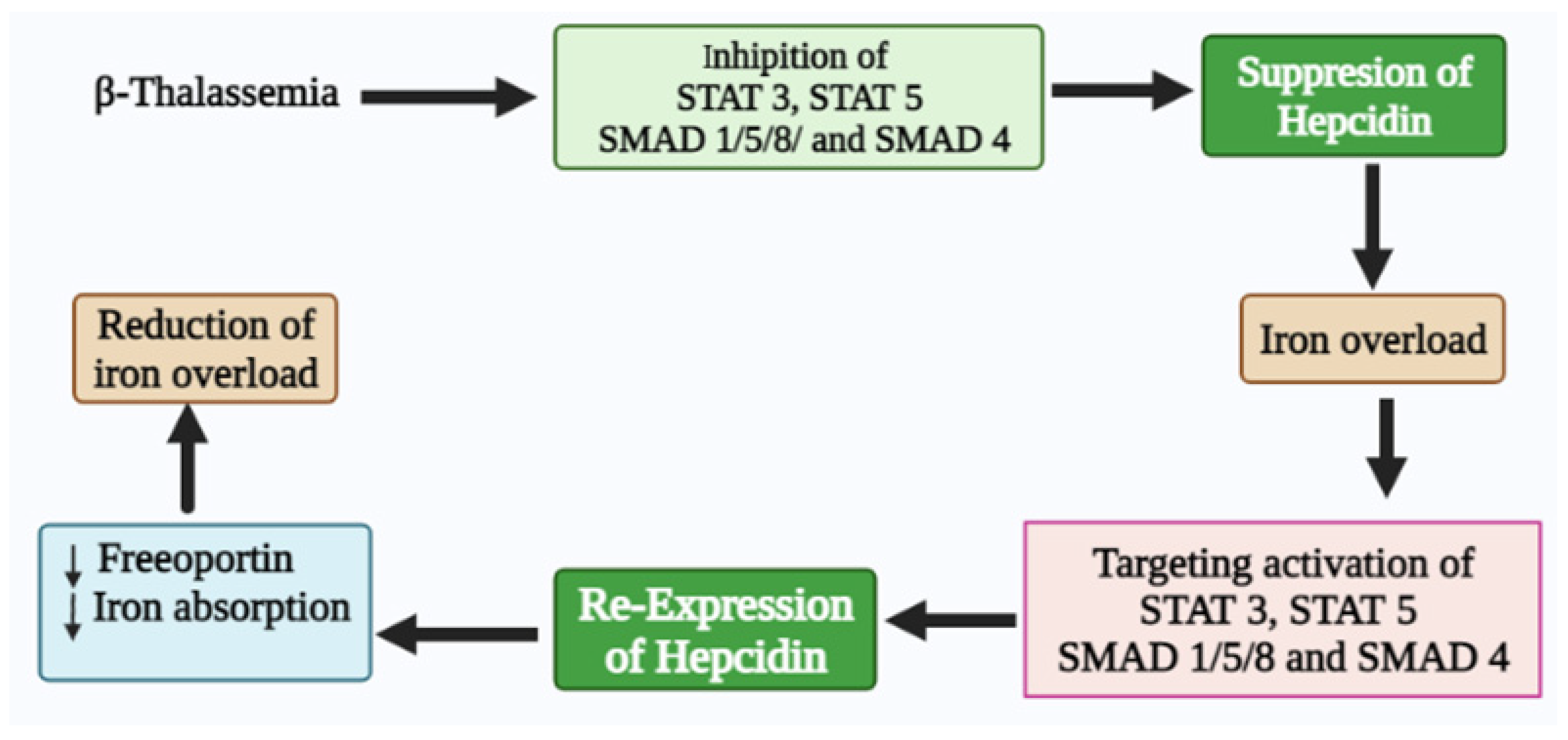
Iron homeostasis is regulated by hepcidin, a hepatic hormone that controls dietary iron absorption and plasma iron concentration. Hepcidin binds to the only known iron export protein, ferroportin (FPN), which regulates its expression. The major factors that implicate hepcidin regulation include iron stores, hypoxia, inflammation, and erythropoiesis. When erythropoietic activity is suppressed, hepcidin expression is hampered, leading to deficiency, thus causing an iron overload in iron-loading anemia, such as β-thalassemia. Iron overload is the principal cause of mortality and morbidity in β-thalassemia patients with or without blood transfusion dependence. In the case of thalassemia major, the primary cause of iron overload is blood transfusion. In contrast, iron overload is attributed to hepcidin deficiency and hyperabsorption of dietary iron in non-transfusion thalassemia. Beta-thalassemia patients showed marked hepcidin suppression, anemia, iron overload, and ineffective erythropoiesis (IE). Recent molecular research has prompted the discovery of new diagnostic markers and therapeutic targets for several diseases, including β-thalassemia.

hepcidin HbE/β-thalassemia iron overload ferroportin iron homeostasis signaling pathways

1. Thalassemia Syndrome

Thalassemia is an inherited autosomal recessive blood disorder that can be divided into either alpha (α) or beta (β) depending on the affected α or β globin chain [1]. The adult hemoglobin (HbA) consists of two α and two β (α2β2) chains in each HbA molecule [2][3]. Alpha-thalassemia occurs if one or more of the four alleles that code for α globin is missing or damaged [4]. On the other hand, β-thalassemia is caused by mutation in the β globin gene leading to a reduction in β globin or production of abnormal hemoglobin.

2. Beta-Thalassemia

Notably, β-thalassemia is highly reported in the Mediterranean and Southeast Asian countries as one of the most common genetic disorders [5]. Nevertheless, a lack of information on knowledge, attitudes, and practices towards β-thalassemia poses a challenge in many countries, including Malaysia [5]. This disease can be categorized into β-thalassemia major, intermedia, or minor [6]. β-thalassemia major and intermedia are genetically homozygous or heterozygous (β0 and β+), whereas β-thalassemia minor is usually heterozygous [6].
The β globin chain is encoded by the β globin gene (HBB), located on chromosome 11 at the short arm position 15.4 [7][8]. Individuals with β-thalassemia major and intermedia inherit the mutation in both copies of the HBB gene, affecting normal β globin chain production [7]. The clinical features of β-thalassemia major are revealed as early as the first two years of the patient’s life and are usually connected to transfusional iron overload, whereas the clinical presentation for β-thalassemia intermedia occurs later in life [2]. In contrast, β-thalassemia minor or trait has one mutation in the HBB gene and is described as a carrier [6]. They are usually symptomless, with a hypochromic microcytic blood picture and mild anemia, and can potentially increase in severity with malnutrition [6].
Beta-thalassemia can be present alongside other diseases associated with an abnormal β globin chain, such as the hemoglobin E (HbE) disease, exhibiting severe anemia [2]. HbE is a hemoglobin (Hb) variant caused by a single base substitution of glutamic acid to lysine at position 26 of the globin chain, commonly found in Southeast Asia [9]. It can be classified into three types: heterozygous, homozygous, or compound heterozygous [9]. When the HbE trait is coinherited with β-thalassemia, it is called compound heterozygous, a condition known as HbE/β-thalassemia, which resembles homozygous β0-thalassemia clinically and hematologically [10].
The phenotypic heterogeneity of HbE/β-thalassemia can range from mild asymptomatic anemia to a severe form that requires regular blood transfusion [11]. A study in Sri Lanka revealed that the HbE/β-thalassemia phenotype is unstable during the first 10 years of life but gradually stabilizes as the patient gets older [11]. This condition is caused by various changes in anemia and erythroid expansion progression during their early life [11]. However, the lack of knowledge on the classification of the disease severity will affect the understanding of HbE/β-thalassemia clinical progression with age [11].

2. Hepcidin Expression in β-Thalassemia

Hepcidin expression in thalassemia was first reported in a mouse model of severe anemia (C57BI/6 Hbbth3/+) [12]. Furthermore, a decline in serum hepcidin levels has been reported in HbE/β-thalassemia patients, β-thalassemia trait, and HbE trait carriers [13]. The decreased serum hepcidin levels in β-thalassemia patients are associated with the downregulation of hepcidin expression in liver cells, resulting in continuous absorption of dietary iron that leads to iron overload [14]. In individuals with thalassemia major and intermedia, liver hepcidin mRNA expression is inversely associated with soluble transferrin receptor (sTfR) and erythropoietin (EPO), but not with iron storage [15]. Suppression of hepcidin in HbE/β-thalassemia patients is linked to increased iron loading, saturated iron-binding proteins, and organ damage [13]. Moreover, hepcidin suppression with enhanced iron absorption was found in the β-thalassemia trait [16].

2.1. Hepcidin Regulation in β-Thalassemia

Hepcidin is suppressed in β-thalassemia patients with increasing iron absorption in response to the iron demand by erythroblasts due to tissue hypoxia erythropoietin (EPO) production and anemia [17]. During the differentiation process, several hepcidin inhibitors are released from erythroblasts to regulate hepcidin expression in β-thalassemia. Growth differentiation factor 15 (GDF15) serum level is inversely correlated with hepcidin expression in hepatocytes of thalassemia patients [18]. Meanwhile, twisted-gastrulation 1 (TWSG1) was upregulated in the bone marrow, spleen, and liver of mice with β-thalassemia major and intermedia, associated with hepcidin suppression and absence of bone morphogenetic protein (BMP). Additionally, human hepatocytes’ TWSG1 indirectly suppressed hepcidin expression through inhibition of BMP-mediated signaling [19].
Erythroferrone (ERFE) hormone functions as a negative regulator of hepcidin synthesis. Elevated ERFE expression is associated with increased erythropoietin and hepcidin suppression in mice models with thalassemia intermediate during stress erythropoiesis. The ERFE-deficient mice failed to suppress hepcidin after hemorrhage and erythropoietin administration [20]. Therefore, increased iron absorption in β-thalassemia is most likely attributed to increased ERFE expression and other hypoxia-related molecules that suppress hepcidin synthesis or increase ferroportin (FPN) expression [21][22].

2.2. Regulatory Effect of Hepcidin Transcription

Hepcidin is regulated by various stimuli, such as inflammation, plasma iron, anemia, and hypoxia. Its expression is inversely correlated with serum ferritin and induced by iron loading and inflammation. Hepcidin dysregulation is the underlying cause of several iron disorders. Erythropoietic activity is the main regulator of hepcidin transcription by stimulating erythropoiesis and increasing iron absorption via hepcidin downregulation. Chromatin immunoprecipitation analysis showed that the binding of CCAT enhancer binding protein (C/EBPa) to the hepcidin promoter was reduced after EPO supplementation. This indicates C/EBPa effects on hepcidin transcription in response to erythropoiesis stimulation [23]. Apart from that, erythropoietin levels increased under hypoxic conditions, involving hypoxia-inducible factor (HIF) in hepcidin regulation [14][19]. Furthermore, higher erythropoiesis activity and GDF15 are responsible for low hepcidin levels instead of high EPO levels [24].
GDF15, TWSG1, and ERFE have been reported as suppressors of hepcidin in β-thalassemia and other iron-containing anemia [14]. GDF15 was initially thought to be a macrophage inhibitory cytokine but it was later proven that its increase indirectly contributes to iron overload in cancer patients and those with sideropenic anemia by downregulating hepcidin expression and increasing iron absorption [25][26]. The tumor suppressor p53 drives GDF15, and its expression in the human body increases under stressful conditions, such as hypoxia, cancer, and tissue damage [27][28]. In addition, pregnancy is associated with low serum hepcidin levels in animal models and humans [29], which positively correlates with GDF15. In contrast, hepcidin is negatively correlated with EPO and hemojuvelin (HJV) during pregnancy [30]. Mutant TFR2 and HJV were associated with hepcidin suppression after hemorrhage and high levels of ERFE mRNA in the th3/+ β-thalassemia mouse model. The significance of ERFE needs to be further evaluated in different conditions of IE and iron loading anemia [31][32].
TWSG1 is higher in immature red cell precursors and mice with β-thalassemia. This erythrokine inhibits hepcidin transcription by inhibiting the BMP 2/4 pathway of SMAD (structurally similar to the small mothers against decapentaplegic in Drosophila) 1/5/8 phosphorylation [19]. Atonal basic helix–loop–helix (bHLH) transcription factor 8 (ATOH8) has been identified as a candidate for activation of liver hepcidin transcription [33]. Hypoxia, hemolysis, hypotransferrinemia, and erythropoietin treatment enhanced erythropoiesis activity and decreased ATOH8 levels in mice. However, erythropoiesis inhibitors increased ATOH8 levels, suggesting the interference between erythropoiesis and hepcidin regulation [33].
Inflammatory cytokines mainly induce hepcidin transcription by activating the STAT3 signaling pathway [34]. The BMP–SMAD signaling pathway also plays an essential role in regulating hepcidin transcription. Binding of BMPs (BMP2,4,5,6) to type I or type II serine or threonine kinase receptors leads to intracellular R-SMADS (SMAD1, 5 and 8) phosphorylation, which, in turn, binds to SMAD4 (Co-SMAD) to promote its nucleus translocation, thus activating the hepcidin transcription. Furthermore, iron management in the body activates BMP/SMAD and hepcidin signaling [35]. Andriopoulos et al. reported that BMP6 physically interacts with HJV and induces hepcidin to lower serum iron in mice [36]. HFE is also involved in hepcidin pathway regulation [37][38]. Mutations in HFE genes involved in the regulation of iron homeostasis cause type I hereditary hemochromatosis (HH) [39]. Additionally, an HFE-deficient mouse develops an iron overload phenotype similar to type I HH in humans [38]. These findings suggest that HFE positively modulates hepcidin expression [40]. Besides, HFE interacts with transferrin receptor 1 (TfR1) and contends with the receptor’s transferrin (Tf) binding site [41], resulting in the activation of downstream signaling pathways, such as the mitogen-activated protein kinase (MAPK) pathway [35]. Moreover, the crosstalk between the activated MAPK pathway and the BMP/SMAD pathway enhances hepcidin expression [42].

2.3. Hepcidin Therapeutics in β-Thalassemia

The current treatment of iron overload in β-thalassemia patients includes the administration of iron chelators, such as deferiprone, deferasirox, and desferrioxamine [43][44]. Chelation therapy is recommended in patients with serum ferritin greater than 1000 ng/mL [45]. The direct scavenging of LPI and NTBI by chelators helps prevent adverse sequelae of iron overload [46]. On the other hand, splenectomy has been recommended when the transfusion requirement increases and worsens anemia [47]. Besides, the allogenic hematopoietic stem cell transplantation can also be a therapeutic option for hereditary β-thalassemia, but 60% of patients lack suitable donors, thus increasing the risk of developing transplant-related complications [48].
The correlation between iron overload and hepcidin has led to new approaches that target the disease pathophysiology, aiming to reduce iron overload and IE [49]. A previous study on β-thalassemia mice indicated that a rise in hepcidin level lowers iron bioavailable to erythroblasts, resulting in decreased heme synthesis and improved erythroid precursor and reticulocyte survival [50]. Furthermore, decreasing hepcidin levels in thalassemia leads to iron overload and restores hepcidin to normal and, hence, is a novel therapeutic approach for thalassemia patients [13]. The ligand of the BMP6 receptor is involved in hepcidin regulation and transcription [51]. Meanwhile, transferrin is a limiting factor and restricts iron availability for erythropoiesis [52]. TMPRSS6 is a negative regulator of hepcidin, and its depletion using small interfering ribonucleic acid siRNA increased hepcidin mRNA and improved erythropoiesis in a β-thalassemia mouse model [53]. Furthermore, the SiRNA therapy decreases TMPRSS6 expression, thus increasing hepcidin expression and improving the incidence of disease-related thalassemia [54]. Moreover, it is reported that the combined administration of iron chelator deferiprone for RNAi targeting TMPRSS6 can significantly reduce iron content in the liver and increase the efficiency of erythropoiesis in β-thalassemia mice [46][55].

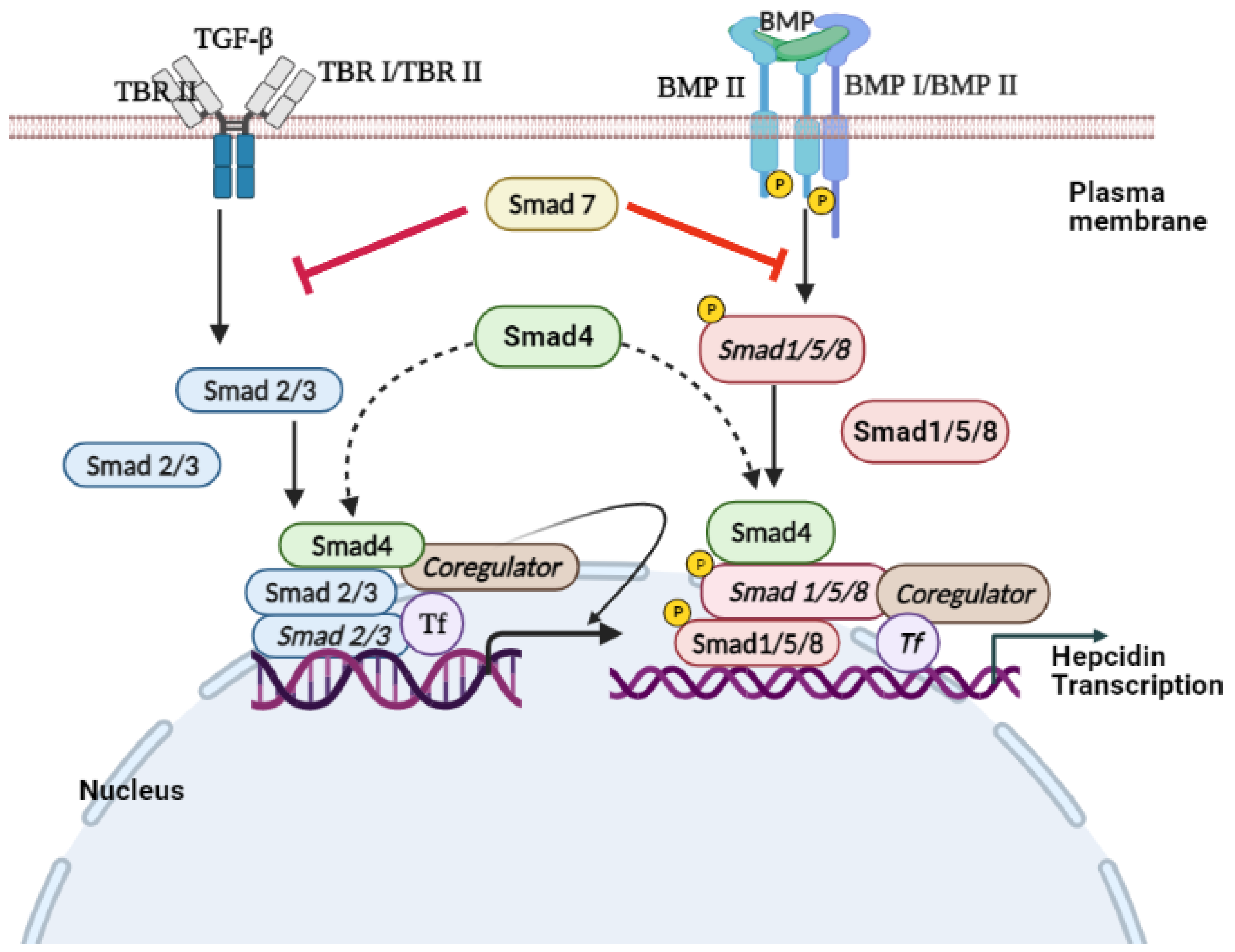
3. TGF-β/SMAD Signaling

SMADs are proteins that are activated by the transforming growth factor β (TGF-β), BMP signaling, to mediate cell proliferation and differentiation [56][57]. Endosome-associated Fab1 (yeast orthologue of PIKfyve, YOTB, vesicle transport protein (Vac1), and EEA1 (FYVE zinc finger domain)-domain protein (endofin)) influences hepcidin expression by regulating SMAD1/5/8 phosphorylation [58]. STAT and SMAD signaling regulate hepcidin expression [59]. Ablation of SMAD4, specifically in the liver, triggers an iron overload in multiple organs due to decreased levels of liver hepcidin [60]. However, SMAD7 acts as an effective inhibitor of hepcidin mRNA expression through a negative regulation effect on TGF-β and BMP/SMAD signaling [61][62]. TGF-β is the prototypical ligand of the TGF-β superfamily, which signals during activation of serine/threonine receptor kinases. This superfamily is subdivided into the TGFβ/activin branch and BMP/growth and differentiation factor (GDF) branch.
TGF-β is expressed in most cell types and translated into a proprotein that is proteolytically cleaved into a noncovalently linked mature TGF-β and latency-associated protein (LAP) [63][64]. The active TGF-β ligand is a 25 kDa dimer, covalently linked by bisulfide bonds between cysteine residues of each monomeric peptide [63][64].
Various mechanisms are used to regulate the bioavailability of TGF-β in vivo. Once the bioavailable TGF-β reaches the target cell’s surface, it bonds with the homodimer of TGF-β type II receptor (TβRII) [65]. The TGF-β–TβRII complex provides a structural interface that forms a stable complex with the homodimer of the TGF-β type I receptor (TβRI) [56]. Subsequently, the active receptor–ligand complex is a heterotetrametric complex composed of TGFβ dimer and homodimer of TβRII and TβRI. In the active receptor complex, TβRII is constitutively activated and stimulates the transphosphorylation of TβRI [56][66]. In the TGF-β pathway, SMAD2 and SMAD3 are receptor-regulated effector proteins (R-SMADs) phosphorylated by activated TβRI on the C-terminal SSXS motif, leading to the nuclear accumulation of R-SMAD [56].
The activated receptor complex bound to the ligand is internalized by endocytosis [67]. Internalization of cell surface receptors can occur through clathrin-mediated or caveolae-mediated endocytosis [68].
Upon ligand stimulation, the SMADs accumulate in the nucleus as R-SMAD/CO-SMAD complex, leading to a decrease in their nuclear export rate [69][70]. The SMAD complex binds to DNA with other transcription factors and interacts with the general transcription machinery to regulate the expression of target genes (Figure 1) [71].
Biomedicines 10 00189 g005
Figure 1. Signal transduction of TGF-β and BMP. The binding of TGF-β to the TβRII dimer allows the ligand to bind to the TβRI dimer and stimulate TβRI kinase activity. In SMAD-mediated TGF-β signal transduction, TβRI phosphorylates cytoplasmic SMAD2 and SMAD3, which interact with SMAD4 after dissociating from TβRI. The two receptors activate the trimeric complex of SMAD2 and SMAD3, and a SMAD4 then enters the nucleus, where it interacts with the DNA-binding transcription factor (TF) and coregulators of the target gene. Similarly, the BMP signals run parallel to the TGF-β signals. In response to the binding of the BMP ligand to the BMPRII heteromeric receptor complex and BMPRI transmembrane kinase, receptor-activated SMAD1 and SMAD5 bind to SMAD4 and are transported to the nucleus to activate or inhibit transcription of hepcidin. ➔: Activation and ⊥: Inhibit.
Signaling pathways such as signal transducers and activators of transcription (STAT) and SMAD regulate the expression of hepcidin. Therefore, it is hypothesized that STAT and SMAD could be dephosphorylated in thalassemia cases, including HbE/β-thalassemia patients, resulting in hepcidin downregulation with the presence of iron accumulation.

4. Conclusions

In summary, iron homeostasis dysregulation has a dominant physiological effect on transfusion-dependent and transfusion-independent β-thalassemia patients. Thus, understanding the expression of hepcidin and its regulation in β-thalassemia patients is vital in developing rational therapeutic interventions to provide safe, effective, and lifelong treatment options for their management. Therefore, recovery of hepcidin in β-thalassemia patients through the activation of STAT 3, STAT 5, SMAD 1/5/8, and SMAD 4 signaling could be a potential therapeutic target for managing iron overload (Figure 2). Therefore, it is highly recommended for future preclinical and clinical studies to evaluate the related risks and benefits of hepcidin-targeted treatment approaches.
Biomedicines 10 00189 g006
Figure 2. Regulation of STAT and SMAD signaling pathway on hepcidin expression.

References

  1. Ayyash, H.; Sirdah, M. Hematological and biochemical evaluation of β-thalassemia major (βTM) patients in Gaza Strip: A cross-sectional study. IJHS 2018, 12, 18.
  2. Fibach, E.; Rachmilewitz, E.A. Pathophysiology and treatment of patients with beta-thalassemia—An update. F1000Research 2017, 6, 1–12.
  3. Aziz, N.; Taib, W.; Kharolazaman, N.; Ismail, I.; Al-Jamal, H.; Jamil, W.; Esa, E.; Ibrahim, H. Evidence of New Intragenic HBB Haplotypes Model for the Prediction of Beta-Thalassemia in the Malaysian Population. Res. Sq. 2021; preprint.
  4. Bender, M.; Yusuf, C.; Davis, T.; Dorley, M.C.; del Pilar Aguinaga, M.; Ingram, A.; Chan, M.S.; Ubaike, J.C.; Hassell, K.; Ojodu, J. Newborn Screening Practices and Alpha-Thalassemia Detection—United States, 2016. Morb. Mortal. Wkly. Rep. 2020, 69, 1269.
  5. Wong, L.P.; George, E.; Tan, J.-A.M.A. Public perceptions and attitudes toward thalassaemia: Influencing factors in a multi-racial population. BMC Public Health 2011, 11, 193.
  6. Galanello, R.; Origa, R. Beta-thalassemia. Orphanet J. Rare Dis. 2010, 5, 11.
  7. Thein, S.L. Molecular basis of β thalassemia and potential therapeutic targets. Blood Cell Mol. Dis. 2018, 70, 54–65.
  8. Abdullah, U.Y.; Ibrahim, H.M.; Mahmud, N.B.; Salleh, M.Z.; Teh, L.K.; Noorizhab, M.N.F.b.; Zilfalil, B.A.; Jassim, H.M.; Wilairat, P.; Fucharoen, S. Genotype-Phenotype Correlation of β-Thalassemia in Malaysian Population: Toward Effective Genetic Counseling. Hemoglobin 2020, 44, 184–189.
  9. Pani, K.; Sharma, S.; Murari, M.; Yadav, M.; Phadke, S.; Agarwal, S. Clinico-hematological Profile of Hb E-β Thalassemia-Prospective Analysis in a tertiary Care Centre. J. Assoc. Phys. India 2018, 66, 42–45.
  10. Fucharoen, S.; Weatherall, D.J. The hemoglobin E thalassemias. Cold Spring Harb. Perspect. Med 2012, 2, a011734.
  11. Olivieri, N.F.; Pakbaz, Z.; Vichinsky, E. Hb E/beta-thalassaemia: A common & clinically diverse disorder. Indian J. Med. Res. 2011, 134, 522.
  12. Adamsky, K.; Weizer, O.; Amariglio, N.; Breda, L.; Harmelin, A.; Rivella, S.; Rachmilewitz, E.; Rechavi, G. Decreased hepcidin mRNA expression in thalassemic mice. Br. J. Hematol. 2004, 124, 123–124.
  13. Saad, H.K.M.; Taib, W.R.W.; Ismail, I.; Johan, M.F.; Al-Wajeeh, A.S.; Al-jamal, H.A.N. Reduced hepcidin expression enhances iron overload in patients with HbE/β-thalassemia: A comparative cross-sectional study. Exp. Ther. Med. 2021, 22, 1–8.
  14. Tanno, T.; Bhanu, N.V.; Oneal, P.A.; Goh, S.-H.; Staker, P.; Lee, Y.T.; Moroney, J.W.; Reed, C.H.; Luban, N.L.; Wang, R.-H. High levels of GDF15 in thalassemia suppress expression of the iron regulatory protein hepcidin. Nat. Med. 2007, 13, 1096–1101.
  15. Kattamis, A.; Papassotiriou, I.; Palaiologou, D.; Apostolakou, F.; Galani, A.; Ladis, V.; Sakellaropoulos, N.; Papanikolaou, G. The effects of erythropoetic activity and iron burden on hepcidin expression in patients with thalassemia major. Haematologica 2006, 91, 809–812.
  16. Jones, E.; Pasricha, S.-R.; Allen, A.; Evans, P.; Fisher, C.A.; Wray, K.; Premawardhena, A.; Bandara, D.; Perera, A.; Webster, C. Hepcidin is suppressed by erythropoiesis in hemoglobin E β-thalassemia and β-thalassemia trait. Blood J. Am. Soc. Hematol. 2015, 125, 873–880.
  17. Pak, M.; Lopez, M.A.; Gabayan, V.; Ganz, T.; Rivera, S. Suppression of hepcidin during anemia requires erythropoietic activity. Blood 2006, 108, 3730–3735.
  18. Weizer-Stern, O.; Adamsky, K.; Amariglio, N.; Levin, C.; Koren, A.; Breuer, W.; Rachmilewitz, E.; Breda, L.; Rivella, S.; Ioav Cabantchik, Z. Downregulation of hepcidin and haemojuvelin expression in the hepatocyte cell-line HepG2 induced by thalassaemic sera. Br. J. Hematol. 2006, 135, 129–138.
  19. Tanno, T.; Porayette, P.; Sripichai, O.; Noh, S.-J.; Byrnes, C.; Bhupatiraju, A.; Lee, Y.T.; Goodnough, J.B.; Harandi, O.; Ganz, T. Identification of TWSG1 as a second novel erythroid regulator of hepcidin expression in murine and human cells. Blood J. Am. Soc. Hematol. 2009, 114, 181–186.
  20. Kautz, L.; Jung, G.; Du, X.; Gabayan, V.; Chapman, J.; Nasoff, M.; Nemeth, E.; Ganz, T. Erythroferrone contributes to hepcidin suppression and iron overload in a mouse model of β-thalassemia. Blood J. Am. Soc. Hematol. 2015, 126, 2031–2037.
  21. Gardenghi, S.; Marongiu, M.F.; Ramos, P.; Guy, E.; Breda, L.; Chadburn, A.; Liu, Y.; Amariglio, N.; Rechavi, G.; Rachmilewitz, E.A. Ineffective erythropoiesis in β-thalassemia is characterized by increased iron absorption mediated by down-regulation of hepcidin and up-regulation of ferroportin. Blood J. Am. Soc. Hematol. 2007, 109, 5027–5035.
  22. Muckenthaler, M.U.; Rivella, S.; Hentze, M.W.; Galy, B. A red carpet for iron metabolism. Cell J. 2017, 168, 344–361.
  23. Pinto, J.P.; Ribeiro, S.; Pontes, H.; Thowfeequ, S.; Tosh, D.; Carvalho, F.; Porto, G. Erythropoietin mediates hepcidin expression in hepatocytes through EPOR signaling and regulation of C/EBPα. Blood J. Am. Soc. Hematol. 2008, 111, 5727–5733.
  24. Liu, Q.; Davidoff, O.; Niss, K.; Haase, V.H. Hypoxia-inducible factor regulates hepcidin via erythropoietin-induced erythropoiesis. J. Clin. Investig. 2012, 122, 4635–4644.
  25. Jiang, F.; Yu, W.-J.; Wang, X.-H.; Tang, Y.-T.; Guo, L.; Jiao, X.-Y. Regulation of hepcidin through GDF-15 in cancer-related anemia. Clin. Chim. Acta 2014, 428, 14–19.
  26. YAŞAR, C.A.; Tarkun, P.; Ateşoğlu, E.B.; Eraldemir, F.C.; Özsoy, Ö.D.; Demirsoy, E.T.; Mehtap, Ö.; Gedük, A.; Hacihanifioğlu, A. The role of hepcidin, GDF15, and mitoferrin-1 in iron metabolism of polycythemia vera and essential thrombocytosis patients. Turk. J. Med. Sci. 2019, 49, 74–80.
  27. Osada, M.; Park, H.L.; Park, M.J.; Liu, J.-W.; Wu, G.; Trink, B.; Sidransky, D. A p53-type response element in the GDF15 promoter confers high specificity for p53 activation. Biochem. Biophys. Res. Commun. 2007, 354, 913–918.
  28. Zimmers, T.A.; Jin, X.; Hsiao, E.C.; McGrath, S.A.; Esquela, A.F.; Koniaris, L.G. Growth differentiation factor-15/macrophage inhibitory cytokine-1 induction after kidney and lung injury. Shock 2005, 23, 543–548.
  29. Jenkitkasemwong, S.; Broderius, M.; Nam, H.; Prohaska, J.R.; Knutson, M.D. Anemic copper-deficient rats, but not mice, display low hepcidin expression and high ferroportin levels. J. Nutr. 2010, 140, 723–730.
  30. Finkenstedt, A.; Widschwendter, A.; Brasse-Lagnel, C.; Theurl, I.; Hubalek, M.; Dieplinger, H.; Tselepis, C.; Ward, D.; Vogel, W.; Zoller, H. Hepcidin is correlated to soluble hemojuvelin but not to increased GDF15 during pregnancy. Blood Cell Mol. Dis. 2012, 48, 233–237.
  31. Vokurka, M.; Krijt, J.; Šulc, K.; Nečas, E. Hepcidin mRNA levels in mouse liver respond to inhibition of erythropoiesis. Physiol. Res. 2006, 55, 667–674.
  32. Kautz, L.; Jung, G.; Valore, E.V.; Rivella, S.; Nemeth, E.; Ganz, T. Identification of erythroferrone as an erythroid regulator of iron metabolism. Nat. Genet. 2014, 46, 678–684.
  33. Patel, N.; Varghese, J.; Masaratana, P.; Latunde-Dada, G.O.; Jacob, M.; Simpson, R.J.; McKie, A.T. The transcription factor ATOH 8 is regulated by erythropoietic activity and regulates HAMP transcription and cellular pSMAD 1, 5, 8 levels. Br. J. Hematol. 2014, 164, 586–596.
  34. Wrighting, D.M.; Andrews, N.C. Interleukin-6 induces hepcidin expression through STAT3. Blood 2006, 108, 3204–3209.
  35. Bayele, H.K.; Srai, S.K.S. Genetic variation in hepcidin expression and its implications for phenotypic differences in iron metabolism. Haematologica 2009, 94, 1185.
  36. Andriopoulos Jr, B.; Corradini, E.; Xia, Y.; Faasse, S.A.; Chen, S.; Grgurevic, L.; Knutson, M.D.; Pietrangelo, A.; Vukicevic, S.; Lin, H.Y. BMP6 is a key endogenous regulator of hepcidin expression and iron metabolism. Nat. Genet. 2009, 41, 482–487.
  37. Bridle, K.R.; Frazer, D.M.; Wilkins, S.J.; Dixon, J.L.; Purdie, D.M.; Crawford, D.H.; Subramaniam, V.N.; Powell, L.W.; Anderson, G.J.; Ramm, G.A. Disrupted hepcidin regulation in HFE-associated haemochromatosis and the liver as a regulator of body iron homoeostasis. Lancet 2003, 361, 669–673.
  38. Zhou, X.Y.; Tomatsu, S.; Fleming, R.E.; Parkkila, S.; Waheed, A.; Jiang, J.; Fei, Y.; Brunt, E.M.; Ruddy, D.A.; Prass, C.E. HFE gene knockout produces mouse model of hereditary hemochromatosis. Proc. Natl. Acad. Sci. USA 1998, 95, 2492–2497.
  39. Feder, J.; Gnirke, A.; Thomas, W.; Tsuchihashi, Z.; Ruddy, D.; Basava, A.; Dormishian, F.; Domingo, R.; Ellis, M.; Fullan, A. A novel MHC class I–like gene is mutated in patients with hereditary haemochromatosis. Nat. Genet. 1996, 13, 399–408.
  40. Schmidt, P.J.; Toran, P.T.; Giannetti, A.M.; Bjorkman, P.J.; Andrews, N.C. The transferrin receptor modulates Hfe-dependent regulation of hepcidin expression. Cell Metab. 2008, 7, 205–214.
  41. Lebrón, J.A.; Bennett, M.J.; Vaughn, D.E.; Chirino, A.J.; Snow, P.M.; Mintier, G.A.; Feder, J.N.; Bjorkman, P.J. Crystal structure of the hemochromatosis protein HFE and characterization of its interaction with transferrin receptor. Cell J. 1998, 93, 111–123.
  42. Ramey, G.; Deschemin, J.-C.; Vaulont, S. Cross-talk between the mitogen activated protein kinase and bone morphogenetic protein/hemojuvelin pathways is required for the induction of hepcidin by holotransferrin in primary mouse hepatocytes. Haematologica 2009, 94, 765–772.
  43. Poggi, M.; Sorrentino, F.; Pugliese, P.; Smacchia, M.P.; Daniele, C.; Equitani, F.; Terlizzi, F.; Guitarrini, M.R.; Monti, S.; Maffei, L. Longitudinal changes of endocrine and bone disease in adults with β-thalassemia major receiving different iron chelators over 5 years. Ann. Hematol. 2016, 95, 757–763.
  44. Farmaki, K.; Tzoumari, I.; Pappa, C. Oral chelators in transfusion-dependent thalassemia major patients may prevent or reverse iron overload complications. Blood Cell Mol. Dis. 2011, 47, 33–40.
  45. Poggiali, E.; Cassinerio, E.; Zanaboni, L.; Cappellini, M.D. An update on iron chelation therapy. Blood Transfus. 2012, 10, 411.
  46. Schmidt, P.J.; Racie, T.; Westerman, M.; Fitzgerald, K.; Butler, J.S.; Fleming, M.D. Combination therapy with a T mprss6 RNA i-therapeutic and the oral iron chelator deferiprone additively diminishes secondary iron overload in a mouse model of β-thalassemia intermedia. Am. J. Hematol. 2015, 90, 310–313.
  47. Sripichai, O.; Munkongdee, T.; Kumkhaek, C.; Svasti, S.; Winichagoon, P.; Fucharoen, S. Coinheritance of the different copy numbers of α-globin gene modifies severity of β-thalassemia/Hb E disease. Ann. Hematol. 2008, 87, 375–379.
  48. Lucarelli, G.; Isgrò, A.; Sodani, P.; Gaziev, J. Hematopoietic stem cell transplantation in thalassemia and sickle cell anemia. Cold Spring Harb. Perspect. Med 2012, 2, a011825.
  49. Camaschella, C.; Nai, A. Ineffective erythropoiesis and regulation of iron status in iron loading anaemias. Br. J. Hematol. 2016, 172, 512–523.
  50. Gardenghi, S.; Ramos, P.; Roy, C.N.; Andrews, N.C.; Nemeth, E.; An, X.; Narla, M.; Ginzburg, Y.; Rachmilewitz, E.A.; Giardina, P. Hepcidin as a Therapeutic Tool to Limit Iron Overload and Improve Anemia In β-Thalassemia. Blood 2010, 116, 1009.
  51. Wu, X.; Yung, L.-M.; Cheng, W.-H.; Yu, P.B.; Babitt, J.L.; Lin, H.Y.; Xia, Y. Hepcidin regulation by BMP signaling in macrophages is lipopolysaccharide dependent. PLoS ONE 2012, 7, e44622.
  52. Nai, A.; Lidonnici, M.R.; Rausa, M.; Mandelli, G.; Pagani, A.; Silvestri, L.; Ferrari, G.; Camaschella, C. The second transferrin receptor regulates red blood cell production in mice. Blood J. Am. Soc. Hematol. 2015, 125, 1170–1179.
  53. Finberg, K.E.; Heeney, M.M.; Campagna, D.R.; Aydınok, Y.; Pearson, H.A.; Hartman, K.R.; Mayo, M.M.; Samuel, S.M.; Strouse, J.J.; Markianos, K. Mutations in TMPRSS6 cause iron-refractory iron deficiency anemia (IRIDA). Nat. Genet. 2008, 40, 569–571.
  54. Bou-Fakhredin, R.; Bazarbachi, A.-H.; Chaya, B.; Sleiman, J.; Cappellini, M.D.; Taher, A.T. Iron overload and chelation therapy in non-transfusion dependent thalassemia. Int. J. Mol. Sci. 2017, 18, 2778.
  55. Casu, C.; Aghajan, M.; Oikonomidou, P.R.; Guo, S.; Monia, B.P.; Rivella, S. Combination of Tmprss6-ASO and the iron chelator deferiprone improves erythropoiesis and reduces iron overload in a mouse model of beta-thalassemia intermedia. Haematologica 2016, 101, e8.
  56. Shi, Y.; Massagué, J. Mechanisms of TGF-β signaling from cell membrane to the nucleus. Cell J. 2003, 113, 685–700.
  57. Massagué, J. TGF-β signaling in development and disease. FEBS Lett. 2012, 586, 1833.
  58. Goh, J.B.; Wallace, D.F.; Hong, W.; Subramaniam, V.N. Endofin, a novel BMP-SMAD regulator of the iron-regulatory hormone, hepcidin. Sci. Rep. 2015, 5, 13986.
  59. Wang, R.-H.; Li, C.; Xu, X.; Zheng, Y.; Xiao, C.; Zerfas, P.; Cooperman, S.; Eckhaus, M.; Rouault, T.; Mishra, L. A role of SMAD4 in iron metabolism through the positive regulation of hepcidin expression. Cell Metab. 2005, 2, 399–409.
  60. Chen, J.; Chloupkova, M. Abnormal iron uptake and liver cancer. Cancer Biol. Ther. 2009, 8, 1699–1708.
  61. Mleczko-Sanecka, K.; Casanovas, G.; Ragab, A.; Breitkopf, K.; Müller, A.; Boutros, M.; Dooley, S.; Hentze, M.W.; Muckenthaler, M.U. SMAD7 controls iron metabolism as a potent inhibitor of hepcidin expression. Blood J. Am. Soc. Hematol. 2010, 115, 2657–2665.
  62. Spasić, M.V.; Sparla, R.; Mleczko-Sanecka, K.; Migas, M.C.; Breitkopf-Heinlein, K.; Dooley, S.; Vaulont, S.; Fleming, R.E.; Muckenthaler, M.U. Smad6 and Smad7 are co-regulated with hepcidin in mouse models of iron overload. Biochim. Biophys. Acta Mol. Basis Dis. 2013, 1832, 76–84.
  63. Annes, J.P.; Munger, J.S.; Rifkin, D.B. Making sense of latent TGFβ activation. J. Cell Sci. 2003, 116, 217–224.
  64. Blobe, G.C.; Schiemann, W.P.; Lodish, H.F. Role of transforming growth factor β in human disease. N. Engl. J. Med. 2000, 342, 1350–1358.
  65. Massague, J. TGF-β signal transduction. Annu. Rev. Biochem. 1998, 67, 753–791.
  66. Salma, U.; Xue, M.; Ali Sheikh, M.S.; Guan, X.; Xu, B.; Zhang, A.; Huang, L.; Xu, D. Role of transforming growth factor-β1 and smads signaling pathway in intrauterine adhesion. Mediat. Inflamm. 2016, 2016, 4158287.
  67. Chen, Y.-G. Endocytic regulation of TGF-β signaling. Cell Res 2009, 19, 58–70.
  68. Di Guglielmo, G.M.; Le Roy, C.; Goodfellow, A.F.; Wrana, J.L. Distinct endocytic pathways regulate TGF-β receptor signalling and turnover. Nat. Cell Biol. 2003, 5, 410–421.
  69. Clarke, D.C.; Betterton, M.; Liu, X. Systems theory of Smad signalling. IEEE Proc. Syst. Biol. 2006, 153, 412–424.
  70. Schmierer, B.; Hill, C.S. Kinetic analysis of Smad nucleocytoplasmic shuttling reveals a mechanism for transforming growth factor β-dependent nuclear accumulation of Smads. Mol.Cell. Biol. 2005, 25, 9845–9858.
  71. Massagué, J.; Seoane, J.; Wotton, D. Smad transcription factors. Genes Dev. 2005, 19, 2783–2810.
More
Information
Contributor MDPI registered users' name will be linked to their SciProfiles pages. To register with us, please refer to https://encyclopedia.pub/register :
View Times: 503
Revisions: 4 times (View History)
Update Date: 28 Jan 2022
1000/1000
Video Production Service