Submitted Successfully!
To reward your contribution, here is a gift for you: A free trial for our video production service.
Thank you for your contribution! You can also upload a video entry or images related to this topic.
Version Summary Created by Modification Content Size Created at Operation
1 + 1532 word(s) 1532 2022-01-20 07:04:09 |
2 format correct + 240 word(s) 1772 2022-01-20 10:32:04 |

Video Upload Options

Do you have a full video?

Confirm

Are you sure to Delete?
Cite
If you have any further questions, please contact Encyclopedia Editorial Office.
Giamas, G. Extracellular Vesicles and the Breast Cancer Microenvironment. Encyclopedia. Available online: https://encyclopedia.pub/entry/18550 (accessed on 06 July 2024).
Giamas G. Extracellular Vesicles and the Breast Cancer Microenvironment. Encyclopedia. Available at: https://encyclopedia.pub/entry/18550. Accessed July 06, 2024.
Giamas, Georgios. "Extracellular Vesicles and the Breast Cancer Microenvironment" Encyclopedia, https://encyclopedia.pub/entry/18550 (accessed July 06, 2024).
Giamas, G. (2022, January 20). Extracellular Vesicles and the Breast Cancer Microenvironment. In Encyclopedia. https://encyclopedia.pub/entry/18550
Giamas, Georgios. "Extracellular Vesicles and the Breast Cancer Microenvironment." Encyclopedia. Web. 20 January, 2022.
Extracellular Vesicles and the Breast Cancer Microenvironment
Edit

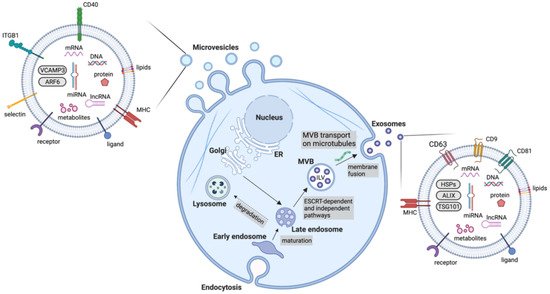
Extracellular vesicles are an important mediator of BC-TME signalling. EVs are non-replicative, lipid bilayer-delimited particles that are naturally released from cells. They have been identified in virtually every physiological fluid and are released by nearly all cell types. EV cargo consists of a number of bioactive molecules, including nucleic acids, lipids and membrane-bound and cytosolic proteins. The uptake of EVs is able to influence cell behaviour and as such, EVs are known to be important signalling particles, as well as diagnostic, predictive and prognostic biomarkers in diseases. Although EVs can be categorised in a number of ways (e.g., based on size, cargo and biological role), they are most often classified based on their biogenesis, with exosomes and microvesicles being the most commonly discussed EV subtypes. Many other subtypes of EVs have been identified, including apoptotic bodies and oncosomes, however, knowledge of their specific roles in cell–cell communication is limited. Due to the lack of consensus on biomarkers for specific subtypes of EVs, this review will use the collective term EV where the biogenesis pathway has not been demonstrated directly, in accordance with the guidelines set by the International Society for Extracellular Vesicles. 

breast cancer therapy resistance tumour microenvironment extracellular vesicles

1. Exosomes

Exosomes are the most studied EV subtype, with the term ‘exosome’ often used interchangeably with ‘extracellular vesicle’ despite the lack of consensus on exosome-specific biomarkers and unavailability of methods to selectively isolate exosomes from other subtypes of EVs [1]. Exosomes are specifically defined as EVs of endosomal origin, formed through the inward budding of the membrane of late endosomes/multivesicular bodies (MVBs) to form intraluminal vesicles (ILVs), followed by the fusion of these MVBs with the plasma membrane which then releases exosomes from the cell [2] (Figure 1). The endosomal sorting complexes required for the transport (ESCRT)-dependent pathway is the main well-characterised mechanism of exosome biogenesis. It consists of five complexes which work to form ILVs in MVBs [3]. Briefly, ESCRT-0, consisting of Hrs (hepatocyte growth factor-regulated tyrosine kinase substrate) and STAM1/2 (signal transducing adaptor molecule 1/2) binds phosphatidylinositol 3-phosphate at the endosomal membrane and ubiquitin on ubiquitinated proteins, enabling ubiquitin-dependent sorting into endosomes [4][5]. ESCRT-0 recruits ESCRT-I (composed of tumour susceptibility gene 101 (Tsg101), human multivesicular body 12 (hMvb12), and the vacuolar protein sorting (Vps) proteins, Vps28 and Vps37) [6]. ESCRT-I interacts with both ESCRT-0 and ESCRT-II. ESCRT-II consists of Vps22, Vps36 and two Vps25 subunits [7]. ESCRT-II has a high affinity for phosphatidylinositol 3-phosphate, enabling localisation to endosomes. ESCRT-III is composed of the charged multivesicular body proteins (CHMPs), CHMP2, CHMP3, CHMP4 and CHMP6 [8]. ESCRT-III monomers exist in an autoinhibited state in the cytoplasm and come together upon activation and recruitment by ESCRT-II [9]. Cargo is then deubiquitinated prior to loading into ILVs and ESCRT-III dissociates from the endosome membrane by the action of ATPase Vps4 and its co-factor, vacuolar protein sorting-associated protein VTA1 homolog (VTA1) which enables the recycling of the ESCRT machinery [3]. Through this process, the endosomal membrane undergoes inward budding before scission occurs and ILVs are released into the MVB [10]. RAB proteins then mediate the transport of the MVBs to the plasma membrane along microtubules where the MVBs then dock and SNARE (soluble N-ethylmaleimide-sensitive fusion protein attachment protein receptor) complexes mediate the fusion of MVBs with the plasma membrane, releasing the ILVs as exosomes [11][12].
Figure 1. Schematic representation of exosome and microvesicle biogenesis, release and cargo. While microvesicles bud directly from the plasma membrane, exosomes are generated within MVB subpopulations that, upon maturation, fuse with the plasma membrane. Alternative MVB pathways include fusion with lysosomes for degradation. (Abbreviations: ALIX, ALG-2 interacting protein X; ARF6, ADP ribosylation factor 6; ER, endoplasmic reticulum; ESCRT, endosomal sorting complexes required for transport; HSPs, heat shock proteins; ILV, intraluminal vesicles; ITGB1, integrin β1; lncRNA, long non-coding RNA; MHC, major histocompatibility complex; miRNA, microRNA; MVB, multivesicular body; TSG101, tumour susceptibility gene 101; VCAMP3, vesicle-associated membrane protein 3).
Additionally, exosomes can be formed in an ESCRT-independent manner. Sphingomyelinase-enriched microdomains containing ceramides within endosome membranes are thought to promote a negative curvature in the membrane, resulting in inward budding [13]. Tetraspanins also participate in cargo selection [14]. Furthermore, RAB31 has been recently shown to control an ESCRT-independent pathway where it works through flotillin proteins to selectively package cargo into ILVs and inhibits RAB7, preventing lysosomal degradation and enhancing the release of ILVs as exosomes [15].

2. Microvesicles

Microvesicles or ectosomes are formed at the plasma membrane by outward budding of the membrane followed by fission through the use of contractile machinery [16] (Figure 1). These are more heterogenous in size than exosomes. Although originally thought to be between 100 nm and 1000 nm, numerous studies have shown the existence of much smaller and larger vesicles sharing a similar biogenesis with microvesicles, for instance large oncosomes, which can reach around 10 μm in size. During microvesicle biogenesis, cargo is selected and trafficked to the membrane to be shed as microvesicles. Ras-related GTPase ADP-ribosylation factor 6 (ARF6)-positive recycling endosomes play an important role in directing cargo to the budding microvesicle. ARF6 has been found to promote the selective packaging of integrin β1, MHC-I (major histocompatibility complex class I) proteins and ARF6 itself into microvesicles [17]. The packaging of miRNA is less well-understood, however, recent evidence indicates that exportin-5 and hnRNPA2B1 play an important role. A number of other proteins participate in cargo delivery to the nascent microvesicle, including RAB22A, SNARE machinery, CD-9 and RNA-binding proteins [16]. ARF6 further promotes scission of the budding microvesicle. ARF6 promotes phospholipase D activation, recruiting ERK (extracellular signal-regulated kinase) to the membrane where it can phosphorylate myosin light chain kinase (MLCK) [18]. This then phosphorylates myosin light chain (MLC), promoting contraction around the budding microvesicle, enabling their release from the plasma membrane [17]. Moreover, RhoA works through the ROCK (rho associated coiled-coil containing protein kinase) signalling pathway to promote contractility, however, much of this pathway remains to be elucidated [19].

3. Extracellular Vesicle Separation

As with many biological components, the production of a pure EV fraction is currently unachievable. However, separation and enrichment of EVs from biological fluids can be carried out, enabling the use of EV-enriched fractions in research, diagnostics and disease monitoring [1]. The separation of EVs from conditioned cell culture media for in vitro studies and from biofluids such as plasma, urine and cerebrospinal fluid (CSF) for diagnostic purposes can be achieved in numerous ways. Among the most common methods are differential ultracentrifugation (DUC), size exclusion chromatography, ultrafiltration, precipitation and immunoaffinity-based techniques [20]. The EV fractions produced by these techniques differ in yield and purity. Importantly, they also produce EV fractions with different proportions of EV subtypes and with different levels of contaminating co-segregating components, such as apolipoproteins A1/2 and B in serum-derived EV fractions and bovine serum albumin (BSA) and secreted proteins in cell culture medium-derived EV fractions [1]. Although no method currently exists to purify different EV subtypes such as exosomes or microvesicles, this does not preclude the use of EVs in diagnostics as the EV fractions as a whole, once separated using one or a combination of these techniques, carry diagnostic, predictive and prognostic relevance in many cancers [21]. Analysis of EVs frequently includes nanoparticle tracking to determine size and quantity of EVs, transmission electron microscopy for observation of lipid bilayer vesicles and Western blotting for detection of membrane-associated and cytosolic EV markers, such as tetraspanins or integrins.

4. Extracellular Vesicle Cargo

A vast wealth of knowledge exists on the specific molecular components of EVs derived from BC cells and TME cells which are discussed in the next section. Briefly, EV cargo consists of proteins, nucleic acids, lipids and other bioactive molecules [1]. Protein cargo can include various molecules involved in signal transduction, for instance receptors and ligands. Some proteins involved in the biogenesis of the different EV subtypes are also enriched in those EVs, for instance ARF6 is found in microvesicles and ESCRT components are frequently detected in exosomes (Table 1). Additionally, immunoregulatory molecules such as immune checkpoint inhibitors and immunosuppressive cytokines can be found in EVs. Nucleic acid cargo varies between EV subtypes. Large DNA fragments have reportedly been detected in apoptotic bodies whereas smaller EV subtypes contain a more limited selection of nucleic acids, mainly consisting of miRNAs, other non-coding RNAs and mRNA [22]. At around a general length of 22 nucleotides, miRNAs are powerful post-transcriptional regulators of gene expression. Typically, miRNAs interact with the 3′ untranslated region (UTR) of mRNA to suppress translation of target genes, resulting in downregulation of proteins [23]. Often, miRNAs within EVs target genes with tumour-suppressive functions, promoting cancer progression. Furthermore, the mRNAs contained within EVs can be translated in target cells [24]. Less research has been carried out into the lipid cargo of EVs, however sphingomyelin, glycosphingolipids and phosphatidylserine are enriched in EVs [25]. Prostaglandins have also been detected in EVs and shown to contribute to signalling in recipient cells [26].
Table 1. Classification and key features of extracellular vesicles.
Type of EV Size (nm) Biogenesis Main Components
Exosomes 30–150 Early endosomes mature into late endosomes. Through the action of ESCRT machinery, MVBs containing ILVs are formed and fuse with the plasma membrane for release Tetraspanins (CD9, CD63, CD81), HSPs, MVB biogenesis components (ALIX, TSG101)
Microvesicles 100–1000 Outward budding of the membrane followed by fission through contractile machinery Cell adhesion molecules (integrins, selectins), Death receptors (CD40), VCAMP3, ARF6
Apoptotic bodies >1000 Cytoplasmic fragmentation during programmed cell death Histones, Annexin V, Caspase 3, Phosphatidylserine
Oncosomes >1000 Cleavage of large cytoplasmic extensions from cell body Cytoskeleton components (cytokeratin 18), Tetraspanins (CD9, CD81), Cell adhesion molecules (integrins, ICAM, CD44)
Abbreviations: ALIX, ALG-2 interacting protein X; ARF6, ADP ribosylation factor 6; ESCRT, endosomal sorting complexes required for transport; EV, extracellular vesicle; HSPs, heat shock proteins; ILV, intraluminal vesicles; MVB, multivesicular body; TSG101, tumour susceptibility gene; 101; VCAMP3, vesicle-associated membrane protein 3.
Changes in EV cargo during oncogenesis enable BC cells and cells of the TME to convey powerful chemical messages to surrounding cells, enhancing tumour progression and they could be used to monitor, detect and classify BC. The isolation of EVs from biofluids including, plasma, serum, urine and ascites, and the analysis of their cargo is emerging as a potential diagnostic and prognostic tool, allowing for the early detection and post-treatment surveillance of BC patients [27]. Due to their stability in biological fluids and their ability to protect and maintain the integrity of their content, preventing degradation and enabling its further study, tumour-associated EVs are currently deeply investigated through several omic techniques in order to identify novel and specific biomarkers that reflect the biological landscape of BC, the state of the tumour, disease progression and the response to cancer treatments. Specific examples of the implementation and clinical implication of these techniques are given later on in the review, highlighting how liquid biopsy, based on the non-invasive sampling and analysis of easily accessible non-solid biological tissue is a promising approach for the detection of good diagnostic, prognostic and therapeutic BC biomarkers [27].

References

  1. Thery, C.; Witwer, K.W.; Aikawa, E.; Alcaraz, M.J.; Anderson, J.D.; Andriantsitohaina, R.; Antoniou, A.; Arab, T.; Archer, F.; Atkin-Smith, G.K.; et al. Minimal information for studies of extracellular vesicles 2018 (MISEV2018): A position statement of the International Society for Extracellular Vesicles and update of the MISEV2014 guidelines. J. Extracell. Vesicles 2018, 7, 1535750.
  2. Zhang, Y.; Liu, Y.; Liu, H.; Tang, W.H. Exosomes: Biogenesis, biologic function and clinical potential. Cell Biosci. 2019, 9, 19.
  3. Henne, W.M.; Buchkovich, N.J.; Emr, S.D. The ESCRT pathway. Dev. Cell 2011, 21, 77–91.
  4. Prag, G.; Watson, H.; Kim, Y.C.; Beach, B.M.; Ghirlando, R.; Hummer, G.; Bonifacino, J.S.; Hurley, J.H. The Vps27/Hse1 complex is a GAT domain-based scaffold for ubiquitin-dependent sorting. Dev. Cell 2007, 12, 973–986.
  5. Raiborg, C.; Bremnes, B.; Mehlum, A.; Gillooly, D.J.; D’Arrigo, A.; Stang, E.; Stenmark, H. FYVE and coiled-coil domains determine the specific localisation of Hrs to early endosomes. J. Cell Sci. 2001, 114, 2255–2263.
  6. Kostelansky, M.S.; Sun, J.; Lee, S.; Kim, J.; Ghirlando, R.; Hierro, A.; Emr, S.D.; Hurley, J.H. Structural and functional organization of the ESCRT-I trafficking complex. Cell 2006, 125, 113–126.
  7. Langelier, C.; von Schwedler, U.K.; Fisher, R.D.; De Domenico, I.; White, P.L.; Hill, C.P.; Kaplan, J.; Ward, D.; Sundquist, W.I. Human ESCRT-II complex and its role in human immunodeficiency virus type 1 release. J. Virol. 2006, 80, 9465–9480.
  8. Babst, M.; Katzmann, D.J.; Estepa-Sabal, E.J.; Meerloo, T.; Emr, S.D. Escrt-III: An endosome-associated heterooligomeric protein complex required for mvb sorting. Dev. Cell 2002, 3, 271–282.
  9. Teo, H.; Perisic, O.; Gonzalez, B.; Williams, R.L. ESCRT-II, an endosome-associated complex required for protein sorting: Crystal structure and interactions with ESCRT-III and membranes. Dev. Cell 2004, 7, 559–569.
  10. Schmidt, O.; Teis, D. The ESCRT machinery. Curr. Biol. 2012, 22, R116–R120.
  11. Sogaard, M.; Tani, K.; Ye, R.R.; Geromanos, S.; Tempst, P.; Kirchhausen, T.; Rothman, J.E.; Sollner, T. A rab protein is required for the assembly of SNARE complexes in the docking of transport vesicles. Cell 1994, 78, 937–948.
  12. Blanc, L.; Vidal, M. New insights into the function of Rab GTPases in the context of exosomal secretion. Small GTPases 2018, 9, 95–106.
  13. Castro, B.M.; Prieto, M.; Silva, L.C. Ceramide: A simple sphingolipid with unique biophysical properties. Prog. Lipid Res. 2014, 54, 53–67.
  14. Perez-Hernandez, D.; Gutierrez-Vazquez, C.; Jorge, I.; Lopez-Martin, S.; Ursa, A.; Sanchez-Madrid, F.; Vazquez, J.; Yanez-Mo, M. The intracellular interactome of tetraspanin-enriched microdomains reveals their function as sorting machineries toward exosomes. J. Biol. Chem. 2013, 288, 11649–11661.
  15. Wei, D.; Zhan, W.; Gao, Y.; Huang, L.; Gong, R.; Wang, W.; Zhang, R.; Wu, Y.; Gao, S.; Kang, T. RAB31 marks and controls an ESCRT-independent exosome pathway. Cell Res. 2021, 31, 157–177.
  16. Tricarico, C.; Clancy, J.; D’Souza-Schorey, C. Biology and biogenesis of shed microvesicles. Small GTPases 2017, 8, 220–232.
  17. Muralidharan-Chari, V.; Clancy, J.; Plou, C.; Romao, M.; Chavrier, P.; Raposo, G.; D’Souza-Schorey, C. ARF6-regulated shedding of tumor cell-derived plasma membrane microvesicles. Curr. Biol. 2009, 19, 1875–1885.
  18. Nguyen, D.H.; Catling, A.D.; Webb, D.J.; Sankovic, M.; Walker, L.A.; Somlyo, A.V.; Weber, M.J.; Gonias, S.L. Myosin light chain kinase functions downstream of Ras/ERK to promote migration of urokinase-type plasminogen activator-stimulated cells in an integrin-selective manner. J. Cell Biol. 1999, 146, 149–164.
  19. Li, B.; Antonyak, M.A.; Zhang, J.; Cerione, R.A. RhoA triggers a specific signaling pathway that generates transforming microvesicles in cancer cells. Oncogene 2012, 31, 4740–4749.
  20. Liangsupree, T.; Multia, E.; Riekkola, M.L. Modern isolation and separation techniques for extracellular vesicles. J. Chromatogr. A 2021, 1636, 461773.
  21. Zhou, E.; Li, Y.; Wu, F.; Guo, M.; Xu, J.; Wang, S.; Tan, Q.; Ma, P.; Song, S.; Jin, Y. Circulating extracellular vesicles are effective biomarkers for predicting response to cancer therapy. EBioMedicine 2021, 67, 103365.
  22. Bergsmedh, A.; Szeles, A.; Henriksson, M.; Bratt, A.; Folkman, M.J.; Spetz, A.L.; Holmgren, L. Horizontal transfer of oncogenes by uptake of apoptotic bodies. Proc. Natl. Acad. Sci. USA 2001, 98, 6407–6411.
  23. O’Brien, J.; Hayder, H.; Zayed, Y.; Peng, C. Overview of MicroRNA Biogenesis, Mechanisms of Actions, and Circulation. Front. Endocrinol. (Lausanne) 2018, 9, 402.
  24. Valadi, H.; Ekstrom, K.; Bossios, A.; Sjostrand, M.; Lee, J.J.; Lotvall, J.O. Exosome-mediated transfer of mRNAs and microRNAs is a novel mechanism of genetic exchange between cells. Nat. Cell Biol. 2007, 9, 654–659.
  25. Skotland, T.; Sagini, K.; Sandvig, K.; Llorente, A. An emerging focus on lipids in extracellular vesicles. Adv. Drug Deliv. Rev. 2020, 159, 308–321.
  26. Subra, C.; Grand, D.; Laulagnier, K.; Stella, A.; Lambeau, G.; Paillasse, M.; De Medina, P.; Monsarrat, B.; Perret, B.; Silvente-Poirot, S.; et al. Exosomes account for vesicle-mediated transcellular transport of activatable phospholipases and prostaglandins. J. Lipid Res. 2010, 51, 2105–2120.
  27. Zhou, B.; Xu, K.; Zheng, X.; Chen, T.; Wang, J.; Song, Y.; Shao, Y.; Zheng, S. Application of exosomes as liquid biopsy in clinical diagnosis. Signal Transduct. Target. Ther. 2020, 5, 144.
More
Information
Subjects: Cell Biology
Contributor MDPI registered users' name will be linked to their SciProfiles pages. To register with us, please refer to https://encyclopedia.pub/register :
View Times: 331
Revisions: 2 times (View History)
Update Date: 20 Jan 2022
1000/1000
Video Production Service