Submitted Successfully!
To reward your contribution, here is a gift for you: A free trial for our video production service.
Thank you for your contribution! You can also upload a video entry or images related to this topic.
Version Summary Created by Modification Content Size Created at Operation
1 + 3098 word(s) 3098 2021-12-23 03:43:33 |
2 format corrected. Meta information modification 3098 2021-12-28 08:00:35 |

Video Upload Options

Do you have a full video?

Confirm

Are you sure to Delete?
Cite
If you have any further questions, please contact Encyclopedia Editorial Office.
Maruyama, H. Genome-Folding Mechanisms in the Tree Domains of Life. Encyclopedia. Available online: https://encyclopedia.pub/entry/17585 (accessed on 27 July 2024).
Maruyama H. Genome-Folding Mechanisms in the Tree Domains of Life. Encyclopedia. Available at: https://encyclopedia.pub/entry/17585. Accessed July 27, 2024.
Maruyama, Hugo. "Genome-Folding Mechanisms in the Tree Domains of Life" Encyclopedia, https://encyclopedia.pub/entry/17585 (accessed July 27, 2024).
Maruyama, H. (2021, December 28). Genome-Folding Mechanisms in the Tree Domains of Life. In Encyclopedia. https://encyclopedia.pub/entry/17585
Maruyama, Hugo. "Genome-Folding Mechanisms in the Tree Domains of Life." Encyclopedia. Web. 28 December, 2021.
Genome-Folding Mechanisms in the Tree Domains of Life
Edit

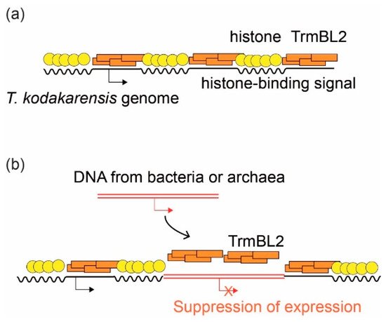
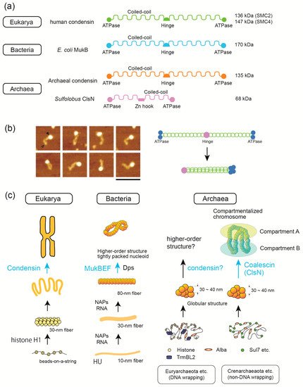
Comparative structural/molecular biology by single-molecule analyses combined with single-cell dissection, mass spectroscopy, and biochemical reconstitution have been powerful tools for elucidating the mechanisms underlying genome DNA folding. All genomes in the three domains of life undergo stepwise folding from DNA to 30–40 nm fibers. Major protein players are histone (Eukarya and Archaea), Alba (Archaea), and HU (Bacteria) for fundamental structural units of the genome. In Euryarchaeota, a major archaeal phylum, either histone or HTa (the bacterial HU homolog) were found to wrap DNA. This finding divides archaea into two groups: those that use DNA-wrapping as the fundamental step in genome folding and those that do not. Archaeal transcription factor-like protein TrmBL2 has been suggested to be involved in genome folding and repression of horizontally acquired genes, similar to bacterial H-NS protein. Evolutionarily divergent SMC proteins contribute to the establishment of higher-order structures. Recent results are presented, including the use of Hi-C technology to reveal that archaeal SMC proteins are involved in higher-order genome folding, and the use of single-molecule tracking to reveal the detailed functions of bacterial and eukaryotic SMC proteins. Here, we highlight the similarities and differences in the DNA-folding mechanisms in the three domains of life. 

higher-order chromosome structure nucleoid atomic force microscopy horizontal gene transfer histone

1. Fundamental Chromosomal/Nucleoid Proteins

The three domains clearly diverged in how they organize their chromosome structures. However, there are proteins that are commonly used in more than one domain such as histone, HU, and SMC proteins. In this section, we focus on the roles of individual chromosomal proteins in understanding the principles of chromosome folding.

1.1. Histone

Histones are the most fundamental building blocks of eukaryotic chromosomes. In 1990, the first archaeal histone was discovered in Methanothermus fervidus, a methanogenic archaeon [1]. Later, it became evident that species in Euryarchaeota (a major phylum in Archaea), except for Thermoplasmata, encode proteins homologous to eukaryotic histones. Species in other phyla, such as Nanoarchaeota, Thaumarchaeota, and Lokiarchaeota, also encode histones [2]. Archaeal histones are smaller (~6 kDa) than eukaryotic histones, and they do not possess the tail region that undergoes post-translational modification and is involved in the regulation of chromosome structure and gene expression in eukaryotes [3]. A recent report of lysine-specific acetylated proteome from Thermococcus gammatolerans suggests that archaeal histones undergo acetylation [4]. However, it needs to be clarified whether this modification really takes place in the cell and has a physiological role. Phylogenetic analysis indicates that there are two paralogues of archaeal histones: histone A and histone B. In contrast to the strict subunit composition of eukaryotic histones (a histone octamer consisting of an H3/H4 tetramer with two H2A/H2B dimers), which is defined by the structure of the four-helix bundle (4HB), archaeal histones can form either homo- or hetero-dimers [2][3]. Studies using AFM have shown that the size of a single archaeal “nucleosome” is approximately 9 nm [5].
Based on early crosslinking and electron microscopic studies, archaeal histones exist as dimers in solution and as stable tetramers in the presence of DNA [6]. However, careful observation of the MNase-digested pattern of DNA from Thermococcus kodakarensis (a minimum of 60 bp fragment and a ladder of 30 bp steps) suggested the presence of structures other than the histone tetramer . This is because if archaeal chromatin is a simple beads-on-a-string structure formed by nucleosomes consisting only of histone tetramers and the linker DNA between them, the size of MNase-protected DNA would be a multiple of 60 bp, without the 30 bp steps that were actually observed [5].
In 2013, analysis of DNA sequence associated with the MNase-protected ladder showed that archaeal ”nucleosome” is not restricted to tetramer, but it is a flexible structure composed of histone dimer units that can pile up to form larger complex [7]. In these structures, the histone dimer is a unit that can bind ~30 bp of DNA. It was proposed that a unit of archaeal histone dimer can stack on each other using its flexible 4HB domain, resulting in the formation of an archaea-specific nucleosome that is flexible in its number of histone dimer units [7].
Since the first proposal of an alternative chromatin structure model specific to archaea [7], a number of experimental studies have proved its correctness. For example, crystallographic analysis of archaeal nucleosomes revealed that certain amino acids within the 4HB domains are responsible for the multiplex stacking of dimers; mutations in these amino acids resulted in the loss of the multiplex ladder pattern in the MNase assay [8]. These mutations have led to changes in the expression of several genes [8], but the biological significance of this structure is not yet fully understood. The flexible multimeric structures formed with archaeal histone have been assigned various names including ”hypernucleosome” [2], ”archaeasome” [9], and ”archaeal histone-based chromatin polymers (AHCP)” [10]. In this review, the term ”hypernucleosome” was adopted.
The structure of the hypernucleosome may dynamically change depending on the state of the cell and the environment, thereby regulating gene expression in response to changes in the environment [8][10]. Another important question is the extent to which hypernucleosomes are present on chromosomes in living cells. Furthermore, since hypernucleosomes are not found in all archaeal species [11], it is possible that they play a role in the evolution of certain lineages of archaea.
Interestingly, although a majority of eukaryotic nucleosomes contain regular histone octamers, several types of non-canonical nucleosomes exist. For example, histone H3 is replaced with the centromere-specific histone CENP-A in eukaryotic centromeres [12]. Moreover, other structural variants of the nucleosome have been found such as overlapping dinucleosomes, which are formed by nucleosome collision during chromatin remodeling, and hexasomes, which are formed by removing one H2A/H2B dimer from a dinucleosome, that are formed during the transcription process [12]. The discovery of a flexible hypernucleosome structure in archaea raises the possibility that the archaeal ancestor of eukaryotes already had the ability to regulate chromatin structure through modulation of histone-based chromatin structure [3].

1.2. Alba

Alba is a 10-kDa DNA/RNA-binding protein found widely in archaea including Euryarchaeota, Crenarchaeota, and newly proposed phyla such as Nanoarchaeota, Korarchaeota, Thaumarchaeota, and Lokiarchaeota [6]. Alba undergoes post-translational modifications, including acetylation and methylation [13], and has several different modes of interaction with DNA such as DNA stiffening or bridging [14]. AFM revealed that Alba forms a ~10 nm fiber without wrapping DNA [5][15].
A comparison of chromosome structures of different archaeal lineages using a combination of in-vitro reconstitution and AFM revealed that Alba-mediated chromosome structures may differ depending on the type of other chromosomal proteins expressed in the cell [15]. For example, Alba and histone are co-expressed in several archaeal lineages, including some euryarchaea [13]. In-vitro reconstitution and competition experiments combined with AFM showed that increasing the amount of Alba decreases the extent of DNA wrapping by histones [15]. This suggests that Alba plays a role in regulating transcription and chromosome structure by modulating the extent of DNA topology determined by histone binding.
Alba superfamily proteins are also found in eukaryotes and have been suggested to be involved in RNA metabolism [13]. It remains to be seen whether eukaryotic Alba plays a role in genome folding.

1.3. HU

HU is a major bacterial NAP that bends DNA. AFM analysis of lysed E. coli cells has shown that its nucleoid is composed of 30–80 nm structures, and RNase A degraded the size of the 30–80 nm structures into 10 nm fibers, indicating that RNA is involved in the formation of the 30–80 nm higher-order structure, but not in the 10 nm fiber [16]. AFM analysis of nucleoid fibers derived from Escherichia coli strains that lack a single NAP gene indicates that there is no single NAP that is responsible for the formation of the 10 nm fiber. However, it is clear that proteins (NAPs or topoisomerase) are involved in the formation of the 10 nm fiber, because naked DNA appears only after proteinase K treatment of the nucleoid [17].
HU protein is associated with the survival of some bacterial species and regulates a variety of cellular processes, such as growth and SOS response [18]. Single-molecule tracking has shown that HU has several different modes of DNA binding and indicated that HU plays a dual function of compacting the nucleoid through specific DNA structure-binding and decondenses the nucleoid through nonspecific and weak interactions with the genomic DNA [19].
In the case of archaea, species in Thermoplasmatales are unique in that, although they belong to Euryarchaeota, they lack histones. Instead, they encode HTa, a protein homologous to the bacterial HU [20]. Early studies have shown that HTa is associated with Thermoplasma genomic DNA and protects about 40 bp of DNA at a minimum [21]. Although phylogenetic analysis suggests that HTa was horizontally transferred from bacteria to archaea at some point [20], it remains unclear whether HTa plays the same role as bacterial HU (i.e., to bend DNA). Several recent studies on HTa have shown that, unlike bacterial HU, HTa in Thermoplasma wraps DNA, forming particles of approximately 6 nm [15][20]. Therefore, DNA wrapping seems to be a common requirement for DNA folding in Euryarcaheaota, regardless of the protein used (histone or HTa).
We propose that, at some point after the horizontal transfer of the bacterial HU to the ancestor of Thermoplasma, HTa acquired the ability to wrap DNA; when and how this shift occurred should be a topic for future studies. After HTa adapted to the archaeal environment, including acquiring the ability to wrap DNA, the histone gene has been lost. For the chromosomes of Euryarchaeota, DNA wrappers may be indispensable for maintaining or relaxing the topological state of DNA, or for other unknown reasons. Therefore, Euryarchaea must encode a protein that fulfills this role, but that protein does not necessarily have to be a histone.

1.4. Suppression of Horizontally Transferred Genes by Global Regulatory Proteins

Horizontal gene transfer (HGT) is fundamental to archaeal and bacterial evolution [22][23]. Various mechanisms of gene flow in archaea have been revealed, for example, transformation in Euryarchaeota, vesicle transport in Thermococcales, transduction (via virus), conjugation in Sulfolobus, cell fusion in Haloarchaea, and chromosomal DNA exchange in Crenarchaea [22][23].
H-NS is an NAP that functions as a global repressor of transcription in bacteria. It preferentially binds to AT-rich sequences and can bridge two DNA strands together [24]. H-NS can suppress the expression of downstream genes by binding to the promoter region and forming a filamentous structure or forming loops [25]. This characteristic of H-NS is involved in virulence of some bacteria, as well as in the suppression of horizontally transferred genes, a function called ”xenogeneic silencing” [26]. When a foreign DNA element is incorporated into the genome, H-NS covers the region, thereby suppressing the expression of potentially harmful genes. H-NS detects foreign DNA by differences in GC content compared to the host genomes [26]. This is important in driving prokaryotic evolution by promoting the horizontal transfer of genes while suppressing their toxic effects [27]. Although HGT frequently occurs between archaea and bacteria, such xenogeneic silencing was not known in archaea.
Recently, a model was proposed that the archaeal chromosomal protein TrmBL2 plays a role similar to that of H-NS in suppressing horizontally transferred genes [28]. TrmBL2 is a transcription factor-like protein with a helix-turn-helix DNA-binding motif, which was initially identified as an abundant chromosomal protein that forms a thick (~14 nm) nucleoprotein filament on DNA without shortening (wrapping) DNA. Interestingly, TrmBL2 binds to both coding and non-coding regions and suppresses gene expression when bound to the promoter region [5]. In contrast to TrmBL2, which can bind to genomic DNA without sequence specificity, the (archaeal) histone binding site on the genomic DNA is more strictly defined by the histone-positioning signal [7][29]. Single-molecule analysis using magnetic tweezers revealed that the TrmBL2 protein of T. kodakarensis competes with archaeal histones for DNA binding [30]. Therefore, TrmBL2 may be located where there is no histone positioning signal on the DNA, and when the binding happens to be in the promoter region, the gene expression is suppressed. Indeed, histone frequency is lower in the promoter region of T. kodakarensis [7][29]. We propose that genes on DNA segments that are horizontally transferred from bacteria or non-histone-coding archaea can be suppressed by TrmBL2 in this manner (Figure 1). These facts support the idea that TrmBL2 is indeed a missing xenogeneic silencer in archaea.
Figure 1. A model of how the transcription factor-like chromosomal protein TrmBL2 represses the expression of horizontally transferred genes in archaea. The chromosome of T. kodakarensis chromosome is shown as an example. (a) The genomic sequence and the state of chromosomal protein binding. Histone localization is determined by the histone binding signals on genomic DNA, and TrmBL2 bind to genomic regions with low histone localization because of its low sequence specificity. (b) When foreign DNA is obtained from bacteria, TrmBL2 essentially covers the entire region, because bacterial DNA does not have a histone-binding signal. This way, the expression of potentially harmful genes is repressed in the early stages of horizontal gene transfer.
As discussed above, proteins that serve as transcriptional repressors and genome folding factors also play an important role in suppressing horizontally transferred genes. Are there any such silencer proteins in eukaryotes? The horizontal transfer of genes from bacteria to eukaryotes is known, and several mechanisms have been proposed for how the expression of these genes is initially regulated, but it is not assumed that transcriptional repressors are involved [31]. In fact, to the best of our knowledge, general xenogeneic silencers such as H-NS in bacteria and TrmBL2 in archaea are absent in eukaryotes. Since eukaryotes have a different gene structure from archaea and bacteria (e.g., eukayotes have introns), they may handle this problem in a way that is different from that of prokaryotes [31]. In summary, the coupling of chromatin structural proteins and the silencing of horizontally transferred genes may be unique to archaea and bacteria.

1.5. SMC Proteins Are Involved in 3D Structure Formation in the Three Domains of Life

SMC family proteins are essential for higher-order chromosome folding in the three domains of life. In general, SMC proteins are composed of ATPase domains, coiled-coil regions, and a hinge domain (Figure 2a). SMC proteins usually form larger functional complexes with accessory proteins [32]. In eukaryotes, condensin and cohesin complexes play roles in mitotic chromosome condensation and sister chromatid cohesion, respectively . Smc5/6 complex plays a role in the cellular response to DNA damage [33]. Single-molecule analysis using AFM revealed that condensin heterodimer forms a head–tail structure and that the ATPase activity of condensin is regulated by the binding of non-SMC trimer to the head of the SMC heterodimer [34](Figure 2b). AFM also showed that condensin but not cohesin induces DNA reannealing through protein–protein assembly [35]. Loop extrusion by condensin and cohesin has been proposed as a mechanism underlying their function in genome organization [36]. Detailed analysis using protein engineering and mutagenesis in conjugation with single-molecule experiments revealed that loop extrusion depends on five DNA binding sites in the case of cohesin [37]. Single-particle tracking using photo activated localization microscopy in live fission yeast revealed that Smc5/6 localize in the nucleus throughout the cell cycle but exhibited more dynamic association with chromatin compared with cohesin [33].
Figure 2. SMC proteins are commonly involved in 3D structure formation in the three domains of life. (a) Comparison of SMC proteins in the three domains. Their domain structures and molecular weight are shown. (b) AFM images of fission yeast condensin heterodimers with a head–tail structure (left). The head (filled triangle) consists of four globular ATPase domains, and the tip of the tail (open triangle) represents the hinge regions. Scale bar, 100 nm. As shown in the hypothetical model (right), the coiled-coil region (green) is folded back at the hinge (purple), and four globular domains (blue) are assembled. The AFM images were taken from Yoshimura et al. [34]. (c) Although various proteins are involved in genome folding at the basic level, SMC proteins (marked in light blue) are commonly involved in the highest level of folding. In eukaryotes, the beads-on-a-string structure folds into 30 nm fibers with the help of linker histone H1. Condensin contributes to the formation of mitotic chromosomes. In bacteria, conserved HU forms 10 nm fibers, and then other NAPs and various types of RNAs contribute to the formation of 30 and 80 nm fibers; MukBEF (SMC complex in E. coli) is involved in higher-order structuring of nucleoid; Dps is responsible with stress-induced compaction. In archaea, regardless of the mode of basic folding of the genome (either DNA wrapping in Euryarchaeota or non-DNA wrapping in Crenarchaeota), the genomes are commonly folded into 30–40 nm globular structures [15]. In Crenarchaeota, the SMC protein ClsN is implicated in chromosome compartmentalization. It needs to be elucidated as to if SMC proteins play a similar role in Euryarchaeota.
Bacterial species encode proteins homologous to eukaryotic SMCs. Bacterial SMCs have a domain structure similar to that of eukaryotic condensin and cohesin (Figure 2a), and they play a role in bacterial nucleoid organization. For example, MukBEF in E. coli and Smc-ScpAB in Bacillus subtitles are involved in the formation of higher-order nucleoid structures [38] (Figure 2c). Single-molecule tracking of B. subtilis nucleoids revealed that SMC operates by different patterns of motion compared to other DNA-condensing NAPs such as gyrase and HBsu (an HU family protein) [39][40]. Electron cryomicroscopy (cryo-EM) single-particle analysis revealed the detailed mechanism of how E. coli MukBEF entraps two distinct DNA strands when bound to the unloader MatP [41].
The condensin complex is highly conserved across all three domains of life and plays an important role in higher-order chromosome folding [42] (Figure 2c). Although proteins homologous to condensin are found in archaea, Sulfolobus species lack condensin. Recently, coalescin (ClsN), a novel Sulfolobus-encoded SMC protein, was shown to be involved in the organization of chromosomes into a two-domain compartment structure resembling the eukaryotic A/B compartments [43]. ClsN is shorter than condensins and has a Zn hock in the middle as in the case of eukaryotic Rad50 (Figure 2a). Comparison of ClsN binding (ChIP-seq) and transcriptional activity (RNA-seq and RNA polymerase localization) indicated that ClsN is associated with the transcriptionally inactive B-compartment [43].
Hi-C analysis of euryarchaeal chromosomes showed slightly different structures. The chromosome of Haloverax volcanii, an extreme halophilic euryarchaeon, and T. kodakarensis, contains multiple large chromatin loops and self-interacting domains similar to bacteria and eukaryotes. Unlike Sulfolobus species, these euryarchaeal chromosomes are not organized in a way that separates transcriptionally active and inactive compartments [44]. It remains to be elucidated whether there is a general rule for chromosome compartmentalization in archaea, the proteins responsible for higher-order genome folding in each archaeal lineage, and the physiological roles of these higher-order structures.
Structurally diverse SMC proteins were found to play similar roles in the higher-order genome folding in the three domains. Interestingly, the absence eukaryotic condensin-like protein and the use of a lineage-specific SMC (i.e., ClsN) coincides with the absence of DNA wrapping at a fundamental level of genome folding in Sulfolobus. Investigating this association between fundamental and higher-order chromosome structures in Sulfolobus and comparing it to other lineages of life may provide clues as to how different genome folding mechanisms have evolved.

References

  1. Kathleen Sandman; J. A. Krzycki; B. Dobrinski; R. Lurz; J. N. Reeve; HMf, a DNA-binding protein isolated from the hyperthermophilic archaeon Methanothermus fervidus, is most closely related to histones.. Proceedings of the National Academy of Sciences 1990, 87, 5788-5791, 10.1073/pnas.87.15.5788.
  2. Bram Henneman; Clara Van Emmerik; Hugo van Ingen; Remus T. Dame; Structure and function of archaeal histones. PLOS Genetics 2018, 14, e1007582, 10.1371/journal.pgen.1007582.
  3. Kathryn M. Stevens; Jacob B. Swadling; Antoine Hocher; Corinna Bang; Simonetta Gribaldo; Ruth A. Schmitz; Tobias Warnecke; Histone variants in archaea and the evolution of combinatorial chromatin complexity. Proceedings of the National Academy of Sciences 2020, 117, 33384-33395, 10.1073/pnas.2007056117.
  4. Béatrice Alpha-Bazin; Aurore Gorlas; Arnaud Lagorce; Damien Joulié; Jean-Baptiste Boyer; Murielle Dutertre; Jean-Charles Gaillard; Anne Lopes; Yvan Zivanovic; Alain Dedieu; et al.Fabrice ConfalonieriJean Armengaud Lysine-specific acetylated proteome from the archaeon Thermococcus gammatolerans reveals the presence of acetylated histones. Journal of Proteomics 2020, 232, 104044, 10.1016/j.jprot.2020.104044.
  5. Hugo Maruyama; Minsang Shin; Toshiyuki Oda; Rie Matsumi; Ryosuke L. Ohniwa; Takehiko Itoh; Katsuhiko Shirahige; Tadayuki Imanaka; Haruyuki Atomi; Shige H. Yoshimura; et al.Kunio Takeyasu Histone and TK0471/TrmBL2 form a novel heterogeneous genome architecture in the hyperthermophilic archaeonThermococcus kodakarensis. Molecular Biology of the Cell 2011, 22, 386-398, 10.1091/mbc.e10-08-0668.
  6. Shawn P. Laursen; Samuel Bowerman; Karolin Luger; Archaea: The Final Frontier of Chromatin. Journal of Molecular Biology 2020, 433, 166791, 10.1016/j.jmb.2020.166791.
  7. Hugo Maruyama; Janet C Harwood; Karen M Moore; Konrad Paszkiewicz; Samuel Durley; Hisanori Fukushima; Haruyuki Atomi; Kunio Takeyasu; Nicholas A Kent; An alternative beads‐on‐a‐string chromatin architecture in Thermococcus kodakarensis. EMBO reports 2013, 14, 711-717, 10.1038/embor.2013.94.
  8. Francesca Mattiroli; Sudipta Bhattacharyya; Pamela N. Dyer; Alison E. White; Kathleen Sandman; Brett W. Burkhart; Kyle R. Byrne; Thomas Lee; Natalie G. Ahn; Thomas J. Santangelo; et al.John N. ReeveKarolin Luger Structure of histone-based chromatin in Archaea. Science 2017, 357, 609-612, 10.1126/science.aaj1849.
  9. Samuel Bowerman; Jeff Wereszczynski; Karolin Luger; Archaeal chromatin ‘slinkies’ are inherently dynamic complexes with deflected DNA wrapping pathways. eLife 2021, 10, e65587, 10.7554/elife.65587.
  10. Travis J. Sanders; Fahad Ullah; Alexandra M. Gehring; Brett W. Burkhart; Robert L. Vickerman; Sudili Fernando; Andrew F. Gardner; Asa Ben-Hur; Thomas J. Santangelo; Extended Archaeal Histone-Based Chromatin Structure Regulates Global Gene Expression in Thermococcus kodakarensis. Frontiers in Microbiology 2021, 12, 681150, 10.3389/fmicb.2021.681150.
  11. Bram Henneman; Thomas B Brouwer; Amanda M Erkelens; Gert-Jan Kuijntjes; Clara van Emmerik; Ramon A van der Valk; Monika Timmer; Nancy C S Kirolos; Hugo van Ingen; John van Noort; et al.Remus T Dame Mechanical and structural properties of archaeal hypernucleosomes. Nucleic Acids Research 2020, 49, 4338-4349, 10.1093/nar/gkaa1196.
  12. Masako Koyama; Hitoshi Kurumizaka; Structural diversity of the nucleosome. The Journal of Biochemistry 2017, 163, 85-95, 10.1093/jb/mvx081.
  13. Manish Goyal; Chinmoy Banerjee; Shiladitya Nag; Uday Bandyopadhyay; The Alba protein family: Structure and function. Biochimica et Biophysica Acta (BBA) - Proteins and Proteomics 2016, 1864, 570-583, 10.1016/j.bbapap.2016.02.015.
  14. Niels Laurens; Rosalie P.C. Driessen; Iddo Heller; Daan Vorselen; Maarten C. Noom; Felix J.H. Hol; Malcolm F. White; Remus T. Dame; Gijs J.L. Wuite; Alba shapes the archaeal genome using a delicate balance of bridging and stiffening the DNA. Nature Communications 2012, 3, 1328, 10.1038/ncomms2330.
  15. Hugo Maruyama; Eloise I. Prieto; Takayuki Nambu; Chiho Mashimo; Kosuke Kashiwagi; Toshinori Okinaga; Haruyuki Atomi; Kunio Takeyasu; Different Proteins Mediate Step-Wise Chromosome Architectures in Thermoplasma acidophilum and Pyrobaculum calidifontis. Frontiers in Microbiology 2020, 11, 1247, 10.3389/fmicb.2020.01247.
  16. Ryosuke L. Ohniwa; Kazuya Morikawa; Sayaka L. Takeshita; Joongbaek Kim; Toshiko Ohta; Chieko Wada; Kunio Takeyasu; Transcription-coupled nucleoid architecture in bacteria. Genes to Cells 2007, 12, 1141-1152, 10.1111/j.1365-2443.2007.01125.x.
  17. Ryosuke L. Ohniwa; Hiroki Muchaku; Shinji Saito; Chieko Wada; Kazuya Morikawa; Atomic Force Microscopy Analysis of the Role of Major DNA-Binding Proteins in Organization of the Nucleoid in Escherichia coli. PLOS ONE 2013, 8, e72954, 10.1371/journal.pone.0072954.
  18. Pavla Stojkova; Petra Spidlova; Jiri Stulik; Nucleoid-Associated Protein HU: A Lilliputian in Gene Regulation of Bacterial Virulence. Frontiers in Cellular and Infection Microbiology 2019, 9, 159, 10.3389/fcimb.2019.00159.
  19. Kelsey Bettridge; Subhash Verma; Xiaoli Weng; Sankar Adhya; Jie Xiao; Single‐molecule tracking reveals that the nucleoid‐associated protein HU plays a dual role in maintaining proper nucleoid volume through differential interactions with chromosomal DNA. Molecular Microbiology 2020, 115, 12-27, 10.1111/mmi.14572.
  20. Antoine Hocher; Maria Rojec; Jacob B Swadling; Alexander Esin; Tobias Warnecke; The DNA-binding protein HTa from Thermoplasma acidophilum is an archaeal histone analog. eLife 2019, 8, e52542, 10.7554/elife.52542.
  21. Dennis G. Searcy; Diana B. Stein; Nucleoprotein subunit structure in an unusual prokaryotic organism: Thermoplasma acidophilum. Biochimica et Biophysica Acta (BBA) - Nucleic Acids and Protein Synthesis 1980, 609, 180-195, 10.1016/0005-2787(80)90211-7.
  22. Purificación López-García; Yvan Zivanovic; Philippe Deschamps; David Moreira; Bacterial gene import and mesophilic adaptation in archaea. Nature Reviews Microbiology 2015, 13, 447-456, 10.1038/nrmicro3485.
  23. Alexander Wagner; Rachel J. Whitaker; David J. Krause; Jan-Hendrik Heilers; Marleen Van Wolferen; Chris Van Der Does; Sonja-Verena Albers; Mechanisms of gene flow in archaea. Nature Reviews Microbiology 2017, 15, 492-501, 10.1038/nrmicro.2017.41.
  24. Thøger Krogh; Jakob Møller-Jensen; Christoph Kaleta; Impact of Chromosomal Architecture on the Function and Evolution of Bacterial Genomes. Frontiers in Microbiology 2018, 9, 2019, 10.3389/fmicb.2018.02019.
  25. Ci Ji Lim; Sin Yi Lee; Linda J. Kenney; Jie Yan; Nucleoprotein filament formation is the structural basis for bacterial protein H-NS gene silencing. Scientific Reports 2012, 2, 509, 10.1038/srep00509.
  26. Charles J. Dorman; H-NS, the genome sentinel. Nature Reviews Genetics 2006, 5, 157-161, 10.1038/nrmicro1598.
  27. Jane Wiedenbeck; Frederick M. Cohan; Origins of bacterial diversity through horizontal genetic transfer and adaptation to new ecological niches. FEMS Microbiology Reviews 2011, 35, 957-976, 10.1111/j.1574-6976.2011.00292.x.
  28. Hugo Maruyama; Nicholas A. Kent; Hiromi Nishida; Taku Oshima; Functions of Archaeal Nucleoid Proteins: Archaeal Silencers are Still Missing. DNA Traffic in the Environment 2019, 1, 29-45, 10.1007/978-981-13-3411-5_2.
  29. Narasimharao Nalabothula; Liqun Xi; Sucharita Bhattacharyya; Jonathan Widom; Ji-Ping Wang; John N Reeve; Thomas J Santangelo; Yvonne N Fondufe-Mittendorf; Archaeal nucleosome positioning in vivo and in vitro is directed by primary sequence motifs. BMC Genomics 2013, 14, 1-13, 10.1186/1471-2164-14-391.
  30. Artem Efremov; Yuanyuan Qu; Hugo Maruyama; Ci J. Lim; Kunio Takeyasu; Jie Yan; Transcriptional Repressor TrmBL2 from Thermococcus kodakarensis Forms Filamentous Nucleoprotein Structures and Competes with Histones for DNA Binding in a Salt- and DNA Supercoiling-dependent Manner. Journal of Biological Chemistry 2015, 290, 15770-15784, 10.1074/jbc.m114.626705.
  31. Filip Husnik; John P. McCutcheon; Functional horizontal gene transfer from bacteria to eukaryotes. Nature Reviews Microbiology 2017, 16, 67-79, 10.1038/nrmicro.2017.137.
  32. Frank Uhlmann; SMC complexes: from DNA to chromosomes. Nature Reviews Molecular Cell Biology 2016, 17, 399-412, 10.1038/nrm.2016.30.
  33. Thomas J Etheridge; Desiree Villahermosa; Eduard Campillo-Funollet; Alex David Herbert; Anja Irmisch; Adam T Watson; Hung Q Dang; Mark A Osborne; Antony W Oliver; Antony M Carr; et al.Johanne M Murray Live-cell single-molecule tracking highlights requirements for stable Smc5/6 chromatin association in vivo. eLife 2021, 10, e68579, 10.7554/elife.68579.
  34. Shige H. Yoshimura; Kohji Hizume; Akiko Murakami; Takashi Sutani; Kunio Takeyasu; Mitsuhiro Yanagida; Condensin Architecture and Interaction with DNA: Regulatory Non-SMC Subunits Bind to the Head of SMC Heterodimer. Current Biology 2002, 12, 508-513, 10.1016/s0960-9822(02)00719-4.
  35. Akiko Sakai; Kohji Hizume; Takashi Sutani; Kunio Takeyasu; Mitsuhiro Yanagida; Condensin but not cohesin SMC heterodimer induces DNA reannealing through protein-protein assembly. The EMBO Journal 2003, 22, 2764-2775, 10.1093/emboj/cdg247.
  36. Iain F. Davidson; Jan-Michael Peters; Genome folding through loop extrusion by SMC complexes. Nature Reviews Molecular Cell Biology 2021, 22, 445-464, 10.1038/s41580-021-00349-7.
  37. Benedikt W. Bauer; Iain F. Davidson; Daniel Canena; Gordana Wutz; Wen Tang; Gabriele Litos; Sabrina Horn; Peter Hinterdorfer; Jan-Michael Peters; Cohesin mediates DNA loop extrusion by a “swing and clamp” mechanism. Cell 2021, 184, 5448-5464.e22, 10.1016/j.cell.2021.09.016.
  38. Sophie Nolivos; David Sherratt; The bacterial chromosome: architecture and action of bacterial SMC and SMC-like complexes. FEMS Microbiology Reviews 2014, 38, 380-392, 10.1111/1574-6976.12045.
  39. Sonja Schibany; Luise A K Kleine Borgmann; Thomas C Rösch; Tobias Knust; Maximilian H Ulbrich; Peter L Graumann; Single molecule tracking reveals that the bacterial SMC complex moves slowly relative to the diffusion of the chromosome. Nucleic Acids Research 2018, 46, 7805-7819, 10.1093/nar/gky581.
  40. Sonja Schibany; Rebecca Hinrichs; Rogelio Hernandez-Tamayo; Peter L. Graumann; The Major Chromosome Condensation Factors Smc, HBsu, and Gyrase in Bacillus subtilis Operate via Strikingly Different Patterns of Motion. mSphere 2020, 5, e00817-e00820, 10.1128/msphere.00817-20.
  41. Frank Bürmann; Louise F.H. Funke; Jason W. Chin; Jan Löwe; Cryo-EM structure of MukBEF reveals DNA loop entrapment at chromosomal unloading sites. Molecular Cell 2021, 81, 4891-4906.e8, 10.1016/j.molcel.2021.10.011.
  42. Tatsuya Hirano; Condensin-Based Chromosome Organization from Bacteria to Vertebrates. Cell 2016, 164, 847-857, 10.1016/j.cell.2016.01.033.
  43. Naomichi Takemata; Rachel Y. Samson; Stephen D. Bell; Physical and Functional Compartmentalization of Archaeal Chromosomes. Cell 2019, 179, 165-179.e18, 10.1016/j.cell.2019.08.036.
  44. Charlotte Cockram; Agnès Thierry; Aurore Gorlas; Roxane Lestini; Romain Koszul; Euryarchaeal genomes are folded into SMC-dependent loops and domains, but lack transcription-mediated compartmentalization. Molecular Cell 2020, 81, 459-472.e10, 10.1016/j.molcel.2020.12.013.
More
Information
Contributor MDPI registered users' name will be linked to their SciProfiles pages. To register with us, please refer to https://encyclopedia.pub/register :
View Times: 429
Revisions: 2 times (View History)
Update Date: 28 Dec 2021
1000/1000
Video Production Service