Submitted Successfully!
To reward your contribution, here is a gift for you: A free trial for our video production service.
Thank you for your contribution! You can also upload a video entry or images related to this topic.
Version Summary Created by Modification Content Size Created at Operation
1 + 3109 word(s) 3109 2021-11-08 09:31:04 |
2 Done Meta information modification 3109 2021-12-17 02:15:31 |

Video Upload Options

Do you have a full video?

Confirm

Are you sure to Delete?
Cite
If you have any further questions, please contact Encyclopedia Editorial Office.
Nakanishi, H. Protein-Based Systems for Translational Regulation of synthetic mRNAs. Encyclopedia. Available online: https://encyclopedia.pub/entry/17201 (accessed on 05 July 2024).
Nakanishi H. Protein-Based Systems for Translational Regulation of synthetic mRNAs. Encyclopedia. Available at: https://encyclopedia.pub/entry/17201. Accessed July 05, 2024.
Nakanishi, Hideyuki. "Protein-Based Systems for Translational Regulation of synthetic mRNAs" Encyclopedia, https://encyclopedia.pub/entry/17201 (accessed July 05, 2024).
Nakanishi, H. (2021, December 16). Protein-Based Systems for Translational Regulation of synthetic mRNAs. In Encyclopedia. https://encyclopedia.pub/entry/17201
Nakanishi, Hideyuki. "Protein-Based Systems for Translational Regulation of synthetic mRNAs." Encyclopedia. Web. 16 December, 2021.
Protein-Based Systems for Translational Regulation of synthetic mRNAs
Edit

Synthetic mRNAs, which are produced by in vitro transcription, have been recently attracting attention because they can express any transgenes without the risk of insertional mutagenesis. Protein-based translational regulation systems enable the context-dependent production of therapeutic proteins and have the potential to further improve the efficacy and safety of synthetic mRNAs.

messenger RNA RNA binding protein translation Protein Modules

1. Introduction

During gene expression, genes are first transcribed from DNA to messenger RNA (mRNA) and then translated from mRNA to protein. Thus, researchers can make cells express exogenous genes by transfecting either DNA or mRNA. Although DNA transfection is the standard method for transgene expression, it can cause insertional mutagenesis of endogenous genes, which is a major drawback in medical applications. In contrast, synthetic mRNAs, which are produced by in vitro transcription, can be used for transgene expression without the risk of insertional mutagenesis. Thus, synthetic mRNAs have recently been attracting attention as tools for gene therapy, cellular reprogramming, and vaccine development [1].

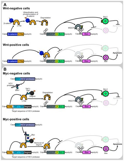
Nevertheless, context-dependent regulation of transgene expression is more difficult in synthetic mRNA transfection than in DNA transfection. Upon DNA transfection, transgene expression can be tuned using transcriptional regulatory sequences, such as drug-inducible or tissue-specific promoters. On the other hand, such transcriptional regulation cannot apply to synthetic mRNA transfection as they must be transcribed in vitro prior to transfection. As some therapeutic genes may cause adverse effects when they are expressed in inappropriate contexts, context-dependent transgene regulation is helpful to cope with low adverse effects and high therapeutic efficacy. For example, cell-selective expression of pro-apoptotic genes [2][3][4][5][6] may enable cancer cell elimination without damaging healthy cells. Similarly, cardiomyocyte-selective expression of proliferation-promoting genes can be helpful to cope with myocardial regeneration without elevating fibrosis and immune response [7]. Thus, the development of translational regulation systems will make synthetic mRNAs more valuable.

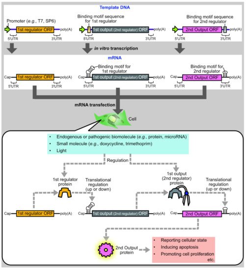
One promising approach is to develop protein-based systems for translational regulation of synthetic mRNAs. There are multiple advantages to protein-based translational regulation systems. First, when proteins are used as translational regulators, the regulators themselves can be translated from synthetic mRNAs, such that all components necessary for regulation can be delivered as synthetic mRNAs. Second, compared to ribozyme-embedded mRNAs [8] or caged mRNAs [9], mRNAs composing protein-based regulation systems have less concern regarding the alteration of mRNA properties during synthesis or storage, such as self-cleavage or decaging. Finally, in protein-based translational regulation systems, the first output protein can be used as the input for the second regulation, allowing layered gene circuits for sophisticated regulation ( Figure 1 ).

Figure 1. Schematic diagram of a protein-based translational regulation system for synthetic mRNAs. Synthetic mRNAs encoding regulator or output proteins are synthesized by in vitro transcription and transfected into cells. After transfection, a regulator protein is translated from an mRNA and regulates the translation of another mRNA encoding an output protein. Then, the output protein induces various biological phenomena such as apoptosis or cell proliferation. In multi-layered gene circuits, the 1st output protein acts as the 2nd regulator to regulate the translation of the 2nd output ORF. For cell state-responsive autonomous regulation, regulator proteins or mRNAs are designed to respond to endogenous or pathogenic biomolecules such as proteins or microRNAs. Regulator proteins can also be designed to respond to external cues such as small molecules or light for deliberate control of translation.

2. Basic Protein Modules for Protein-Based Translational Regulation Systems

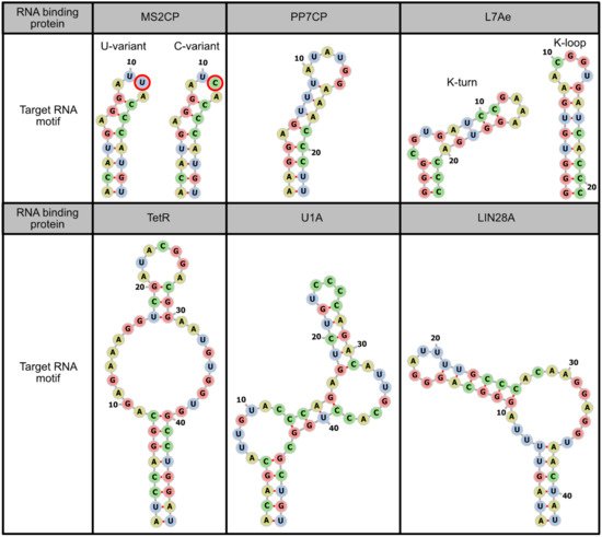
2.1. Binding to Target mRNAs

Motif-specific RNA binding proteins (RBPs) are mainly used for two purposes. One is to fuse RBPs with other protein modules, so that other modules can act on synthetic mRNAs. The other is to repress translation only by the binding of RBPs to the 5' untranslated regions (UTRs) of synthetic mRNAs, without being combined with other protein modules (Figure 2, the 1st row).
Figure 2. List of protein modules used in translational regulation of synthetic mRNAs.
In order to regulate the translation of only synthetic mRNAs while avoiding the effect on endogenous mRNAs, it is desirable to use RBPs that have high specificity for target RNA motifs, and these motifs are not present in endogenous mRNAs. Thus, microbial RBPs are primarily used in mRNA-based mammalian synthetic biology. The representative microbial RBPs used to regulate synthetic mRNAs are coat proteins derived from the bacteriophages MS2 (MS2CP) [2][3][4][5][6][10][11][12][13][14][15][16][17][18][19][20][21] and PP7 (PP7CP) [6][14][21], the archaeal ribosomal protein L7Ae [2][3][4][6][7][11][12][21][22][23][24][25], and the tetracycline-responsive repressor protein (TetR) from Escherichia coli [23][26]. Among these, TetR is unique in its ability to conditionally dissociate from the target RNA motif by doxycycline addition. In addition to these microbial proteins, mammalian RBPs can also be used, such as U1A, a spliceosomal protein [10][13][21][27], and LIN28A, a pre-microRNA binding protein [21][27] (Figure 3). However, when these mammalian proteins and their target RNA motifs are used in mammalian cells, it should be noted that they may interact with endogenous RNAs or proteins. Such interactions can induce unintended effects on both the regulation of synthetic mRNAs and endogenous pathways.
Figure 3. Target RNA motifs of representative RNA binding proteins. Sequences and secondary structures of target RNA motifs of MS2CP [28], PP7CP [21], L7Ae [11], TetR [26], U1A [29], and LIN28A [27] are shown. The secondary structures are visualized by forna [30].

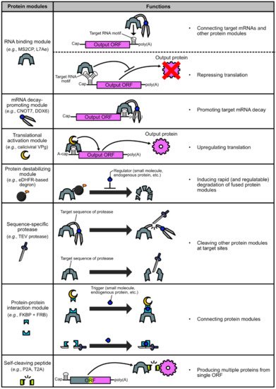
2.2. Promoting Target mRNA Decay

Fusion of motif-specific RBPs and mRNA decay-promoting proteins, such as dead box helicase 6 (DDX6) [23] and CCR4-NOT transcription complex subunit 7 (CNOT7) [2][12][14], are used to induce targeted mRNA decay and eventual translational shut-off. DDX6 is a protein that interacts with decapping coactivators and the CCR4-NOT complex, which has a role in mRNA deadenylation and translational repression [31], whereas CNOT7 is a deadenylase module of the CCR4-NOT complex [32]. Since 5’ cap structures and poly(A) tails have a crucial role in both mRNA stability and translation, targeted binding of these proteins to synthetic mRNAs can promote their decay and translational shut-off (Figure 2, the 2nd row). In addition, studies using pDNA transfection reported that mRNA decay can also be promoted by targeted binding of nonsense-mediated mRNA decay (NMD)-related proteins (e.g., Y14, RNPS1, and Upf1, 2, 3a, and 3b) [16][17][18][19], Staufen1, which can induce Staufen-mediated mRNA decay (SMD) [16][17], and the RNAi-related protein Ago2 [33].
Although translational repression can be achieved by only RBPs, combining mRNA decay-promoting proteins has two advantages. First, in the case that an RBP itself cannot sufficiently repress translation, the fusion of an mRNA decay-promoting protein may be helpful to improve the translational repression efficiency. Wagner et al. reported that the TetR-DDX6 fusion protein can be used for doxycycline-controllable translational repression of an N1-methyl-pseudouridine-containing mRNA, whereas TetR only cannot [23]. The second advantage of combining mRNA decay-promoting proteins is that the insertion sites of target RNA motifs can be placed in 3’ UTRs [2][12][14][16][17][18][19][33]. In the case of translational repression using only RBPs, target motifs need to be inserted in 5' UTRs, but the presence of stable stem structures in 5' UTRs is itself disadvantageous for translation [34]. Therefore, designing mRNAs for high protein production efficiency is relatively easy when target motifs are placed in 3’ UTRs instead of 5’ UTRs.
It is stated that the regulation by CNOT7 is more effective in RNA replicons than in non-replicative mRNAs [6][14]. RNA replicons, which are also called self-amplifying mRNAs, replicate themselves in the cytoplasm through the activity of RNA-dependent RNA polymerase (also called RNA replicase) that is expressed from RNA replicons themselves. Thus, while the amount of protein produced by conventional synthetic mRNAs decreases over time due to mRNA decay by the endogenous machinery, RNA replicons replenish themselves and enable continuous protein production by single transfection [35][36]. Nevertheless, caution is required when mRNA decay-promoting proteins are combined with RNA replicons. If RNA replicons are completely removed by mRNA decay-promoting proteins, the replenishment of RNA replicons cannot occur. Although there is a report that RNA replicons can partially be restored after the termination of promoted decay [14], designing RNA replicon-based gene circuits containing mRNA decay-promoting proteins should be done with caution, especially when there is a long duration of promoted mRNA decay.

2.3. Activating Target mRNA Translation

Translation of a eukaryotic mRNA is typically initiated by the binding of the eukaryotic initiation factor (eIF) 4F complex to the cap structure at the 5' end of the mRNA, which in turn recruits the 40S subunit of the ribosome via the eIF4F-eIF3-40S subunit interaction [37]. Therefore, two components are needed to achieve conditional activation of mRNA translation by protein-based systems. One is an mRNA lacking the canonical 5' cap (typically, a translationally inactive cap analog, termed "A-Cap", is used instead of the canonical cap), and the other is a translational activator protein that can directly or indirectly recruit ribosomes to mRNAs even in the absence of the canonical 5' cap.
One of the proteins that can recruit ribosomes is the viral protein genome-linked (VPg) from calicivirus. Caliciviral VPg is a relatively small (e.g., 111 amino acids in the case of feline calicivirus) protein that is fused to the 5' end of the caliciviral RNA. Since caliciviral RNAs lack the 5' cap, caliciviruses use VPg to recruit ribosomes to their RNAs via a VPg-eIF4F interaction [38][39][40]. Researchers showed that the fusion protein of MS2CP and the feline caliciviral VPg, named caliciviral VPg-based translational activator (CaVT), can activate the translation of A-capped mRNAs containing the MS2CP-target motif in their 5' UTRs [5][20] (Figure 2, the 3rd row).
Another example of a translational activator protein is a fusion protein composed of MS2CP and the C-terminal region (amino acids 623-1600) of eIF4G. eIF4G is a component of the eIF4F complex, and its C-terminal region has a role in the interaction with eIF3 and the 40S subunit of the ribosome. Paek et al. reported that connecting the C-terminal region of eIF4G to the 3’ UTRs of A-capped mRNAs can activate their translation [15]. It should be noted, however, that in the study, the MS2CP-eIF4G fusion protein was expressed not from a synthetic mRNA, but a cytomegalovirus promoter-embedded plasmid DNA (pDNA). Compared to synthetic mRNA, pDNA containing a strong promoter shows a higher maximum level of expression, although the variability in the expression level among cells is also higher [41]. Therefore, future studies are needed to determine whether synthetic mRNAs can produce an adequate amount of MS2CP-eIF4G to activate the translation of A-capped mRNAs. The translational activating function of eIF4E, another component of the eIF4F complex, was also shown in experiments using pDNA transfection [42].

2.4. Destabilizing Proteins

Destabilizing domains (also called degrons) that induce rapid degradation of fused proteins are used to regulate the abundance of translational regulator proteins (Figure 2, the 4th row). For example, rapid degradation of a translational repressor protein results in an enhancement of its target mRNA translation [4][6][14][20][23][25]. Conversely, rapid degradation of a translational activator protein diminishes the translation of its target mRNA [20]. In many cases, the purpose is to achieve deliberate or cell state-responsive translational regulation by using degrons whose destabilizing activity can be altered by small molecules or endogenous biomolecules.
There are several small molecule-responsive degrons, such as the FK506-binding protein (FKBP)-based degron [43] and the auxin-inducible degron [44], and the Escherichia coli-derived dihydrofolate reductase (eDHFR)-based degron [45] is one of the most popular. Importantly, the destabilizing activity of the eDHFR-based degron can be inhibited by trimethoprim, an antibiotic that is used clinically. Translational regulator proteins fused with the eDHFR-based degron are rapidly degraded in the absence of trimethoprim, but can be stabilized by the addition of trimethoprim, allowing them to repress or activate the translation of their target mRNAs [4][6][20][23][25]. In addition, researchers recently showed that the combination of the eDHFR-based degron and photocaged trimethoprim, which can bind the degron only after light irradiation, enables photo-regulatable translation [20].
In mammalian cells, the degradation of some proteins is known to be promoted in response to cell state, and such proteins can be used as cell state-responsive degrons. One example used in translational regulation is β-catenin, which becomes a target for ubiquitination and phosphorylation to promote degradation in Wnt-negative cells. Utilizing this feature of β-catenin, Yang and Ding achieved Wnt-positive cell-selective translational repression by the β-catenin-fused translational repressor protein [4] (Figure 4A). They also used another type of degron whose destabilizing activity is activated when its N-terminal region is removed by a sequence-specific protease [4][6][46]. Therefore, the protease can relieve mRNAs from translational repression caused by the degron-fused translational repressor protein.
Figure 4. Representatives of endogenous protein-responsive translational regulation systems. (A) Wnt-responsive apoptosis induction system. In Wnt-negative cells, β-catenin-fused L7Ae is rapidly degraded due to ubiquitination and phosphorylation of β-catenin. Therefore, the L7Ae target motif (K-turn)-embedded mRNA encoding MS2CP and Bcl-2 is not translationally repressed. The translated MS2CP represses the translation of Bax, whereas Bcl-2 prevents apoptosis caused by leaky expression of Bax (top). In contrast, in Wnt-positive cells, β-catenin-fused L7Ae is not rapidly degraded and represses the translation of MS2CP and Bcl-2. As MS2CP translation is repressed, Bax is not translationally repressed by MS2CP and induces apoptosis (bottom). (B) Myc-responsive apoptosis induction system. In Myc-negative cells, L7Ae is rapidly degraded due to the fused degron. Similar to the case of the Wnt-responsive system shown above, the rapid degradation of L7Ae results in the prevention of apoptosis (top). In Myc-positive cells, TAF1-TBP that is fused to L7Ae attracts BIN1-fused HCV protease. Then, HCV protease cleaved its target sequence inserted between L7Ae and TAF1-TBP-degron. Due to the cleavage, the degron is removed from L7Ae, which stabilizes L7Ae. Then, stabilized L7Ae translationally represses MS2CP and Bcl-2, thereby induces Bax-mediated apoptosis (bottom).

2.5. Cleaving Proteins

Some viruses such as tobacco etch virus (TEV), tobacco vein mottling virus (TVMV) [47], and hepatitis C virus (HCV) [48] have sequence-specific proteases that recognize and cleave the target sequences of five to seven amino acid residues. When multiple proteins are connected by a linker containing a target sequence of such protease, the protease can separate these proteins and alter the property of the fusion construct (Figure 2, the 5th row). One example is abolishing promoted mRNA decay by separating the RNA binding module and the mRNA decay-promoting module [12][14]. A fusion protein consisting of an RBP (MS2CP or PP7CP) and CNOT7 can normally promote the decay of mRNAs that have target RNA motifs. However, if the linker connecting these two proteins contains a target sequence of protease, the corresponding protease can separate CNOT7 from the RBP, thereby abolishing CNOT7-mediated mRNA decay. Similarly, when a protease target sequence is inserted between the degron and the translational regulator protein, the corresponding protease can stabilize the translational regulator by removing the degron from it [4][6].
If a target sequence can be inserted without impairing the function of the protein, sequence-specific proteases can also be used to cleave the protein that is not constructed by fusing multiple proteins. For example, Cella et al. showed that L7Ae with a TEV protease target sequence can repress translation in the absence of TEV protease, and translational repression can be released by cleavage of L7Ae by TEV protease [12].
When multiple proteases are used simultaneously in a single system, the orthogonality of these proteases needs to be checked, as unexpected cleavage by non-corresponding proteases can cause unexpected output. For example, Cella et al. reported that the turnip mosaic virus (TUMV) protease has moderate cleavage activity against the target sequences of TEV, TVMV, and sunflower mosaic virus (SuMMV) [12].

2.6. Combining Separate Proteins

Protein–protein interaction modules that respond to specific cues (e.g., small molecules or light) are useful to achieve deliberate or cell state-responsive autonomous translational regulation. Representative protein–protein interaction modules are FKBP-FRB and its variant [5][6][14][49][50], ABI-PLY [4][6][14], and CRY2-CIBN [6][14][51]. By fusing such protein–protein interaction modules to two proteins with different functions, one can control the effect on mRNAs or other protein modules (Figure 2, the 6th row). One example is the small molecule-controllable translational activation system. By fusing small molecule-responsive hetero-dimerization domains to MS2CP and VPg, researchers have succeeded in inducing translational activation in the small molecule-dependent manner [5][20]. Furthermore, the addition of a photo-cage to such a trigger molecule enabled photo-controllable translational activation [20]. Similarly, intracellular proteins can also be used as triggers to control protein–protein interactions. Yang and Ding reported the system that selectively induces translational repression in Myc-positive cells [4]. This system is composed of two fusion proteins. One is an engineered translational repressor protein composed of L7Ae, an HCV protease target sequence, TAF1-TBP, and a degron. The other is an HCV protease fused with BIN1. As both BIN1 and TAF1-TBP bind Myc, in Myc-positive cells, the BIN1-fused HCV protease is attracted to the engineered translational repressor by the BIN1–Myc–TAF1-TBP interaction. Then, HCV protease removes the degron from the translational repressor to abolish the degron-induced destabilization of L7Ae. Thus, target mRNA translation was repressed in Myc-positive cells (Figure 4B). Another example is the selective translation system for viral protein-expressing cells, using a translational repressor containing a TEV protease target sequence [12]. In that study, Cella et al. designed two fusion proteins. One is an intrabody-fused L7Ae which has a cleavage sequence of TEV protease. The other is an intrabody-fused TEV protease. Both intrabodies bind the viral protein NS3, but their target epitopes are different. Thus, in cells expressing NS3, the TEV protease is attracted to L7Ae via the intrabody–NS3–intrabody interaction. Then, the TEV protease cleaves L7Ae, thereby abolishing L7Ae-mediated translational repression.
Protein–protein interaction modules can also be used to reconstitute a full-length protein from split fragments. Liu et al. developed various split proteases that are fused with small molecule- or light-responsive protein–protein interaction modules. These split protease fragments do not cleave their target sequences until they interact with one another. The corresponding cues (a small molecule or light) induce the interaction of these split fragments to reconstitute the full-length proteases [14]. When using split proteins, the split-site should be carefully selected because, depending on the split-site, fragments may spontaneously assemble independently of the protein–protein interaction modules or may not function even if it binds via the interaction modules. The tool to predict suitable split sites may be helpful to design split proteins [52].

2.7. Producing Multiple Proteins from a Single ORF

Self-cleaving peptides such as porcine teschovirus-1-derived 2A (P2A) [53] can be utilized to link the production of multiple proteins. For example, a translational regulator protein and a fluorescent protein can be produced as two separate proteins from an mRNA containing a single ORF, in which the translational regulator and fluorescent protein genes are paired via a self-cleaving peptide gene [2][23]. Since the translational level of these two proteins must be identical, the fluorescence signal can be used to monitor the expression level of the translational regulator in living cells. This method is particularly useful when the expression level of multiple proteins should be linked, but the function of each protein is affected if fused.

References

  1. Gómez-Aguado, I.; Rodríguez-Castejón, J.; Vicente-Pascual, M.; Rodríguez-Gascón, A.; Solinís, M.Á.; del Pozo-Rodríguez, A. Nanomedicines to Deliver MRNA: State of the Art and Future Perspectives. Nanomaterials 2020, 10, 364.
  2. Wroblewska, L.; Kitada, T.; Endo, K.; Siciliano, V.; Stillo, B.; Saito, H.; Weiss, R. Mammalian Synthetic Circuits with RNA Binding Proteins for RNA-Only Delivery. Nat. Biotechnol. 2015, 33, 839–841.
  3. Matsuura, S.; Ono, H.; Kawasaki, S.; Kuang, Y.; Fujita, Y.; Saito, H. Synthetic RNA-Based Logic Computation in Mammalian Cells. Nat. Commun. 2018, 9, 4847.
  4. Yang, J.; Ding, S. Engineering L7Ae for RNA-Only Delivery Kill Switch Targeting CMS2 Type Colorectal Cancer Cells. ACS Synth. Biol. 2021, 10, 1095–1105.
  5. Nakanishi, H.; Saito, H. Caliciviral Protein-Based Artificial Translational Activator for Mammalian Gene Circuits with RNA-Only Delivery. Nat. Commun. 2020, 11, 1297.
  6. Yang, J.; Ding, S. Chimeric RNA-Binding Protein-Based Killing Switch Targeting Hepatocellular Carcinoma Cells. Mol. Ther. - Nucleic Acids 2021, 25, 683–695.
  7. Magadum, A.; Singh, N.; Kurian, A.A.; Munir, I.; Mehmood, T.; Brown, K.; Sharkar, M.T.K.; Chepurko, E.; Sassi, Y.; Oh, J.G.; et al. Pkm2 Regulates Cardiomyocyte Cell Cycle and Promotes Cardiac Regeneration. Circulation 2020, 141, 1249–1265.
  8. Yokobayashi, Y. Aptamer-Based and Aptazyme-Based Riboswitches in Mammalian Cells. Curr. Opin. Chem. Biol. 2019, 52, 72–78.
  9. Zhang, D.; Zhou, C.Y.; Busby, K.N.; Alexander, S.C.; Devaraj, N.K. Light-Activated Control of Translation by Enzymatic Covalent MRNA Labeling. Angew. Chem. 2018, 130, 2872–2876.
  10. Stripecke, R.; Oliveira, C.C.; McCarthy, J.E.; Hentze, M.W. Proteins Binding to 5’ Untranslated Region Sites: A General Mechanism for Translational Regulation of MRNAs in Human and Yeast Cells. Mol. Cell. Biol. 1994, 14, 5898–5909.
  11. Endo, K.; Stapleton, J.A.; Hayashi, K.; Saito, H.; Inoue, T. Quantitative and Simultaneous Translational Control of Distinct Mammalian MRNAs. Nucleic Acids Res. 2013, 41, e135.
  12. Cella, F.; Wroblewska, L.; Weiss, R.; Siciliano, V. Engineering Protein-Protein Devices for Multilayered Regulation of MRNA Translation Using Orthogonal Proteases in Mammalian Cells. Nat. Commun. 2018, 9, 4392.
  13. Parr, C.J.C.; Wada, S.; Kotake, K.; Kameda, S.; Matsuura, S.; Sakashita, S.; Park, S.; Sugiyama, H.; Kuang, Y.; Saito, H. N1-Methylpseudouridine Substitution Enhances the Performance of Synthetic MRNA Switches in Cells. Nucleic Acids Res. 2020, 48, e35.
  14. Liu, K.; Yang, J.; Ding, S.; Gao, Y. Daisy Chain Topology Based Mammalian Synthetic Circuits for RNA-Only Delivery. ACS Synth. Biol. 2020, 9, 269–282.
  15. Paek, K.Y.; Hong, K.Y.; Ryu, I.; Park, S.M.; Keum, S.J.; Kwon, O.S.; Jang, S.K. Translation Initiation Mediated by RNA Looping. Proc. Natl. Acad. Sci. 2015, 112, 1041–1046.
  16. Hosoda, N.; Kim, Y.K.; Lejeune, F.; Maquat, L.E. CBP80 Promotes Interaction of Upf1 with Upf2 during Nonsense-Mediated MRNA Decay in Mammalian Cells. Nat. Struct. Mol. Biol. 2005, 12, 893–901.
  17. Kim, Y.K.; Furic, L.; DesGroseillers, L.; Maquat, L.E. Mammalian Staufen1 Recruits Upf1 to Specific MRNA 3′UTRs so as to Elicit MRNA Decay. Cell 2005, 120, 195–208.
  18. Lykke-Andersen, J.; Shu, M.-D.; Steitz, J.A. Communication of the Position of Exon-Exon Junctions to the MRNA Surveillance Machinery by the Protein RNPS1. Science 2001, 293, 1836–1839.
  19. Lykke-Andersen, J.; Shu, M.-D.; Steitz, J.A. Human Upf Proteins Target an MRNA for Nonsense-Mediated Decay When Bound Downstream of a Termination Codon. Cell 2000, 103, 1121–1131.
  20. Nakanishi, H.; Yoshii, T.; Kawasaki, S.; Hayashi, K.; Tsutsui, K.; Oki, C.; Tsukiji, S.; Saito, H. Light-Controllable RNA-Protein Devices for Translational Regulation of Synthetic MRNAs in Mammalian Cells. Cell Chem. Biol. 2021, 28, 662–674.e5.
  21. Ono, H.; Kawasaki, S.; Saito, H. Orthogonal Protein-Responsive MRNA Switches for Mammalian Synthetic Biology. ACS Synth. Biol. 2020, 9, 169–174.
  22. Stapleton, J.A.; Endo, K.; Fujita, Y.; Hayashi, K.; Takinoue, M.; Saito, H.; Inoue, T. Feedback Control of Protein Expression in Mammalian Cells by Tunable Synthetic Translational Inhibition. ACS Synth. Biol. 2012, 1, 83–88.
  23. Wagner, T.E.; Becraft, J.R.; Bodner, K.; Teague, B.; Zhang, X.; Woo, A.; Porter, E.; Alburquerque, B.; Dobosh, B.; Andries, O.; et al. Small-Molecule-Based Regulation of RNA-Delivered Circuits in Mammalian Cells. Nat. Chem. Biol. 2018, 14, 1043–1050.
  24. Shen, C.-C.; Hsu, M.-N.; Chang, C.-W.; Lin, M.-W.; Hwu, J.-R.; Tu, Y.; Hu, Y.-C. Synthetic Switch to Minimize CRISPR Off-Target Effects by Self-Restricting Cas9 Transcription and Translation. Nucleic Acids Res. 2019, 47, e13.
  25. Mc Cafferty, S.; De Temmerman, J.; Kitada, T.; Becraft, J.R.; Weiss, R.; Irvine, D.J.; Devreese, M.; De Baere, S.; Combes, F.; Sanders, N.N. In Vivo Validation of a Reversible Small Molecule-Based Switch for Synthetic Self-Amplifying MRNA Regulation. Mol. Ther. J. Am. Soc. Gene Ther. 2021, 29, 1164–1173.
  26. Goldfless, S.J.; Belmont, B.J.; de Paz, A.M.; Liu, J.F.; Niles, J.C. Direct and Specific Chemical Control of Eukaryotic Translation with a Synthetic RNA–Protein Interaction. Nucleic Acids Res. 2012, 40, e64.
  27. Kawasaki, S.; Fujita, Y.; Nagaike, T.; Tomita, K.; Saito, H. Synthetic MRNA Devices That Detect Endogenous Proteins and Distinguish Mammalian Cells. Nucleic Acids Res. 2017, 45, e117.
  28. Lowary, P.T.; Uhlenbeck, O.C. An RNA Mutation That Increases the Affinity of an RNA-Protein Interaction. Nucleic Acids Res. 1987, 15, 10483–10493.
  29. Nagai, K.; Oubridge, C.; Ito, N.; Avis, J.; Evans, P. The RNP Domain: A Sequence-Specific RNA-Binding Domain Involved in Processing and Transport of RNA. Trends Biochem. Sci. 1995, 20, 235–240.
  30. Kerpedjiev, P.; Hammer, S.; Hofacker, I.L. Forna (Force-Directed RNA): Simple and Effective Online RNA Secondary Structure Diagrams. Bioinformatics 2015, 31, 3377–3379.
  31. Mugridge, J.S.; Coller, J.; Gross, J.D. Structural and Molecular Mechanisms for the Control of Eukaryotic 5′–3′ MRNA Decay. Nat. Struct. Mol. Biol. 2018, 25, 1077–1085.
  32. Inada, T.; Makino, S. Novel Roles of the Multi-Functional CCR4-NOT Complex in Post-Transcriptional Regulation. Front. Genet. 2014, 5, 135.
  33. Pillai, R.S.; Artus, C.G.; Filipowicz, W. Tethering of Human Ago Proteins to MRNA Mimics the MiRNA-Mediated Repression of Protein Synthesis. RNA 2004, 10, 1518–1525.
  34. Babendure, J.R.; Babendure, J.L.; Ding, J.-H.; Tsien, R.Y. Control of Mammalian Translation by MRNA Structure near Caps. RNA 2006, 12, 851–861.
  35. Petrakova, O.; Volkova, E.; Gorchakov, R.; Paessler, S.; Kinney, R.M.; Frolov, I. Noncytopathic Replication of Venezuelan Equine Encephalitis Virus and Eastern Equine Encephalitis Virus Replicons in Mammalian Cells. J. Virol. 2005, 79, 7597–7608.
  36. Yoshioka, N.; Gros, E.; Li, H.-R.; Kumar, S.; Deacon, D.C.; Maron, C.; Muotri, A.R.; Chi, N.C.; Fu, X.-D.; Yu, B.D.; et al. Efficient Generation of Human IPSCs by a Synthetic Self-Replicative RNA. Cell Stem Cell 2013, 13, 246–254.
  37. Fukao, A.; Tomohiro, T.; Fujiwara, T. Translation Initiation Regulated by RNA-Binding Protein in Mammals: The Modulation of Translation Initiation Complex by Trans-Acting Factors. Cells 2021, 10, 1711.
  38. Herbert, T.P.; Brierley, I.; Brown, T.D. Identification of a Protein Linked to the Genomic and Subgenomic MRNAs of Feline Calicivirus and Its Role in Translation. J. Gen. Virol. 1997, 78, 1033–1040.
  39. Goodfellow, I.; Chaudhry, Y.; Gioldasi, I.; Gerondopoulos, A.; Natoni, A.; Labrie, L.; Laliberté, J.-F.; Roberts, L. Calicivirus Translation Initiation Requires an Interaction between VPg and EIF4E. EMBO Rep. 2005, 6, 968–972.
  40. Chaudhry, Y.; Nayak, A.; Bordeleau, M.-E.; Tanaka, J.; Pelletier, J.; Belsham, G.J.; Roberts, L.O.; Goodfellow, I.G. Caliciviruses Differ in Their Functional Requirements for EIF4F Components. J. Biol. Chem. 2006, 281, 25315–25325.
  41. Matsui, A.; Uchida, S.; Ishii, T.; Itaka, K.; Kataoka, K. Messenger RNA-Based Therapeutics for the Treatment of Apoptosis-Associated Diseases. Sci. Rep. 2015, 5, 15810.
  42. De Gregorio, E.; Baron, J.; Preiss, T.; Hentze, M.W. Tethered-Function Analysis Reveals That ElF4E Can Recruit Ribosomes Independent of Its Binding to the Cap Structure. RNA 2001, 7, 106–113.
  43. Banaszynski, L.A.; Chen, L.; Maynard-Smith, L.A.; Lisa Ooi, A.G.; Wandless, T.J. A Rapid, Reversible, and Tunable Method to Regulate Protein Function in Living Cells Using Synthetic Small Molecules. Cell 2006, 126, 995–1004.
  44. Nishimura, K.; Fukagawa, T.; Takisawa, H.; Kakimoto, T.; Kanemaki, M. An Auxin-Based Degron System for the Rapid Depletion of Proteins in Nonplant Cells. Nat. Methods 2009, 6, 917–922.
  45. Iwamoto, M.; Björklund, T.; Lundberg, C.; Kirik, D.; Wandless, T.J. A General Chemical Method to Regulate Protein Stability in the Mammalian Central Nervous System. Chem. Biol. 2010, 17, 981–988.
  46. Taxis, C.; Stier, G.; Spadaccini, R.; Knop, M. Efficient Protein Depletion by Genetically Controlled Deprotection of a Dormant N-Degron. Mol. Syst. Biol. 2009, 5, 267.
  47. Tözsér, J.; Tropea, J.E.; Cherry, S.; Bagossi, P.; Copeland, T.D.; Wlodawer, A.; Waugh, D.S. Comparison of the Substrate Specificity of Two Potyvirus Proteases. FEBS J. 2005, 272, 514–523.
  48. Pethe, M.A.; Rubenstein, A.B.; Khare, S.D. Data-Driven Supervised Learning of a Viral Protease Specificity Landscape from Deep Sequencing and Molecular Simulations. Proc. Natl. Acad. Sci. USA 2019, 116, 168–176.
  49. Bayle, J.H.; Grimley, J.S.; Stankunas, K.; Gestwicki, J.E.; Wandless, T.J.; Crabtree, G.R. Rapamycin Analogs with Differential Binding Specificity Permit Orthogonal Control of Protein Activity. Chem. Biol. 2006, 13, 99–107.
  50. Liberles, S.D.; Diver, S.T.; Austin, D.J.; Schreiber, S.L. Inducible Gene Expression and Protein Translocation Using Nontoxic Ligands Identified by a Mammalian Three-Hybrid Screen. Proc. Natl. Acad. Sci. USA 1997, 94, 7825–7830.
  51. Kennedy, M.J.; Hughes, R.M.; Peteya, L.A.; Schwartz, J.W.; Ehlers, M.D.; Tucker, C.L. Rapid Blue-Light–Mediated Induction of Protein Interactions in Living Cells. Nat. Methods 2010, 7, 973–975.
  52. Dagliyan, O.; Krokhotin, A.; Ozkan-Dagliyan, I.; Deiters, A.; Der, C.J.; Hahn, K.M.; Dokholyan, N.V. Computational Design of Chemogenetic and Optogenetic Split Proteins. Nat. Commun. 2018, 9, 4042.
  53. Kim, J.H.; Lee, S.-R.; Li, L.-H.; Park, H.-J.; Park, J.-H.; Lee, K.Y.; Kim, M.-K.; Shin, B.A.; Choi, S.-Y. High Cleavage Efficiency of a 2A Peptide Derived from Porcine Teschovirus-1 in Human Cell Lines, Zebrafish and Mice. PLoS ONE 2011, 6, e18556.
More
Information
Contributor MDPI registered users' name will be linked to their SciProfiles pages. To register with us, please refer to https://encyclopedia.pub/register :
View Times: 520
Entry Collection: Biopharmaceuticals Technology
Revisions: 2 times (View History)
Update Date: 17 Dec 2021
1000/1000
Video Production Service