Submitted Successfully!
To reward your contribution, here is a gift for you: A free trial for our video production service.
Thank you for your contribution! You can also upload a video entry or images related to this topic.
Version Summary Created by Modification Content Size Created at Operation
1 + 2870 word(s) 2870 2021-09-19 14:58:51 |
2 format correct Meta information modification 2870 2021-10-08 08:27:07 |

Video Upload Options

Do you have a full video?

Confirm

Are you sure to Delete?
Cite
If you have any further questions, please contact Encyclopedia Editorial Office.
Peinado, J.R. Proteomics in CSF of AD. Encyclopedia. Available online: https://encyclopedia.pub/entry/14660 (accessed on 27 July 2024).
Peinado JR. Proteomics in CSF of AD. Encyclopedia. Available at: https://encyclopedia.pub/entry/14660. Accessed July 27, 2024.
Peinado, Juan Ramon. "Proteomics in CSF of AD" Encyclopedia, https://encyclopedia.pub/entry/14660 (accessed July 27, 2024).
Peinado, J.R. (2021, September 28). Proteomics in CSF of AD. In Encyclopedia. https://encyclopedia.pub/entry/14660
Peinado, Juan Ramon. "Proteomics in CSF of AD." Encyclopedia. Web. 28 September, 2021.
Proteomics in CSF of AD
Edit

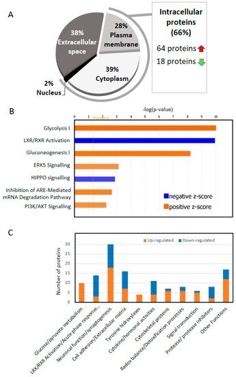
The fact that cerebrospinal fluid (CSF) deeply irrigates the brain together with the relative simplicity of sample extraction from patients make this biological fluid the best target for biomarker discovery in neurodegenerative diseases. Biomarker discovery has been especially fruitful for the identification new proteins that appear in the CSF of Alzheimer’s disease (AD) patients together with amyloid-β (Aβ42), total tau (T-tau), and phosphorylated tau (P-tau). Thus, several proteins have been already stablished as important biomarkers, due to an increase (i.e., CHI3L1) or a decrease (i.e., VGF) in AD patients’ CSF. Notwithstanding this, only a deep analysis of a database generated with all the changes observed in CSF across multiple proteomic studies, and especially those using state-of-the-art methodologies, may expose those components or metabolic pathways disrupted at different levels in AD. Deep comparative analysis of all the up- and down-regulated proteins across these studies revealed that 66% of the most consistent protein changes in CSF correspond to intracellular proteins.

Alzheimer’s disease proteomics cerebrospinal fluid CSF biomarkers

1. Two Thirds of the of the Proteins That Change in the CSF of AD Are Intracellular

While the identification of biomarkers in CSF is crucial to the understanding of AD, unveiling the source of the change is essential to reveal the biological mechanisms that underlie this pathology. Contrary to what we initially expected, given the reduced number of cells present in CSF (approximately 5 cells per mL that mainly correspond to lymphocytes and monocytes); [1], our study reveals that 66% of the CSF altered proteins identified across studies are intracellular proteins, mostly cytoplasmatic (39%; Figure 1A). Amongst them, approximately 80% of the proteins (64) were increased in AD vs. controls. Taking into consideration the widely known neural death which occurs in AD [2], we would expect that a significant origin of these intracellular proteins in CSF may arise from remains of cellular debris that translocate to the CSF [3]. Furthermore, an increased infiltration of neutrophils has been observed during the development of AD. Indeed, increased expression of CD11b positive neutrophils, which are directly related to neutrophil migration, positively correlated with the severity of AD. In this line, a higher number of neutrophils were found in brain vessels of AD patients when compared with controls of similar age [4].
Figure 1. Information regarding proteins that consistently emerge across proteomic studies. (A) Schematic distribution of intracellular (plasma membrane and cytoplasm) and extracellular proteins (extracellular space). (B) Molecular pathways identified using IPA. Only those pathways with a –log(p-value) over 2 and a z-score of + or − 2 were considered. Positive z-scores are represented in orange. Negative z-scores are represented in blue. (C) Classification of the proteins according to their function.
*: Proteins identified with inverted pattern of expression in 1 additional article; &: Proteins identified with inverted pattern of expression in two additional articles; $: β-Amyloid-interacting proteins #: Proteins that bind copper; Δ: Proteins that bind metals. Down/up: protein down-up-regulated in the number of articles indicated; ε: Proteins that were identified at least in one article that used a less novel technique such as two-dimensional gel electrophoresis (2-DE); C: cytokine; H: hormone.
Platelets are considered biomarkers for early diagnosis of AD (as reviewed in [5]). Treatment of platelets with Aβ led to platelet activation and enhanced generation of reactive oxygen species (ROS) and membrane scrambling, suggesting enhanced platelet apoptosis [6]. Interestingly, among the top five pathways detected using Reactome, three of them were related to platelets. Specifically, this study has identified a total of 17 proteins directly related to platelet degranulation (p-value 2.1 × 10⁻¹¹) and or platelet aggregation (p-value 1.1 × −10−07). Whether this finding is related to increased apoptosis requires further investigation.
Together with the cells discussed above an additional source for membrane proteins are exosomes. In this sense, it was previously shown that 1 mL of human CSF contains ~2 μg of endogenous exosomes [7]. The finding that exosomes isolated from human CSF or brain samples sequestered oligomeric Aβ in the brain has led to propose their protective role in AD pathogenesis [7]. Indeed, several proteins of our database such as CHL1, KNG1, or APOA1 were found to be constituents of human CSF exosomes [8].

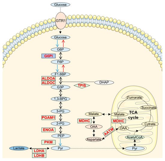
2. Increased Glucose/Pyruvate Metabolism in AD CSF

A variety of proteins related to glucose metabolism were found altered in CSF to the extent that glycolysis and gluconeogenesis constitute the two metabolic pathways most represented in our study (p-value 9.5 × 10⁻¹¹ and 6 × 10⁻⁹, respectively; Figure 1B,C). Furthermore, all the proteins related to glucose metabolism appeared increased in AD CSF. Most of the identified proteins are common enzymes for both processes, glycolysis and gluconeogenesis (Figure 2).
Figure 2. Alteration of glucose/pyruvate metabolism in AD CSF. Schematic representation of glycolysis, gluconeogenesis and pyruvate metabolism. Proteins that increase in AD CSF are labelled in red and green indicates decreased proteins. G6P: glucose-6-phosphate; F6P: Fructose-6-phosphate; G3P: glyceraldehide-3-phosphate; 1,3-BPG: 1,3-Biphosphoglycerate; 3PG: 3-phosphoglyucerate; 2PG: 2-phosphoglycerate; PEP: phosphoenolpyruvate; Pyr: pyruvate; OAA: oxalacetate. Background images were created using templates from Servier Medical Art, which are licensed under a Creative Commons Attribution 3.0 Unported License (http://smart.servier.com/ accessed on 5 July 2021).
It is also important to mention the glycolytic enzyme pyruvate kinase M1/2 (KPYM), that catalyzes the synthesis of pyruvate from phosphoenolpyruvate [9] and the lactate dehydrogenase (LDH), which reduces pyruvate to lactate through a reversible reaction, thus allowing cells to generate or consume lactate depending on their metabolic profile.
Impaired glucose metabolism has been widely recognized as an early feature in the brain of subjects with AD since alteration of brain aerobic glycolysis is frequently observed in the course of AD [10][11]. It has also been proposed that reduced glucose availability in AD would force the brain to rely on gluconeogenesis (de novo synthesis of glucose). Interestingly, despite the low brain glucose uptake in AD, most post-mortem studies show consistent upregulation in glycolytic enzyme proteins [11][12], thus suggesting a compensatory mechanism for the low glucose supply in order to overcome a compromised mitochondrial function. In conclusion, evaluation of an increase of proteins directly related to glucose metabolism in CSF may reveal what takes place in surrounding tissues during AD progression.

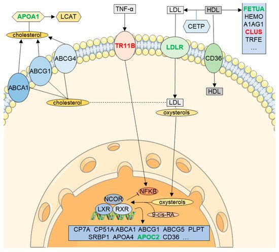
3. RXR Signaling in CSF (LXR/RXR Activation Pathway)

One of the most relevant findings of this study is the significant reduction (p-value = 1.1 × 10⁻¹⁰) of proteins participating in the Liver X Receptor (LXR)/Retinoid X receptor (RXR) pathway in CSF from AD patients (z-score 2.7; Figure 1B,C). LXR/RXR activation pathway (Figure 3) is involved in a variety of processes associated with cholesterol metabolism, inflammation, oxidative stress, etc. [13][14]. Although LXR/RXR pathway represents a relevant pathway altered in AD [13][15], as far as we know, this is the first relevant mention of an overall reduction of LXR/RXR activation in AD CSF. In line with our observations, previous works have detected a reduction in the expression of LXR-β in plasma of AD patients compared to control samples [16]. Furthermore, the LXR/RXR pathway was also reduced in plasma samples of PSAPP and hTau mice models [17]. Retinoids modulate the expression of different key proteins in AD, as presenilin 1 (PS1), metalloprotease 10 (ADAM 10) or β-secretase [18][19]. Moreover, retinoic acid (RA) may have a central role in the pathophysiology of AD and reduced brain levels of this metabolite would constitute a risk factor for the development of the disease. Different mutations on the RA receptors can misregulate AD candidate genes such as PS1, ADAM 10, PS2 or APP [13].
Figure 3. Alteration of LXR/RXR activation pathway in AD CSF. Summarized representation of the LXR/RXR activation pathway. Proteins that increase in AD CSF are labelled in red and green indicates decreased proteins. Background images were created using templates from Servier Medical Art, which are licensed under a Creative Commons Attribution 3.0 Unported License (http://smart.servier.com/ accessed on 5 July 2021).
As we pointed out, a reduction in the LRX activation was predicted with high confidence. This decrease might be related to the advanced development of the pathology, since LXR activation plays a key role in plaque reduction by increasing clearance, thereby improving cognitive impairment [20][21]. Furthermore, genetic loss of LXR in APP/PS1 mice induced a greater accumulation of Aβ plaques [22], while loss of LXR in healthy mice triggered neurodegeneration [23]. Therefore, decreased LXR/RXR pathway components in CSF may inversely correlate with Aβ deposition in tissues.
Another function of the LXR/RXR pathway comprises the activation of apolipoproteins that may serve as cholesterol acceptors [24]. Apolipoproteins constitute a family of proteins with a key role in transport and delivery of lipids, cholesterol homeostasis, and central nervous system (CNS) remodeling [25]. In this study, we observed decreased levels of different apolipoproteins (APOA1, APOC2, and APOL1) in CSF. APOA1, one of the most abundant proteins in human CSF, has been identified as part of senile plaques of AD patients’ brains [26]. A link between the presence of APOC2 and familial Alzheimer’s was observed decades ago [27], although, to date, its specific role in late-onset AD (LOAD) is still unknown. Similarly, APOL1 has been related to other pathologies such as kidney disease [28], while a direct link with AD has not been established yet.

4. Neuronal Function/Synaptogenesis

The organization of the proteins according to their function (Figure 1C) revealed that a high number of them were directly related to neuronal function and specially, synaptogenesis. Synaptogenesis is a dynamic process by which the formation and stabilization of synapses occur in the CNS [29]. In neurodegenerative diseases, such as AD, synaptic degeneration and synapse loss have been described as early events that precede neuronal death [30][31]. Indeed, several studies have proposed a role for synaptic proteins as specific biomarkers for AD in CSF [32][4][5].
The clearest reflection in CSF of the synaptic degeneration that occurs in AD is the family of ‘long’ neuronal pentraxins (NPTX1, NPTX2) and its receptor NPTXR since they constitute the most consistently decreased proteins in CSF and they display a direct function in neural differentiation [33], synaptogenesis [34] and synaptic plasticity [35][36][37]. Specifically, these proteins form mixed NPTX complexes which traffic to the extracellular surface at excitatory synapses where they interact with postsynaptic glutamate receptors [38][39]. Taken together, these three proteins were found to decrease in 8 different proteomic studies. The parallel and consistent decrease of these proteins in CSF would not fit with our previous suggestion of increased neuronal cell death. However, different studies have already described a down-regulation of NPTX2 in AD brains [40][41]. It has also been described that NPTX1 is accumulated in dystrophic neurites and surround plaques in postmortem AD brains [42][43][44], which would explain their decreased levels in CSF. Therefore, the inverse correlation of pentraxins amount in plaques/CSF could be a good indicator of neuronal death and/or synaptic loss.
Similarly, neurexins (NRX), one of the best-characterized families of presynaptic organizers, appeared reduced in 6 different studies. Likewise NPTX1, two of the three family members (NRX1 and NRX2) have been proposed as direct targets of Aβ oligomers [45]. In the same line, calsyntenin-3 (CSTN3), a direct NRX interactor [46][47] was also found to decrease in CSF. This transmembrane protein of the cadherin superfamily is distributed in postsynaptic membranes throughout the adult brain [48]. As occur with NPTX1, CSTN3 accumulates in dystrophic neurite surrounding Aβ plaques [49].
In view of these studies, we have to take into consideration that several proteins may not be released to the CSF and, oppositely, they might somehow be accumulated into the plaque in a process that could be carried out by Aβ and tau aggregation, thus ultimately driving a reduction of certain proteins in CSF. This may explain the variability observed across proteins not only directly involved in neuronal function but also those related to cell adhesion and components of the cell-matrix (Figure 1C). Although at different levels, 21 of these proteins decreased while 25 increased in CSF. The best example that reflects this heterogeneity raises from the members of the SPRC family. Based on our results, four members of this family are differently altered in AD CSF. This family of proteins comprises six members that present calcium-binding domains and regulate cell interaction with the microenvironment [50]. It is worth highlighting the potential role of SPRC on vascular pathology in AD. In the brain, SPRC is also expressed in endothelial cells, wherein it affects trans-endothelial permeability [50]. Moreover, since it acts as a chaperone of collagen IV through the SPRC-collagen binding domain, it has been observed a direct relationship between increased SPRC, collagen IV, and the thickening of the basal lamina of the cerebral vasculature, a feature commonly observed in AD brains [50][51][52].
Notwithstanding this, according to our study, SMOC1 was consistently upregulated in AD CSF in seven different studies, thus making it a potential biomarker. Among its functions, SMOC1 promotes endothelial proliferation [53], and although it is overexpressed in AD brains, wherein it colocalizes with Aβ plaques [54][55], its specific role in AD is still unknown. Conversely, testican-1 (TICN1) was consistently downregulated in AD CSF. Different studies have shown a link between TICN1 overexpression in the brain and AD [56]. It surrounds Aβ plaques in brains of AD patients [56] and regulates proteins related to Aβ production and degradation, such as MMP2 or cathepsin-L [57][58]. Finally, it has been linked to APP sorting. Therefore, as we have previously proposed for other proteins, lower CSF levels of this protein may indicate an accumulation in the brain during the development of the pathology [56].

5. 14-3-3 Proteins Are up Regulated in CSF

The 14-3-3 family consists of seven highly homologous molecules that were first reported as regulators of tyrosine hydroxylase (TH) activity [59], four of which were consistently upregulated in the CSF of AD. These proteins have recently been linked to a variety of processes such as regulation of protein interaction and localization or transcription since they have a nuclear localization sequence [60]. It has been reported that 14-3-3 proteins regulate neuronal differentiation, morphogenesis and migration [61]. The expression of these proteins increases in cortical regions of AD patients and, even though they have not been observed in Aβ plaques, some evidence connects them with neurofibrillary tangles [60]. However, 14-3-3 family members cannot be considered suitable markers for differential diagnosis of AD [62][63] since they have also been detected in CSF of all dementia patients, thus suggesting their role as common markers for neurodegenerative diseases.

6. Cytokines and Hormones up and down Regulated in CSF

Five of the proteins in this section correspond to members of the granin family of proteins (chromogranins, secretogranins and VGF). These precursors of biologically active peptides and the products of their proteolitical cleavage have been proposed as biomarkers of different neurological diseases, included AD [64][65].
As expected, among them, VGF was the most consistent downregulated protein among the identified secreted proteins with biological activities. This protein is nowadays considered one of the best AD markers in CSF as it is consistently reduced in CSF of these patients [32].
Another remarkable protein that was consistently reduced in CSF was somatostatin (SMS). A long time ago it was proposed that diminished levels of somatostatin in CSF may be a specific AD signature compared to other neurodegenerative pathologies as Parkinson’s [66][67]. Whereas the reason for this reduction as a consequence of the pre-propeptide processing remains elusive, it has been recently reported that its deficiency has a direct link with a loss of integrity of the BBB in Aβ-induced toxicity [68].
On the contrary, among the four proteins in this group that appear increased, phosphoprotein 1 (SPP1, also called osteopontin) was found to be the most consistent upregulated protein (increased in seven different studies). This extracellular phosphoprotein is expressed in response to stress and injury and regulates macrophage infiltration and cytokine production [69][70]. In the past years, SPP1 has been linked to inflammation-associated neurological disease. Indeed, higher SPP1 levels have already been described in brain and CSF in AD patients [71][72].

7. Importance of Cofactors in CSF

Herein, we explored common affinities of the identified proteins for given cofactors. Interestingly, copper (Cu2+) emerged as the most common cofactor for several of the identified proteins. Several meta-analysis have identified changes in Cu2+ concentration in brain and serum (reviewed by [73]), although conflicting evidence of copper’s role in AD has been pointed ([74][75], reviewed by [76]). It is well known that Cu2+ and Zn2+ interact with Aβ peptides with high affinity and these interactions have been proposed to accelerate Aβ1-40 and Aβ1-42 aggregation in vitro, thus contributing to their toxicity, ROS generation, and the development of Aβ neurotoxicity [77]. Nonetheless, the mechanism that Cu2+ utilizes to reach the brain is partially understood. Together with albumin (ALBU), the main protein needed for passive diffusion of Cu2+ through the blood-CSF barrier to the brain is ceruloplasmin (CERU) [78]. The fact that both proteins seem to behave inversely in CSF AD, showing decreased levels of ALBU and increased CERU, indicate that the equilibrium of Cu2+ transport in CSF is altered in AD.
Further evidence of the implication of Cu2+ in AD would be represented by metallothionein 3 (MT3), which regulates Cu2+ and Zn2+ transport and storage in CNS and inhibits their toxicity, thus representing one of the major players in metal homeostasis [79]. Conversely, the peptidylglycine α-amidating monooxygenase (AMD), a copper-dependent enzyme that regulates the secretory pathway, was found increased in CSF from AD patients. In mammals, AMD is essential to catalyze α-amidation, a necessary step to confer full biological activity to many neuropeptides [80][81]. Herein, we report for the first time the potential use of AMD as a consistent AD marker in CSF. Furthermore, to our knowledge, there is only one study investigating AMD in CSF where a reduction in enzyme activity in AD samples as compared to healthy, age-matched control was proposed, thus suggesting neuronal dysfunction within the CNS in AD patients [82].

References

  1. Rahimi, J.; Woehrer, A. Overview of cerebrospinal fluid cytology. In Neuropathology; Kovacs, G., Alafuzoff, I., Eds.; Elsevier: Amsterdam, The Netherlands, 2018; pp. 563–571.
  2. Guo, T.; Zhang, D.; Zeng, Y.; Huang, T.Y.; Xu, H.; Zhao, Y. Molecular and cellular mechanisms underlying the pathogenesis of Alzheimer’s disease. Mol. Neurodegener. 2020, 15, 40.
  3. Reiber, H. Dynamics of brain-derived proteins in cerebrospinal fluid. Clin. Chim. Acta 2001, 310, 173–186.
  4. Stock, A.J.; Kasus-Jacobi, A.; Pereira, H.A. The role of neutrophil granule proteins in neuroinflammation and Alzheimer’s disease. J. Neuroinflamm. 2018, 15, 240.
  5. Talib, L.L. Platelet biomarkers in Alzheimer’s disease. World J. Psychiatry 2012, 2, 95.
  6. Arai, K.; Gowert, N.S.; Donner, L.; Chatterjee, M.; Eisele, Y.S.; Towhid, S.T.; Münzer, P.; Walker, B.; Ogorek, I.; Borst, O.; et al. Blood Platelets in the Progression of Alzheimer’s Disease. PLoS ONE 2014, 9, e0090523.
  7. An, K.; Klyubin, I.; Kim, Y.; Jung, J.; Mably, A.J.; O’Dowd, S.T.; Lynch, T.; Kanmert, D.; Lemere, C.A.; Finan, G.M.; et al. Exosomes neutralize synaptic-plasticity-disrupting activity of Aβ assemblies in vivo. Mol. Brain 2013, 6, 47.
  8. Li, M.; Huang, L.; Chen, J.; Ni, F.; Zhang, Y.; Liu, F. Isolation of Exosome Nanoparticles from Human Cerebrospinal Fluid for Proteomic Analysis. ACS Appl. Nano Mater. 2021, 4, 3351–3359.
  9. Zahra, K.; Dey, T.; Ashish; Mishra, S.P.; Pandey, U. Pyruvate Kinase M2 and Cancer: The Role of PKM2 in Promoting Tumorigenesis. Front. Oncol. 2020, 10, 159.
  10. Bell, S.M.; Burgess, T.; Lee, J.; Blackburn, D.J.; Allen, S.P.; Mortiboys, H. Peripheral Glycolysis in Neurodegenerative Diseases. Int. J. Mol. Sci. 2020, 21, 8924.
  11. Le Douce, J.; Maugard, M.; Veran, J.; Matos, M.; Jégo, P.; Vigneron, P.-A.; Faivre, E.; Toussay, X.; Vandenberghe, M.; Balbastre, Y.; et al. Impairment of Glycolysis-Derived l-Serine Production in Astrocytes Contributes to Cognitive Deficits in Alzheimer’s Disease. Cell Metab. 2020, 31, 503–517.e508.
  12. Bigl, M.; Bruckner, M.K.; Arendt, T.; Bigl, V.; Eschrich, K. Activities of key glycolytic enzymes in the brains of patients with Alzheimer’s disease. J. Neural. Transm. 1999, 106, 499–511.
  13. Goodman, A.B.; Pardee, A.B. Evidence for defective retinoid transport and function in late onset Alzheimer’s disease. Proc. Natl. Acad. Sci. USA 2003, 100, 2901–2905.
  14. Ray, S.; Das, B.; Dasgupta, S. Potential therapeutic roles of retinoids for prevention of neuroinflammation and neurodegeneration in Alzheimer’s disease. Neural Regen. Res. 2019, 14, 1880.
  15. Kölsch, H.; Lütjohann, D.; Jessen, F.; Popp, J.; Hentschel, F.; Kelemen, P.; Friedrichs, S.; Maier, T.A.W.; Heun, R. RXRA gene variations influence Alzheimer’s disease risk and cholesterol metabolism. J. Cell. Mol. Med. 2009, 13, 589–598.
  16. Xiong, H.; Callaghan, D.; Jones, A.; Walker, D.G.; Lue, L.-F.; Beach, T.G.; Sue, L.I.; Woulfe, J.; Xu, H.; Stanimirovic, D.B.; et al. Cholesterol retention in Alzheimer’s brain is responsible for high β- and γ-secretase activities and Aβ production. Neurobiol. Dis. 2008, 29, 422–437.
  17. Ojo, J.O.; Crynen, G.; Reed, J.M.; Ajoy, R.; Vallabhaneni, P.; Algamal, M.; Leary, P.; Rafi, N.G.; Mouzon, B.; Mullan, M.; et al. Unbiased Proteomic Approach Identifies Unique and Coincidental Plasma Biomarkers in Repetitive mTBI and AD Pathogenesis. Front. Aging Neurosci. 2018, 10, 405.
  18. Sodhi, R.K.; Singh, N. Retinoids as potential targets for Alzheimer’s disease. Pharmacol. Biochem. Behav. 2014, 120, 117–123.
  19. Obulesu, M.; Dowlathabad, M.R.; Bramhachari, P.V. Carotenoids and Alzheimer’s Disease: An insight into therapeutic role of retinoids in animal models. Neurochem. Int. 2011, 59, 535–541.
  20. Fitz, N.F.; Nam, K.N.; Koldamova, R.; Lefterov, I. Therapeutic targeting of nuclear receptors, liver X and retinoid X receptors, for Alzheimer’s disease. Br. J. Pharmacol. 2019, 176, 3599–3610.
  21. Terwel, D.; Steffensen, K.R.; Verghese, P.B.; Kummer, M.P.; Gustafsson, J.A.; Holtzman, D.M.; Heneka, M.T. Critical Role of Astroglial Apolipoprotein E and Liver X Receptor- Expression for Microglial A Phagocytosis. J. Neurosci. 2011, 31, 7049–7059.
  22. Zelcer, N.; Khanlou, N.; Clare, R.; Jiang, Q.; Reed-Geaghan, E.G.; Landreth, G.E.; Vinters, H.V.; Tontonoz, P. Attenuation of neuroinflammation and Alzheimer’s disease pathology by liver x receptors. Proc. Natl. Acad. Sci. USA 2007, 104, 10601–10606.
  23. Andersson, S.; Gustafsson, N.; Warner, M.; Gustafsson, J.A. Inactivation of liver X receptor leads to adult-onset motor neuron degeneration in male mice. Proc. Natl. Acad. Sci. USA 2005, 102, 3857–3862.
  24. Zelcer, N. Liver X receptors as integrators of metabolic and inflammatory signaling. J. Clin. Investig. 2006, 116, 607–614.
  25. Dominiczak, M.H.; Caslake, M.J. Apolipoproteins: Metabolic role and clinical biochemistry applications. Ann. Clin. Biochem. Int. J. Lab. Med. 2011, 48, 498–515.
  26. Paula-Lima, A.C.; Tricerri, M.A.; Brito-Moreira, J.; Bomfim, T.R.; Oliveira, F.F.; Magdesian, M.H.; Grinberg, L.T.; Panizzutti, R.; Ferreira, S.T. Human apolipoprotein A–I binds amyloid-β and prevents Aβ-induced neurotoxicity. Int. J. Biochem. Cell Biol. 2009, 41, 1361–1370.
  27. Schellenberg, G.; Bird, T.; Wijsman, E.; Orr, H.; Anderson, L.; Nemens, E.; White, J.; Bonnycastle, L.; Weber, J.; Alonso, M.; et al. Genetic linkage evidence for a familial Alzheimer’s disease locus on chromosome 14. Science 1992, 258, 668–671.
  28. Friedman, D.J.; Pollak, M.R. APOL1and Kidney Disease: From Genetics to Biology. Annu. Rev. Physiol. 2020, 82, 323–342.
  29. Cohen-Cory, S. The developing synapse: Construction and modulation of synaptic structures and circuits. Science 2002, 298, 770–776.
  30. Masliah, E.; Mallory, M.; Alford, M.; DeTeresa, R.; Hansen, L.A.; McKeel, D.W.; Morris, J.C. Altered expression of synaptic proteins occurs early during progression of Alzheimer’s disease. Neurology 2001, 56, 127–129.
  31. Selkoe, D.J. Alzheimer’s Disease Is a Synaptic Failure. Science 2002, 298, 789–791.
  32. Pedrero-Prieto, C.M.; García-Carpintero, S.; Frontiñán-Rubio, J.; Llanos-González, E.; Aguilera García, C.; Alcaín, F.J.; Lindberg, I.; Durán-Prado, M.; Peinado, J.R.; Rabanal-Ruiz, Y. A comprehensive systematic review of CSF proteins and peptides that define Alzheimer’s disease. Clin. Proteom. 2020, 17, 1–24.
  33. Boles, N.C.; Hirsch, S.E.; Le, S.; Corneo, B.; Najm, F.; Minotti, A.P.; Wang, Q.; Lotz, S.; Tesar, P.J.; Fasano, C.A. NPTX1 Regulates Neural Lineage Specification from Human Pluripotent Stem Cells. Cell Rep. 2014, 6, 724–736.
  34. Sia, G.-M.; Béïque, J.-C.; Rumbaugh, G.; Cho, R.; Worley, P.F.; Huganir, R.L. Interaction of the N-Terminal Domain of the AMPA Receptor GluR4 Subunit with the Neuronal Pentraxin NP1 Mediates GluR4 Synaptic Recruitment. Neuron 2007, 55, 87–102.
  35. Cummings, D.M.; Benway, T.A.; Ho, H.; Tedoldi, A.; Fernandes Freitas, M.M.; Shahab, L.; Murray, C.E.; Richard-Loendt, A.; Brandner, S.; Lashley, T.; et al. Neuronal and Peripheral Pentraxins Modify Glutamate Release and may Interact in Blood–Brain Barrier Failure. Cereb. Cortex 2017, 27, 3437–3448.
  36. Lee, S.-J.; Wei, M.; Zhang, C.; Maxeiner, S.; Pak, C.; Calado Botelho, S.; Trotter, J.; Sterky, F.H.; Südhof, T.C. Presynaptic Neuronal Pentraxin Receptor Organizes Excitatory and Inhibitory Synapses. J. Neurosci. 2017, 37, 1062–1080.
  37. Wang, Z.; Wang, X.; Zou, H.; Dai, Z.; Feng, S.; Zhang, M.; Xiao, G.; Liu, Z.; Cheng, Q. The Basic Characteristics of the Pentraxin Family and Their Functions in Tumor Progression. Front. Immunol. 2020, 11, 1757.
  38. Yue, W.; Wang, T.; Zachariah, E.; Lin, Y.; Yang, C.S.; Xu, Q.; DiPaola, R.S.; Tan, X.-L. Transcriptomic analysis of pancreatic cancer cells in response to metformin and aspirin: An implication of synergy. Sci. Rep. 2015, 5, 1–11.
  39. Karagkounis, G.; Thai, L.; DeVecchio, J.; Gantt, G.A.; Duraes, L.; Pai, R.K.; Kalady, M.F. NPTX2 is associated with neoadjuvant therapy response in rectal cancer. J. Surg. Res. 2016, 202, 112–117.
  40. Lee, G.W.; Lee, T.H.; Vilcek, J. TSG-14, a tumor necrosis factor- and IL-1-inducible protein, is a novel member of the pentaxin family of acute phase proteins. J. Immunol. 1993, 150, 1804–1812.
  41. Xiao, M.-F.; Xu, D.; Craig, M.T.; Pelkey, K.A.; Chien, C.-C.; Shi, Y.; Zhang, J.; Resnick, S.; Pletnikova, O.; Salmon, D.; et al. NPTX2 and cognitive dysfunction in Alzheimer’s Disease. eLife 2017, 6, e23798.
  42. De Groot, J.; Sontheimer, H. Glutamate and the biology of gliomas. Glia 2011, 59, 1181–1189.
  43. Radin, D.P.; Patel, P. A current perspective on the oncopreventive and oncolytic properties of selective serotonin reuptake inhibitors. Biomed. Pharmacother. 2017, 87, 636–639.
  44. Abad, M.A.; Enguita, M.; DeGregorio-Rocasolano, N.; Ferrer, I.; Trullas, R. Neuronal Pentraxin 1 Contributes to the Neuronal Damage Evoked by Amyloid- and Is Overexpressed in Dystrophic Neurites in Alzheimer’s Brain. J. Neurosci. 2006, 26, 12735–12747.
  45. Brito-Moreira, J.; Lourenco, M.V.; Oliveira, M.M.; Ribeiro, F.C.; Ledo, J.H.; Diniz, L.P.; Vital, J.F.S.; Magdesian, M.H.; Melo, H.M.; Barros-Aragão, F.; et al. Interaction of amyloid-β (Aβ) oligomers with neurexin 2α and neuroligin 1 mediates synapse damage and memory loss in mice. J. Biol. Chem. 2017, 292, 7327–7337.
  46. Pettem, K.L.; Yokomaku, D.; Luo, L.; Linhoff, M.W.; Prasad, T.; Connor, S.A.; Siddiqui, T.J.; Kawabe, H.; Chen, F.; Zhang, L.; et al. The Specific α-Neurexin Interactor Calsyntenin-3 Promotes Excitatory and Inhibitory Synapse Development. Neuron 2013, 80, 113–128.
  47. Um, J.W.; Pramanik, G.; Ko, J.S.; Song, M.Y.; Lee, D.; Kim, H.; Park, K.S.; Südhof, T.C.; Tabuchi, K.; Ko, J. Calsyntenins Function as Synaptogenic Adhesion Molecules in Concert with Neurexins. Cell Rep. 2014, 6, 1096–1109.
  48. Hintsch, G. The Calsyntenins—A Family of Postsynaptic Membrane Proteins with Distinct Neuronal Expression Patterns. Mol. Cell. Neurosci. 2002, 21, 393–409.
  49. Uchida, Y.; Gomi, F. The role of calsyntenin-3 in dystrophic neurite formation in Alzheimer’s disease brain. Geriatr. Gerontol. Int. 2016, 16, 43–50.
  50. Pilozzi, A.; Carro, C.; Whalen, M.; Huang, X. Blood-Brain Barrier Degradation and the Implication of SPARC Protein as a Potential Therapeutic Target for Alzheimer’s Disease. In Alzheimer’s Disease: Drug Discovery; Huang, X., Ed.; Exon Publications: Brisbane, Australia, 2020.
  51. McCurdy, S.; Baicu, C.F.; Heymans, S.; Bradshaw, A.D. Cardiac extracellular matrix remodeling: Fibrillar collagens and Secreted Protein Acidic and Rich in Cysteine (SPARC). J. Mol. Cell. Cardiol. 2010, 48, 544–549.
  52. Giannoni, P.; Arango-Lievano, M.; Neves, I.D.; Rousset, M.-C.; Baranger, K.; Rivera, S.; Jeanneteau, F.; Claeysen, S.; Marchi, N. Cerebrovascular pathology during the progression of experimental Alzheimer’s disease. Neurobiol. Dis. 2016, 88, 107–117.
  53. Awwad, K.; Hu, J.; Shi, L.; Mangels, N.; Abdel Malik, R.; Zippel, N.; Fisslthaler, B.; Eble, J.A.; Pfeilschifter, J.; Popp, R.; et al. Role of secreted modular calcium-binding protein 1 (SMOC1) in transforming growth factor β signalling and angiogenesis. Cardiovasc. Res. 2015, 106, 284–294.
  54. Bai, B.; Wang, X.; Li, Y.; Chen, P.-C.; Yu, K.; Dey, K.K.; Yarbro, J.M.; Han, X.; Lutz, B.M.; Rao, S.; et al. Deep Multilayer Brain Proteomics Identifies Molecular Networks in Alzheimer’s Disease Progression. Neuron 2020, 105, 975–991.e977.
  55. Sathe, G.; Albert, M.; Darrow, J.; Saito, A.; Troncoso, J.; Pandey, A.; Moghekar, A. Quantitative proteomic analysis of the frontal cortex in Alzheimer’s disease. J. Neurochem. 2020, 156, 988–1002.
  56. Barrera-Ocampo, A.; Arlt, S.; Matschke, J.; Hartmann, U.; Puig, B.; Ferrer, I.; Zürbig, P.; Glatzel, M.; Sepulveda-Falla, D.; Jahn, H. Amyloid-β Precursor Protein Modulates the Sorting of Testican-1 and Contributes to Its Accumulation in Brain Tissue and Cerebrospinal Fluid from Patients with Alzheimer Disease. J. Neuropathol. Exp. Neurol. 2016, 75, 903–916.
  57. Schechter, I.; Ziv, E. Cathepsins S, B and L with aminopeptidases display beta-secretase activity associated with the pathogenesis of Alzheimer’s disease. Biol. Chem. 2011, 392, 555–569.
  58. Merlo, S.; Sortino, M.A. Estrogen activates matrix metalloproteinases-2 and -9 to increase beta amyloid degradation. Mol. Cell. Neurosci. 2012, 49, 423–429.
  59. Ichimura, T.; Isobe, T.; Okuyama, T.; Yamauchi, T.; Fujisawa, H. Brain 14-3-3 protein is an activator protein that activates tryptophan 5-monooxygenase and tyrosine 3-monooxygenase in the presence of Ca2+,calmodulin-dependent protein kinase II. FEBS Lett. 1987, 219, 79–82.
  60. Cho, E.; Park, J.-Y. Emerging roles of 14-3-3γ in the brain disorder. BMB Rep. 2020, 53, 500–511.
  61. Cornell, B.; Toyo-oka, K. 14-3-3 Proteins in Brain Development: Neurogenesis, Neuronal Migration and Neuromorphogenesis. Front. Mol. Neurosci. 2017, 10, 318.
  62. Foote, M.; Zhou, Y. 14-3-3 proteins in neurological disorders. Int. J. Biochem. Mol. Biol. 2012, 3, 152–164.
  63. Kelly, J.; Moyeed, R.; Carroll, C.; Albani, D.; Li, X. Gene expression meta-analysis of Parkinson’s disease and its relationship with Alzheimer’s disease. Mol. Brain 2019, 12, 1–10.
  64. Bartolomucci, A.; Pasinetti, G.M.; Salton, S.R.J. Granins as disease-biomarkers: Translational potential for psychiatric and neurological disorders. Neuroscience 2010, 170, 289–297.
  65. Bartolomucci, A.; Possenti, R.; Mahata, S.K.; Fischer-Colbrie, R.; Loh, Y.P.; Salton, S.R.J. The Extended Granin Family: Structure, Function, and Biomedical Implications. Endocr. Rev. 2011, 32, 755–797.
  66. Serby, M.; Richardson, S.B.; Twente, S.; Siekierski, J.; Corwin, J.; Rotrosen, J. CSF somatostatin in Alzheimer’s disease. Neurobiol. Aging 1984, 5, 187–189.
  67. Sagar, S.M.; Flint Beal, M.; Marshall, P.E.; Landis, D.M.D.; Martin, J.B. Implications of neuropeptides in neurological diseases. Peptides 1984, 5, 255–262.
  68. Paik, S.; Somvanshi, R.K.; Kumar, U. Somatostatin-Mediated Changes in Microtubule-Associated Proteins and Retinoic Acid–Induced Neurite Outgrowth in SH-SY5Y Cells. J. Mol. Neurosci. 2019, 68, 120–134.
  69. Sodhi, C.P.; Phadke, S.A.; Batlle, D.; Sahai, A. Hypoxia Stimulates Osteopontin Expression and Proliferation of Cultured Vascular Smooth Muscle Cells. Diabetes 2001, 50, 1482–1490.
  70. Rabenstein, M.; Vay, S.U.; Flitsch, L.J.; Fink, G.R.; Schroeter, M.; Rueger, M.A. Osteopontin directly modulates cytokine expression of primary microglia and increases their survival. J. Neuroimmunol. 2016, 299, 130–138.
  71. Wung, J.; Perry, G.; Kowalski, A.; Harris, P.R.; Bishop, G.; Trivedi, M.; Johnson, S.; Smith, M.; Denhardt, D.; Atwood, C. Increased Expression of the Remodeling- and Tumorigenic-Associated Factor Osteopontin in Pyramidal Neurons of the Alzheimers Disease Brain. Curr. Alzheimer Res. 2007, 4, 67–72.
  72. Comi, C.; Carecchio, M.; Chiocchetti, A.; Nicola, S.; Galimberti, D.; Fenoglio, C.; Cappellano, G.; Monaco, F.; Scarpini, E.; Dianzani, U. Osteopontin is Increased in the Cerebrospinal Fluid of Patients with Alzheimer’s Disease and Its Levels Correlate with Cognitive Decline. J. Alzheimer’s Dis. 2010, 19, 1143–1148.
  73. Sensi, S.L.; Granzotto, A.; Siotto, M.; Squitti, R. Copper and Zinc Dysregulation in Alzheimer’s Disease. Trends. Pharmacol. Sci. 2018, 39, 1049–1063.
  74. Mold, M.; Ouro-Gnao, L.; Wieckowski, B.M.; Exley, C. Copper prevents amyloid-β1–42 from forming amyloid fibrils under near-physiological conditions in vitro. Sci. Rep. 2013, 3, 1256.
  75. Singh, I.; Sagare, A.P.; Coma, M.; Perlmutter, D.; Gelein, R.; Bell, R.D.; Deane, R.J.; Zhong, E.; Parisi, M.; Ciszewski, J.; et al. Low levels of copper disrupt brain amyloid-β homeostasis by altering its production and clearance. Proc. Natl. Acad. Sci. USA 2013, 110, 14771–14776.
  76. Bagheri, S.; Squitti, R.; Haertlé, T.; Siotto, M.; Saboury, A.A. Role of Copper in the Onset of Alzheimer’s Disease Compared to Other Metals. Front. Aging Neurosci. 2018, 9, 446.
  77. Roberts, B.R.; Ryan, T.M.; Bush, A.I.; Masters, C.L.; Duce, J.A. The role of metallobiology and amyloid-beta peptides in Alzheimer’s disease. J. Neurochem. 2012, 120 (Suppl. 1), 149–166.
  78. Twomey, P.J.; Viljoen, A.; House, I.M.; Reynolds, T.M.; Wierzbicki, A.S. Relationship between serum copper, ceruloplasmin, and non-ceruloplasmin-bound copper in routine clinical practice. Clin. Chem. 2005, 51, 1558–1559.
  79. Vašák, M.; Meloni, G. Mammalian Metallothionein-3: New Functional and Structural Insights. Int. J. Mol. Sci. 2017, 18, 1117.
  80. Eipper, B.A.; Mains, R.E. Peptide α-Amidation. Annu. Rev. Physiol. 1988, 50, 333–344.
  81. Eipper, B.A.; Stoffers, D.A.; Mains, R.E. The biosynthesis of neuropeptides: Peptide alpha-amidation. Annu. Rev. Neurosci. 1992, 15, 57–85.
  82. Wand, G.S.; May, C.; May, V.; Whitehouse, P.J.; Rapoport, S.I.; Eipper, B.A. Alzheimer’s disease: Low levels of peptide alpha-amidation activity in brain and CSF. Neurology 1987, 37, 1057–1061.
More
Information
Contributor MDPI registered users' name will be linked to their SciProfiles pages. To register with us, please refer to https://encyclopedia.pub/register :
View Times: 532
Revisions: 2 times (View History)
Update Date: 08 Oct 2021
1000/1000
Video Production Service