Submitted Successfully!
To reward your contribution, here is a gift for you: A free trial for our video production service.
Thank you for your contribution! You can also upload a video entry or images related to this topic.
Version Summary Created by Modification Content Size Created at Operation
1 + 7829 word(s) 7829 2021-09-19 15:27:57 |
2 format correct Meta information modification 7829 2021-10-08 08:05:23 |

Video Upload Options

Do you have a full video?

Confirm

Are you sure to Delete?
Cite
If you have any further questions, please contact Encyclopedia Editorial Office.
Neagu, M. CM Epigenetics. Encyclopedia. Available online: https://encyclopedia.pub/entry/14656 (accessed on 28 April 2024).
Neagu M. CM Epigenetics. Encyclopedia. Available at: https://encyclopedia.pub/entry/14656. Accessed April 28, 2024.
Neagu, Monica. "CM Epigenetics" Encyclopedia, https://encyclopedia.pub/entry/14656 (accessed April 28, 2024).
Neagu, M. (2021, September 28). CM Epigenetics. In Encyclopedia. https://encyclopedia.pub/entry/14656
Neagu, Monica. "CM Epigenetics." Encyclopedia. Web. 28 September, 2021.
CM Epigenetics
Edit

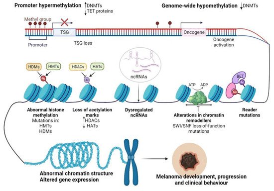
Cutaneous melanoma (CM) is an aggressive neoplasm that evolves from the malignant transformation of neural crest stem cell-derived melanocytes. The etiology of melanoma is multifactorial, and the most prominent factors include genetic predisposition, light skin color, multiple naevi, and excessive exposure to UV. Epigenetic alterations have emerged as essential contributors in the pathogenesis of various human diseases, including CM. Epigenetics is another layer of instructions apart from the genetic code that controls how genes are read and expressed, involving a change in the cell phenotype without changes in the genotype. Epigenetic regulation is an umbrella term that encompasses several mechanisms such as DNA methylation, histone post-translational modifications (PTMs), nucleosome remodeling, histone variants, and RNA-mediated post-transcriptional regulation. Influenced by lifestyle and environmental factors, epigenetic changes are highly dynamic and reversible and thus easy to regulate. 

cutaneous melanoma epigenetic regulation inflammation drug resistance biomarkers therapeutic targets

1. CM Epigenetics

Recent advancements in next-generation sequencing (NGS) technologies and their coupling with chromatin-immunoprecipitation (CHIP), altogether RNA interference (RNAi) screening methods, and matrix-assisted laser desorption/ionization time-of-flight (MALDI-TOF) proteomic tools have allowed the dissection of the epigenome for various cancers [1]. In particular, for CM, epigenomic interrogation revealed aberrant DNA methylation in gene promoters [2], histone PTMs [3], alteration of epigenetic regulators [4], and dysregulated ncRNAs [5] (Figure 1). It seems that in CM these epigenetic alterations may allow melanocytes to overcome senescence and metastasize at a distance [6][7], support the immune escape of CM [8], but also the transcriptomic reprogramming of cancer cells to overcome the cancer therapy-induced apoptosis [9].
Figure 1. Epigenetic changes regulating CM progression and clinical behavior. Abbreviations: Me—methylation, Ac—acetylation, DNMTs—DNA methyltransferases, TET—ten-eleven-translocation enzyme, TSG—tumor suppressor gene, HDMs—histone demethylases, HMTs—histone methyltransferases, HDACs—histone deacetylases, HATs—histone acetyltransferases, SWI/SNF—switch/sucrose non-fermentable chromatin remodeling complex, BET—bromodomain and extra-terminal domain protein, ncRNAs—non-coding RNAs, ATP—adenosine triphosphate, ADP—adenosine diphosphate. Figure created with https://biorender.com/.
Inflammation and epigenetic alterations play pivotal roles in CM initiation and development. However, in recent years, considerable research efforts have been devoted to identifying a potential link between these processes in the context of cancer. Current literature confirms that these two phenomena are interconnected and mutually regulate each other [10][11]. Epigenetics can modulate tumor antigen presentation and immune cell functions, therefore impacting tumor development and clinical behavior [12]. Conversely, inflammation can induce epigenetic alterations in resident skin cells, promoting immune evasion and tumorigenesis [11]. We will focus herein mainly on inflammation-induced epigenetic changes in CM, as it is a less studied topic in the field. In the first instance, inflammation can disrupt epigenetic programs by altering the metabolic state of a cell [13]. Their activation determines alteration of immune cells’ metabolism and activated immune cells further disrupt the metabolic processes in neighboring tissues. Since the activity of many epigenetic enzymes depends on cellular metabolism intermediates, a dysfunctional metabolism will significantly impact the molecular processes within the cell [13]. For example, DNMTs and HMTs use SAM as a cofactor, while HDMs and TET proteins require α-ketoglutarate produced in the tricarboxylic acid cycle for their activity [14]. Moreover, it has been reported that increased production of cytokines, chemokines, and ROS, including hydrogen peroxides, can orchestrate dramatic epigenetic changes in resident epithelial cells. For instance, DNA damage triggered by ROS exposure can interfere with the ability of certain epigenetic regulators to bind to DNA, leading to abnormal DNA methylation patterns and altered gene expression [15]. Additionally, long-term production and accumulation of cytokines and ROS/reactive nitrogen species (RNS) have been correlated with the activation of STAT3 and NF-κB oncogenic pathways in epithelial cells [16][17]. In parallel, antigen-presenting cells (APCs) and cytokines can activate T cells, involving transcription factors that participate in their transcriptomic reprogramming, which includes epigenetic changes, among others [11]. Epigenetic alterations are usually pro-tumorigenic, facilitating the suppression of tumor suppressor genes and the activation of oncogenes. These reversible changes may also help tumors escape the immune response by reducing the expression of genes involved in the antigen processing and presentation or viral defense pathways. In line with these observations, myeloid-derived suppressor cells (MDSCs) may differentiate into tumor-associated macrophages (TAMs) or interfere with T cell activity, promoting tumorigenesis as transcription factors induce alternative transcriptional programs resulting in epigenetic alterations [11]. Here, we will describe the current status of knowledge regarding the roles of DNA methylation and chromatin modifications in CM as a problematic inflammatory malignancy.

2. Epigenetic Alterations Driving CM initiation and Progression

2.1. DNA Methylation in CM Development

Disruption of DNA methylation is a common event in cancer. Both focal hypermethylation at CpG islands and global hypomethylation are constant hallmarks of the cancer genome and often coexist in tumor cells, impacting tumor biology and behavior. As with other cancers, CM initiation and progression have been associated with loss of tumor suppressors and oncogene activation (Figure 1) [18].
Inactivation of tumor suppressor genes (TSGs) due to specific DNA methylation in the promoter regions was the first epigenetic alteration studied in CM more than 10 years ago [19]. So far, dozens of genes are known to be regulated by this mechanism. These genes appear to be involved in various signaling pathways, usually disrupted in CM, such as the phosphatidylinositol 3-kinase/protein kinase B (PI3K/Akt) and MAPK pathways, cell cycle, DNA repair, retinoblastoma (RB) and Wnt signaling [18]. Furthermore, it was also observed that three TSGs are frequently inactivated by methylation: RASSF1A (55%), RAR-β2 (70%), and MGMT (34%) can also be identified in the circulating tumor DNA of CM patients, which makes them useful diagnostic and prognostic biomarkers in the clinical setting [20][21]. In several cancers, but also in a significant proportion of melanomas, a gradual increase in DNA hypermethylation was observed along with tumor aggressiveness; this phenomenon, called CpG methylator phenotype (CIMP), was reported for the first time in colorectal cancers, a finding that highlighted a tight correlation between altered DNA methylation patterns and the clinical outcome of the affected patients. Tanemura at al. demonstrated that during tumor progression, several tumor-related genes and loci, including WIF1, SOCS1, RASSF1A, TFPI2, MINT17, and MINT31, gain methylation with advancing stages. These genes have been suggested to constitute CM’s CIMP [22]; however, recent research highlights that CIMP is usually associated with an NRAS-mutant phenotype, which is more aggressive than a non-NRAS-mutant tumor [23]. Therefore, future approaches should correlate CIMP with CM patients’ clinical outcomes and mutational profiles. This information would be essential for developing novel tools for prognosis and response to therapy in the clinical cohorts of melanoma patients.
Epigenetic silencing of tumor suppressor genes has been one of the best-studied phenomena in CM; however, although less studied, DNA hypomethylation is equally important in the initiation and development of CM. It was shown that in many cancers, hypomethylation contributes to tumor progression by inducing genome instability via the demethylation of transposons and pericentromeric repeats or the activation of certain oncogenes [24]Figure 1 depicts hypomethylation mechanisms as a hallmark of melanomagenesis. LINE-1 elements are one of the most abundant classes of mobile DNAs within the human genome [25]. LINE-1 hypomethylation, detected in both tissues and plasma circulating DNA of melanoma patients seems to be a hallmark of the metastatic capacity of primary melanomas [26][27]. Other reports highlighted that LINE-1 hypomethylation may predict the OS in stage III CM patients [28][29]. In parallel, DNA hypomethylation has been described as one of the main mechanisms regulating the expression of cancer-testis-antigens (CTAs) in human melanomas [30]. CTAs are a specific group of tumor-associated antigens (TAAs) whose expression in normal tissues is generally restricted to the gametogenic tissues of the testis and fetal ovaries [31]; nonetheless, CTAs were found to be re-expressed via hypomethylation in CM, regulating vital cellular processes such as tumor cell division, differentiation, invasion and drug resistance [30][32][33][34].
Several studies have revealed that DNA methylome analysis may help discriminate between normal melanocytes, nevi, and melanomas. For instance, Fujiwara et al. identified several novel genes that were hypermethylated in melanomas compared to melanocytes, such as KRTCAP3, AGAP2, ZNF490, and TTC22, in addition to those previously documented, such as COL1A2, GPX3, and NPM2 [35]. Among those genes, they found that NPM2 showed distinct immunohistochemical (IHC) staining in normal melanocytes, whereas its expression was lost in CM samples [35]. Moreover, Gao et al. reported a diagnostic algorithm based on the methylation patterns of CLDN11, CDH11, and PPP1R3C genes that can differentiate between dysplastic nevi and primary melanomas with a specificity of 89% and a sensitivity of 67% [36]. Other reports highlighted that several methylation subgroups might be associated with different clinical characteristics of the disease, potentiating that the evaluation of DNA methylomes may have prognostic applications in CM. A study led by Lauss et al. revealed three methylation clusters: MS1, MS2, and MS3, which differ significantly in terms of promoter methylation, proliferation, and presence in immune cells [37]. The MS1 group has the highest methylation level, especially at CpG islands and poised promoters, enriched in polycomb repressive complex (PRC2) target genes. The MS1 subtype also showed an increased frequency of homologous deletions of CDKN2A and IDH1R132 hotspot mutations. The MS3 group had the lowest methylation levels, similar to peripheral blood leukocytes, and MS2 was intermediate. No correlations were identified between methylation clusters and clinicopathological variables or actionable mutations such as BRAF or NRAS. However, the tumors bearing MS1-signature were associated with the lower patient OS (20 months for MS1 vs. 60 months for MS3). Interestingly, genetic analysis revealed methylation clusters are associated with different biological and clinical behaviors. The MS1 subtype termed “proliferative” was associated with the upregulation of TP53, MDM2, CDK4, CDK6, CCND1, CCNE1, and E2F3, as well as epigenetic modifiers TET1, JARID1B, SWI/SNF chromatin remodelers, and DNMT3A; in contrast, the MS3 subtype harbored an “immune high” signature, possibly explaining the better survival of patients appending to the MS3 cluster [37]. Similarly, Yamamoto et al. stratified 51 CM into two risk groups based on the promoter region’s methylation status. The high-methylated subgroup was positively associated with a thicker tumor progression and hence a worse clinical prognosis. Among the 27 genes proposed to distinguish between the two subtypes, TFPI2 was the most frequently hypermethylated gene in the aggressive subtype [38]. In addition, altered methylation patterns of the homeobox D cluster were linked with melanoma metastasizing to the brain [39].
Further complicating this scenario, genome-wide mapping of CM revealed that 5 hmC levels are progressively lost during tumor progression from benign nevus to malignant melanoma, via IDH2 and TET family downregulation [40]. Elevated levels of 5 hmC were subsequently validated by IHC staining as predictors of metastasis-free survival and overall survival in CM patients [41]. Taken together, all this information supports the further development of 5-hmC IHC expression as a prognostic biomarker that can add some precision to the American Joint Committee on Cancer (AJCC) staging system [42].

2.2. Histone-Modifying Enzymes and PTMs in CM Development

In addition to the undeniable role of DNA methylation in melanomagenesis, histone PTMs mediated by several writers, readers and erasers may also alter some transcriptional processes closely associated with CM initiation and progression [43].
The involvement of histone modifications in CM development has been suggested since benign nevi, which usually carry the BRAF V600E oncogenic mutation, rarely become melanoma, as this conversion requires additional events. Yet, valuable insights on the importance of histone-modifying enzymes and PTMs in melanomagenesis were obtained using zebrafish melanoma models [44]. Patton et al. developed the first experimental model of BRAF V600E driven melanoma using a zebrafish model expressing BRAF V600E under the control of the mitfa promoter in a p53 loss-of-function background [45]. Only a fraction of zebrafish developed melanocytic tumors, highlighting the existence of additional molecular events operating in concert with genetic alterations in melanoma. To examine these processes in more detail, the researchers developed a triple transgenic zebrafish model (p53/BRAF/crestin: EGFP), in which the crestin/EGFP gene marks neural crest stem and progenitor cells, from which melanocytes originate [45]. Melanocytic tumors reported in zebrafish models re-expressed crestin-EGFP gene, suggesting that these cancer cells are maintaining their neural crest identity. Notably, they identified enrichment of H3K27ac marks in super-enhancers at the sox10 locus, a major regulator of neural crest formation and melanomagenesis, suggesting that epigenetic regulation of SOX10 is an important step in melanoma initiation [45]. Later on, Scahill at al. revealed that loss of kdm2aa, an orthologue of KDM2A, triggered the spontaneous formation of melanomas at a high frequency in zebrafish [46]. These tumors were generated independently of BRAF V600E and other melanoma-related mutations in oncogenes and tumor suppressors. Finally, gene expression analysis revealed altered levels of genes involved in DNA replication, translation, and chromatin regulation after kdm2aa silencing, confirming on an alternative pathway that histone methylation may have vital roles in melanomagenesis [46].

2.2.1. Histone Modifications “Writers”

H3K4 Methyltransferases (KMT2D)

One of the chromatin’s writer enzymes that has been identified to function aberrantly in melanomas is the KMT2D, also known as MLL2. KMT2D is associated with gene promoter and enhancer regions and catalyzes H3K4 monomethylation [47][48][49]. Several recent in vitro genetic screen studies have revealed important details regarding the roles of KMT2D in CM tumorigenesis [50][4]. By performing the first in vivo genetic screen with shRNA libraries targeting fundamental epigenetic players in CM, Bossi et al. observed multiple genes involved in melanomagenesis [50]. Among them, KMT2D orchestrates a migratory transcriptional program in NRAS melanomas. The authors also reported some interpatient heterogeneity in their study [50]. Interestingly, KMT2D silencing resulted in the inactivation of a subset of KMT2D-bound enhancers (reduced H3K4me1 and H3K27ac) and downregulation of MFGE8 and RPL39L cell motility genes. Notably, the closest genes to these enhancers were the KMT2D target genes, suggesting that KMT2D can deregulate enhancer activity to promote tumorigenesis [50]. Therefore, a better understanding of the roles of KMT2D in CM may help expand the number of biomarkers and druggable genes in the clinical management of CM patients.
After the first published results in 2016, KMT2D was reported as frequently mutated in a variety of solid and hematologic tumors, including melanomas (15%) [4]. Recently, Maitituoheti at al. identified KMT2D serving tumor suppressor roles in CM. In addition to KMT2D, the authors identified seven more epigenetic regulators in CM cell lines (KDM1A, APOBEC2, HDAC6, KMT2F, SETD4, KAT4, and KDM5B) whose loss accelerates CM tumor progression. Among them, KMT2D, KDM5B, KMT2F and KDM1A were mainly associated with H3K4 methylation. However, the most potent phenotypes were linked with mutations in KMT2D. To investigate CM genesis in more detail, the authors developed a genetically engineered mouse model (GEMM) based on conditional and melanocyte-specific ablation of KMT2D [4]. It has been further observed that H3K4me1-marked enhancer reprogramming by KMT2D loss is associated with a drastic alteration of the central metabolic pathways in the tumor cells. Furthermore, the authors observed a preferential dependence of glycolysis in deficient KMT2D tumors compared to WT cells, most likely to provide cancerous tumors’ energy and biomass needs. Interestingly, pharmacological inhibition of glycolysis and the IGF signaling pathway reduced the proliferation of KMT2D-deficient tumor cells in both murine models and human melanoma cell lines [4]. Thus, this study highlights exciting aspects of the biology of mutant KMT2D tumors and identifies new potential therapeutic vulnerabilities concerning them.

The Roles of H3K4 Methylation Marks

H3K4me1, H3K4me2 and H3K4me3 are generally associated with active gene transcription [51]. Although partially overlapping with H3K4me3 at the 5’-end level of transcribing genes, H3K4me2 decorates genomic regions independently of H3K4me3 [52]. H3Kme2’s role as a biomarker in the diagnosis and prognosis of CM patients has been suggested by several studies. For example, Uzdensky at al. found elevated levels of H3K4me2 in tumor samples compared to paired normal skin [53]. Later on, Kampilafkos at al. observed that H3K4me2 and H3K27me3 levels were lower in metastatic compared to primary melanomas and that combined IHC analysis of H3K4me2, H3K27me3, and EZH2 may help identify cancer cells with stem cell-like behaviors, particularly at the invasion front of CM [3]. This study highlighted that the combination of several genetic alterations may be more relevant for characterizing and predicting complex events such as metastasis and that all these epigenetic changes can be integrated as a code that can provide valuable information about the biology of melanocytic tumors.
H3K4me3 is a chromatin landmark of promoters of transcriptionally active genes or genes poised for activation in mammalian cells [51]. In particular, for CM, Cheng at al. observed that human metastatic tissues are highly heterogeneous in terms of H3K4me3 levels [49]. Further analysis showed that metastatic lesions that displayed low levels of H3K4me3 were associated with repressed genes in embryonic stem cells (ESCs) and PRC2-target genes. In contrast, elevated H3K4me3 levels correlated with interferon and inflammatory response genes [49]. However, Terranova at al. found that metastatic melanomas harbor exceptionally wide H3K4me3 domains [54]. These domains can span tens of thousands of kilobases, and appear to be involved in the regulation of several EMT transcription factors (POU3F2, SOX9, and PDGFRA) as well as melanocyte-specific master regulators (MITF, ZEB2, and TFAP2A) [55]. Terranova et al. finally highlighted that particular events such as BRAF or NRAS mutations may employ specific chromatin states (bivalent states and broad H3K4me3 domains) to orchestrate transcriptional changes unique to a genotype, suggesting that epigenetic mechanisms play important roles in regulating CM behaviors [54].

H3K27 Methyltransferases (EZH2)

Histone lysine methyltransferase EZH2, responsible for H3K27 trimethylation, was also found to be dysregulated during the development of human melanomas. EZH2 is the catalytic subunit of the PRC2 complex and appears overexpressed in various tumors, including melanoma [56]. PRC2 trimethylates H3K27, orchestrating the repression of transcriptional programs [56]. Particularly for melanoma, PRC2 levels have been reported to increase gradually over the progression from benign nevi to malignant melanomas, suggesting that this protein plays a key role in CM initiation and progression [3]. Moreover, EZH2 and H3K27me3 are overexpressed in highly invasive melanoma cells and metastatic melanomas, leading to TSGs inactivation [3][7][56][57]. Finally, other studies have revealed that increased EZH2 levels are associated with poor prognosis in CM patients [58].
Regarding the roles played by EZH2 in the pathogenesis of CM, several mechanisms by which it supports tumor growth and metastasis have been proposed. Some authors have shown that EZH2 expression is associated with the suppression of senescence in human melanoma cells. For example, Fan at al. have shown that EZH2 can support unlimited melanocyte proliferation by repressing CDKN1A, which is not mediated by H3K27me3 deposition [59]. Conversely, EZH2 silencing inhibits cell proliferation, restoring senescent phenotype and p21/CDKN1A expression in a p53-independent manner. It was further observed that depletion of EZH2 removes HDAC1 from the transcriptional start site of CDKN1A, resulting in increased H3 acetylation and transcriptional activation. These observations confirm the existence of a synergistic relationship between EZH2, as part of PRC2 and HDAC, in mediating the suppression of certain senescence-related genes in melanoma cells [59]. In parallel, De Donatis at al. showed that EZH2 oncogenic activation is mediated by the non-canonical NF-kB signaling pathway; interestingly, NF-kB2 silencing was associated with reconversion to the senescent phenotype, suggesting the pivotal roles of the NF-kB2/EZH2 axis in CM initiation and development [60]. Other studies have shown that induction of EZH2 in benign BrafV600E- or NrasQ61K-expressing melanocytes facilitates tumor metastasis and invasiveness by silencing genes relevant for cell surface organelle primary cilium integrity and by activating Wnt/β-catenin oncogenic signaling [8]. Finally, it is also worth mentioning that EZH2 gain-of-function mutations usually co-occur with BRAF V600E mutations in CM, promote aggressive cell morphologies and enhance melanoma tumor growth in vitro [61].

H3K9 Methyltransferases

Another histone-modifying enzyme involved in CM pathogenesis is bifurcated domain SUV39/SET 1 (SETDB1), which belongs to the SUV39 family of histone lysine methyltransferases [62]. This enzyme is involved in the trimethylation of H3K9, which is a specific signature of transcriptionally repressive chromatin [63].
In human melanoma samples, SETB1 often appears amplified in association with another histone methyltransferase, EHMT1 [64]. Subsequently, a positive association between SETB1 expression and several prognostic factors such as increased mitotic index, advanced Clark levels, and epidermal involvement in tissue biopsies of CM patients was also described [65]. A recent study, led by Orouji at al., revealed that the expression and amplification rate of SETDB1 may serve as an individual prognostic biomarker in CM, with increased levels of SETB1 protein being associated with metastasis and lower patient survival rates [66]. Compared to normal melanocytes, melanoma cells showed 8–13.9 times higher levels of SETDB1. Interestingly, they found that all those SETDB1 highly amplified human cell lines were BRAF V600E mutants [66]. Functional studies have shown that SETDB1 exerts its oncogenic effects in CM by modulating the expression of thrombospondin 1, a molecule known for its involvement in cell migration and invasiveness [66]. Surprisingly, it was found that SETDB1 regulates not only H3K9 methylation patterns but also H3K4me1 deposition, emphasizing that SETDB1’s involvement in tumorigenesis is much broader than previously thought. Furthermore, treatment with an H3K9me-specific histone methyltransferase inhibitor is highly effective in this context, leading to a considerable decrease in tumor cell viability. Interestingly, melanoma cells harboring low levels of SETDB1 were not affected by treatment with epigenetic inhibitors, underscoring SETDB1’s role as a valuable therapeutic target in CM [66].

2.2.2. Histone Modifications “Readers”

Protein readers can recognize specific chromatin changes or combinations of PTMs and histone variants to further direct the transcriptional outcome [67]. Some of the best-studied families of chromatin readers are the bromodomain and extra-terminal domain (BET) family of adapter proteins (BRD2, BRD3, BRD4, and BRDT). BET proteins have an increased affinity for acetylated histone residues, enabling transcriptional activation by interaction with the transcriptional machinery [68]. Notably, the SWI/SNF complex, which uses energy from ATP hydrolysis to reshape the structure of chromatin, is also dependent on the presence of bromodomain-containing domains to be fully functional [69]. BET protein involvement in melanomagenesis was demonstrated by Segura at al. when it was shown that BRD2 and BRD4 are overexpressed in human melanoma cell lines and tissues, controlling the expression of certain genes involved in cell cycle progressions and survival [70]. Gene expression and IHC analysis of human tissue biopsies have confirmed higher levels of BRD2 and BRD4 in primary and metastatic tumors relative to melanocytes and nevi, suggesting that these BET proteins are involved in melanoma tumorigenesis [70]. Treatment with BET inhibitors (BETi) significantly reduced tumor growth and metastasis in both in vitro and in vivo models, and those effects were recapitulated by individual silencing of BRD4. Notably, the pharmacological capabilities of BETi have not been influenced by the mutational status of BRAF or NRAS, offering new promises for the treatment of patients that do not harbor actionable mutations [70]. Collectively, all these observations potentiate the pivotal role of the BRD4 protein in CM tumorigenesis, propelling it as a potential prognosis biomarker and therapeutic target in CM.

2.2.3. Histone Modifications “Erasers”

Histone Deacetylases (HDACs)

One of the most important classes of chromatin erasers associated with CM pathogenesis is HDACs, which catalyze the deletion of acetyl groups from histone tails [71]. At least 18 mammalian HDACs were identified and subdivided into four main classes, depending on their location and functional characteristics. Given the significant differences reported between the acetylation patterns of benign nevi and the malignant tissues of patients diagnosed with CM, it was thought that aberrant histone deacetylation could play important roles in the pathobiology of CM [72]. Subsequent studies with patient-derived cell cultures have indeed shown that there is a loss of acetylation marks (H3K27Ac, H2BK5Ac, and H4K5Ac) and H3K4me2/3 during the transition from premalignant to the malignant phenotype, resulting in alterations of some essential signaling pathways in CM formation, including PI3K, interferon (IFN) -Ɣ, and TRAIL- and platelet-derived growth factor (PDGF) signaling [72].
Many other studies have shown that various HDACs are involved in skin tumorigenesis and can be exploited as biomarkers or therapeutic targets in melanomas. For instance, Wilmott et al. identified that increased expression of nuclear HDAC3 and cytoplasmic HDAC8 may serve as indicators of better prognosis in stage IV melanoma patients [73]. They also revealed an increase in HDAC8 levels in BRAF-mutant tumors [73]. Additionally, HDAC6 expression has recently been correlated with advanced stages and with an unfavorable prognosis [74]. In line with these observations, certain in vitro studies have shown that HDAC5 and HDAC6 are overexpressed in melanoma cell lines versus normal skin cells, being required for melanoma proliferation and metastasis through different signaling pathways [75]. Interestingly, HDAC6 inhibition inhibited tumor cell proliferation, and when knocked down cells were inoculated in animal models a decreased PD-L1 production and an augmented T-cell-mediated immune response was obtained [75].
Another epigenetic player investigated for its involvement in CM initiation is SIRT1 (class III HDAC proteins), which was found overexpressed in human melanoma cells and tissues in comparison to normal skin and melanocytes [76]. Further studies suggested that SIRT1 is upregulated in metastatic tumors compared to primary tumors, most likely due to its ability to support EMT programs via autophagic degradation of E-cadherin [77]. SIRT1, SIRT3 and SIRT6 were also proposed to support tumor growth in CM [78][79]. Interestingly, a recent study showed that dual inhibition of SIRT1 and SIRT3 mediated by 4′-bromo-resveratrol inhibits melanoma cell proliferation and growth [80]. Thus, all this information suggests that pro-tumorigenic sirtuins have not only the value of prognosis biomarkers but also of potential therapeutic targets in CM and that inhibition of multiple sirtuins may be a promising strategy for improving clinical management of CM.

Histone Demethylases (HDMs)

The scientific progress made in recent years in deciphering the cancer epigenome has revealed the critical roles of histone H3 lysine 4 (H3K4) JARID1B/KDM5B demethylase in the tumorigenesis of various human tumors, including melanoma [81]. One of the pioneering studies in the field revealed that H3K4 demethylase JARID1B is overexpressed in melanocyte nevi and almost absent in melanoma samples [82]. However, subsequent studies have revealed increased heterogeneity of KDM5B in CM, and its expression levels have been documented to define distinct cellular states, even with antithetical effects on cellular tumor fate depending on the biological and clinical context [83]. KDM5B was found to mark a slow-cycling subpopulation of tumor cells which is essential for continuous tumor growth and resistance to therapy [84]. This population displays similar behaviors to that of cancer stem cells and give rise to a heterogeneous population of melanoma cells, being a major contributor to the increased heterogeneity observed in CM tumors [64]. Therefore, the assessment of KDM5B expression and H3K4 deposition patterns can provide valuable information about the clinical behavior of these tumors and may lead to more personalized therapies for CM patients.
H3K9me3 is an epigenetic mark of heterochromatin, which is often present on distal regions of genes [64]. H3K9 methyl groups may be erased by members of the lysine-specific histone demethylase (LSD) family. LSD1, often referred to as KDM1A, has the ability to demethylate histone 3 on lysine residues at position 4 (H3K4- gene promoter) and 9 (H3K9- distal) [64]. Interestingly, Yu et al. reported that oncogene-induced senescence of melanocytes relies on the deposition of H3K9me3 at the promoters of proliferation-related genes [6]. This is in accordance with their findings highlighting that benign naevi displayed increased senescence-associated H3K9me3 levels, with almost no detectable activity of H3 lysine 9 demethylases LSD1 and Jumonji Domain-Containing Protein 2D (JMJD2C/KDM4C), whilst human melanoma tissues generally harbored increased expression of LSD1 and JMJD2C and reduced H3K9me3 reactivity [6]. To gain a broader understanding of the molecular mechanisms that are involved in this process, the authors induced the expression of LSD1 and JMJD2C in mouse and zebrafish models. It was further shown that these two enzymes cooperated to overcome the oncogenic Ras G12V/BRAF V600E-induced senescence by preventing H3K9me3 deposition at E2F target gene promoters, which further augmented melanomagenesis [6]. Of note, targeted inhibition of LSD1 and JMJD2C demethylases restored cellular senescence and growth arrest, potentiating LSD1 and JMJD2C regulation as a potential anti-cancer therapeutic strategy [6].

3. Epigenetic Alterations Involved in CM Drug Resistance

3.1. Resistance to MAPK Inhibitors (MAPKi)

Genomic profiling of CMs revealed several actionable mutations in tumors that may be matched with targeted therapies. Recurrent driver alterations such as BRAF V600, NRAS, and NF-1 facilitated the design of BRAF inhibitors (BRAFi: vemurafenib and dabrafenib) and MEKi (trametinib, cobimetinib, and binimetinib) that have significantly improved patient OS [85][86]. Although 60–80% of BRAF-mutated CM patients respond well to targeted therapy, a significant proportion of them develop resistance, which results in life-threatening metastases and death [43][87][88]. The phenomenon of MAPKi resistance is complex and multifactorial and involves, among others, alterations of the BRAF V600E gene (amplification, aberrant splicing) [89][90][91], which leads to MAPK pathway hyperactivation, mutations that activate alternative survival pathways [92], modifications in apoptotic machinery [93], RTK hyperactivation [94][95], and the presence of slow-cycling populations [84], to which are added other MITFs, c-AMP, and NF-kB related mechanisms [96]. In CM, tumor refractoriness has been extensively linked with genetic alterations in many cancer-related genes; however, in some cases, the cause of the resistance appears to be non-genetic in nature [97]. Peculiarities such as the rapid kinetics and the transient nature of refractory phenotypes suggest the existence of an epigenetic basis for drug resistance in CM, pointing out that epigenetic remodeling is a fundamental feature of tumor development and adaptation to therapy [97][98]. Therefore, new therapeutic targets and therapies are critically necessary to improve the therapeutic management of CM. In this section, epigenetic alterations associated with MAPKi resistance and how they can be exploited in the future to become therapeutic targets and biomarkers in CM will be highlighted.

3.1.1. DNA Methylation and MAPKi Resistance

Studies highlighting the involvement of DNA methylation in CM targeted therapy resistance are relatively few. Al Emran et al. reported DNMT3A, DNMT3B, and DNMT1 as differentially expressed in the BRAF V600E melanoma cells refractory to MAPKi, which resulted in low DNA methylation levels. However, genome-wide integrated epigenetic analyses revealed that altered histone methylation patterns, rather than DNA methylation, are involved in the transition from the normal state toward the resistant phenotype [99]. In parallel, Hugo at al. observed that drug resistance programs are associated with dramatic transcriptomic and methylomic alterations in MAPKi-treated CM patients. Transcriptomic analyses indicated dysregulated mRNA levels of LEF1, TAP1, CD8, and DUSP4 genes in MAPKi-resistant tumors, which correlated with differential methylation at CpG islands, suggesting the critical roles of DNA methylation in transcriptomic reprogramming of melanoma cells to support MAPKi resistance [100].

3.1.2. Histone-Modifying Enzymes and PTMs Involved in MAPKi Resistance

One of the clinical observations that have postulated the link between epigenetic regulation and resistance to cancer therapy is the so-called “drug holiday” concept, which refers to intermittent treatment programs or treatment breaks. This strategy is often applied to delay the onset of resistance to therapy but is not potent on a genetically resistant phenotype [101]. Particularly for CM, it was observed that rechallenging patients with BRAFi after a free period of treatment and tumor progression resulted in a significant clinical response upon BRAFi and BRAF + MEKi treatments [102]. Recently, several studies have shown that the administration of a third-line BRAF-targeted therapy following first-line targeted therapy and second-line immunotherapy may be an effective strategy in CM metastatic patients [103][104]. Targeted therapy rechallenge in subjects who previously progressed on targeted therapies and immunotherapy was associated with a 2.7–5.9 month median progression-free survival (PFS), 9.3–19 month median OS and a 34–35% disease control rate. Notably, the time between treatment initiation and rechallenge did not seem to impact treatment responses [103][104].
Another aspect that advocates for epigenetically mediated drug-resistance phenotypes is that of slow-cycling cell populations, which appear to be endowed with reversible drug tolerance. One of the most important observations in this regard is that a very small fraction of cells can survive following exposure to drug concentrations 100-fold higher than IC50 [105]. These cells were found in a quiescent state and G1 arrest and continued to be viable in the presence of the drug. The induction of a “drug holiday”, however, resensitized these cells to initial therapy, potentiating the plasticity of the drug-tolerant phenotype of these cells. These refractory tumor populations showed an altered chromatin state, with elevated KDM5A expression levels and a dramatic depletion of H3K4me2/3 marks. Notably, RNA-mediated KDM5A silencing confirmed that this histone demethylase allows for the maintenance of a reversible drug-tolerant state in human melanoma cells [105]. The critical role of the KDM5B epigenetic regulator in the generation of cell subpopulations with distinct drug sensitivity profiles was recently confirmed in the study of Liu et al. [9]. They found in mouse melanomas two cell subpopulations, CD34+ and CD34−, endowed with the characteristics of stem and progenitor cells, which may differ considerably in their clinical behaviors. It was further observed that the CD34+ and CD34− subpopulations displaying the BRAFV600E mutation may respond differently to targeted BRAFi. Interestingly, KDM5B overexpression reprogrammed melanoma cells to a CD34−, more drug-tolerant, phenotype, while KDM5B loss shifted melanoma cells to a more BRAFi-responsive CD34+ state, potentiating the pivotal role of KDM5A in modulating intratumoral heterogeneity in CM [9]. Moreover, KDM5B, another H3K4 demethylase, has been observed to play similar roles in the responsiveness of CM tumors to targeted therapies [106].
Further complicating the drug-resistance scenario, several studies have highlighted those particularities of the tumor microenvironment such as hypoxia and nutrient starvation alongside genotoxic pressure exerted by drugs can give rise to induced drug-tolerant cells (IDTCs) rather than a selection of a pre-existing subpopulation. One of these studies revealed that continuous exposure of melanoma cells to sub-lethal BRAFi concentrations induced surviving cells to adopt a less differentiated state and become refractory to 20-fold higher BRAFi concentrations, as well as to other MEKi and platinum salts [107]. At the molecular level, it has been observed that IDTCs display exacerbated expression of drug efflux ATB-binding cassette transporters and melanoma stem cell markers and loss of differentiation markers such as melan-A and Tyrosinase, which are MITF-target genes. Depletion of histone marks H3K4me3, H3K27me3 alongside a remarkable increase in H3K9me3 was observed in IDTCs cells. The authors also reported an overexpression of several histone-modifying enzymes including the H3K27-specific demethylases, KDM6A, KDM6B, and the H3K4-specific demethylases, KDM1B, KDM5A, and KDM5B, in the IDTC states. Interestingly, as was observed for the KDM5A-enriched subpopulation, IDTCs regained their therapeutic sensitivity seven days after treatment interruption [107]. Hypoxic conditions and nutrient starvation were also associated with the transition to an H3K4me3low/H3K27me3low/H3K9me3high phenotype and the IDTCs generated in this manner exhibited increased refractoriness to BRAFi, suggesting an epigenetically regulated drug-independent stress response that allows cancer cells to cope with difficult environmental conditions [107]. All these observations enhance the role of tumor heterogeneity of CM as the main determinant of resistance to targeted therapies.
It is also well documented that epigenetic alterations can interfere with the MAPKi mechanism of action. MAPK inhibitors cause cellular apoptosis by adjusting the balance between members of the Bcl-2 family, more precisely, by inducing the pro-apoptotic factors Bim and Bmf and by reducing Mcl-1 expression [108][109]. In contrast, MAPKi-resistant melanoma cells showed overexpression of Mcl-1, concomitantly with Noxa downregulation, which counteracted the MAPKi-induced cell death [93][110]. Notably, inhibition of EZH2 expression has been associated with the release of apoptosis-inducing factor (AIFM1) from mitochondria and the induction of caspase-independent apoptosis in human melanoma cell lines [111]. Therefore, all this information suggests that using EZH2 inhibition in conjunction with MAPKi may be a promising strategy to combat the hurdle of drug resistance in CM. Moreover, it is well documented that the BET family of histone reader proteins also turns melanoma cells against apoptosis. There are at least two BET proteins in melanoma, in this case BRD2 and BRD4, that are documented to be overexpressed during melanoma progression. Interestingly, several studies have potentiated that inhibition of BET proteins is associated with an increase in BIM, which synergizes with the induction of BIM after MAPK inhibition, and that the combination of BETi and MAPKi may be an effective pharmacological approach in CM [70][112]. Equally exciting results were obtained by combining HDACi with BETi in CM cell lines, leading to deregulation of anti-apoptotic proteins and components of the AKT and Hippo/YAP signaling pathways [113].
Despite the remarkable progress made in understanding the biology of melanocytic tumors, drug resistance remains a major problem in CM therapeutic management. Epigenetic reprogramming has the potential to reshape the metabolic and signaling networks in cancers, facilitating the emergence of tumor cell subpopulations with distinct behavior and antigenic profile [114]. This intratumor heterogeneity drives new resistance mechanisms to escape the genotoxic pressure or the immune system, facilitating metastasis and disease relapse [43]. However, novel omics technologies such as single-cell analysis and clustered regularly interspaced short palindromic repeats (CRISPR)-associated protein 9 (Cas9) genome editing tool are expected to revolutionize CM research and partially solve the issue of intratumoral heterogeneity, either by screening for novel therapeutic targets or by functional genome/epigenome editing.

3.2. Resistance to Immunotherapy

The development of immunotherapy, which has transformed the management of metastatic tumors, has undoubtedly been fostered by a comprehensive understanding of the tumor microenvironment (TME) and its immunophenotype. Tumors can be reduced to two main compartments that are closely intertwined: the malignant cells and TME. TME is composed of a variety of stromal cells embedded in an extracellular matrix irrigated by a complex network of blood and lymphatic vessels [115]. The cells within the stromal compartment can include immune cells (macrophages, B lymphocytes and cytotoxic T lymphocytes (CTLs), natural killer cells (NKCs), neutrophils, and dendritic cells), mesenchymal cells (fibroblasts, myofibroblasts, cancer stem cells- CSCs, mesenchymal stem cells- MSCs, adipocytes, and endothelial cells), and MDSCs [116][117]. Moreover, in melanoma’s TME, TAMs are abundant and due to their pro-tumoral M2 phenotype these “tumor hijacked” cells sustain therapy resistance [118]. Stromal cells are in close communication with tumor cells and help them adapt to a changing microenvironment, survive, and replicate. As melanomas have a clear immune fate since immunosurveillance favors efficient tumor elimination and immunotolerance promotes tumor survival [116], therapy resistance has a clear immunological background. Additionally, it is well known that melanomas have an increased mutational rate and express a plethora of antigens, for example CTAs, which attracts immune cells that can eradicate the tumor or can be diverted towards pro-tumoral activity [119].
Natural immunosuppression has emerged as a physiological mechanism, but in TME immunosuppression usually interferes with CTL activity and functions [120][121]. Tumor cells mediate immunosuppression taking over inhibitory checkpoint proteins such as PD-1, T cell immunoglobulin and mucin domain 3 (TIM-3), lymphocyte activation gene 3 (LAG-3), and CTLA-4 expressed on the surface of T lymphocytes [122][123]. PD-1 and CTLA-4 bind to specific ligands such as PD-L1 and PD-L2, or CD80 and CD86, respectively, to negatively regulate T cell activity, leading to immune cell escape [123]. Therefore, the task of immunotherapy is much more challenging than it seems, as its goal is not to induce apoptosis but to modulate TME to induce a state of immunosurveillance that destroys cancer cells [43]. Anti-CTLA-4 antibody (Ipilimumab/Tramelimumab), approved for clinical management of CM more than five years ago [124] and followed closely by the approval of anti-PD1 (Nivolumab) [125], is the main immune therapeutical player that changed the fate of melanoma patients. Recently published data regarding the long-term therapy with individual and/or combined therapies in melanoma patients has shown that complete response is witnessed in 28% of patients and that there is still a great percentage of patients with incomplete response, patients that gain resistance and/or patients that due to immune-related adverse effect have to cease their immune therapy [126].
To date, the proposed mechanisms for CM resistance to immunotherapy are downregulation of MHC molecules, loss of antigenic expression, T-cell exhaustion, aberrant expression of PD-L1 in response to IFN-γ production by T cells, along with the altered expression of chemokines such as CCL3, CXCL1, and CCL4 [96]. Although it is well known that epigenetic regulation has critical roles in shaping the identity and differentiation of immune cells, the administration of epigenetic therapy should be done with caution in immunotherapy-resistant CMs, as these agents can affect other cells within TME in addition to tumor cells [101].

3.2.1. DNA Methylation and Resistance to Immunotherapy

The discovery that the immune system can be harnessed to fight cancer and improve clinical outcomes in CM was recognized with a Nobel prize in 2018 [127]. Nevertheless, further studies drove research toward elucidating how DNA methylation can impact the function and activity of immune system components [128]. Earlier studies have shown that DNA methylation appears to be involved in regulating T cell differentiation and exhaustion [129][130], but also in modulating immune checkpoint genes [131], which are the main biomarkers for the response to immunotherapy.
Several authors have reported a mechanistic link between DNA methylation status and immune checkpoint gene expression that may have important predictive and monitoring implications for immunotherapy-treated CM patients. For instance, methylation of immune checkpoint CTLA4 has recently been associated with worse response and progression-free survival (PFS) in stage IV CM patients treated with ipilimumab [132]. The same study also highlighted an inverse correlation between CTLA4 promoter methylation and the presence of tumor-infiltrating lymphocytes (TILs), which play a critical role in tumor control and response to immunotherapy. Therefore, melanoma samples with a low level of methylation are likely to have an increased immune cell infiltration, and an increased number of TILs is an indicator of a good clinical response. However, no significant correlation was reported between CTLA4 promoter methylation and CTLA-4 protein expression, suggesting that the level of protein expression of CTLA4 cannot be used as a predictive biomarker in CM [132]. Other authors have shown that the pattern of PD-L1 methylation may also be suggestive of the response to immunotherapy. Briefly, it has been shown that the degree of DNA methylation of melanoma cells facilitates the stratification of CM patients into four subgroups based on the expression of PD-L1 and TILs and that this information may provide clues about the therapeutic response and survival rates of these CM patients [133][134]. Earlier studies have shown that highly responsive patients showed elevated levels of TILs and PD-L1, while the nonresponsive group displayed low levels of TILs and PD-L1 [133]. In the meantime, while clinical information has been gathered about this clear-cut PD-L1 high expression, efficient immune therapy has been shaken due to various newly discovered molecular mechanisms [135]. DNA methylome analysis of 52 stage III patients from TCGA revealed that low/absent PD-L1 expression is associated with high DNA methylation, differential expression of immune-related genes and worse survival [136]. In parallel, Micevic at al. confirmed that PD-L1 methylation regulates PD-L1 expression and identified for the first time the existence of methylated CpG loci at the PD-L1 promoter [137]. The authors further stratified melanomas into 2 groups based on PD-L1 status and observed that PD-L1 hypomethylation is associated with increased PD-L1 expression and superior OS in CM patients regardless of the diagnosed stages [137]. Moreover, studies on melanoma cells showed that treatment with the hypomethylating agent 5-azacytidine can orchestrate transcriptional derepression in hypermethylated PD-L1 tumors, leading to amplification of PD-L1 expression [137]. Finally, some other studies confirmed the epigenetic regulation of immune checkpoint gene LAG3 via DNA methylation in CM [138]. LAG3 is a molecule involved in blocking tumor cell proliferation and regulating the production of IFN-γ and TNFα cytokines. Interestingly, it has been shown that LAG3 promoter hypomethylation positively associates with increased levels of tumor-infiltrating immune cells and better PFS in CM patients [138].
Given that anti-PD-L1 and anti-PD-1 antibodies are the most extensively used immunotherapies in the clinical setting, and PD-L1 hypermethylation renders CM resistant to ICIs, applying DNMTi treatments appears a tempting strategy to reverse CM immunotherapy resistance. Interestingly, it has been noted that DNMTi hs the ability to activate endogenous retroviruses (ERVs) and virus defense-related pathways in melanoma cells [139]. In the TCGA, the expression levels of viral defense genes may help in stratifying primary samples from multiple tumors, including CM, into two risk groups where a high defense signature positively associates with improved OS and more durable clinical response. Moreover, combining anti-CTLA-4 with low doses of DNMTi proved to be an effective strategy in augmenting the immunotherapy efficiency in a mouse melanoma model [139]. Therefore, the use of immune checkpoint inhibitors in conjunction with DNMTi may be a promising strategy for maximizing the therapeutic benefit in CM patients. Certainly, we will soon find out more about the efficacy and safety of combining DNMTi with immunotherapy, given that ongoing clinical trials are studying the oral use of azacitidine with pembrolizumab in patients with metastatic CM (NCT02816021).

3.2.2. Histone-Modifying Enzymes and PTMs Involved in Immunotherapy Resistance

To date, information on histone enzymes and PTMs involved in immunotherapy resistance are even vaguer than in the case of epigenetic regulation by DNA methylation. Several studies have suggested a mechanistic link between EZH2 activity and resistance to immunotherapy. For example, Zingg at al. observed that anti-CTLA-4 or IL-2 immunotherapy leads to TNFα amplification and T cell infiltration, resulting in EZH2 overexpression and loss of tumor control in melanoma mouse models [140]. Mechanistically, EZH2 catalyzes the deposition of H3K27me3 marks and the suppression of a plethora of immune-related genes. Notably, EZH2 inactivation reversed the drug-resistant phenotype and amplified the effects of anti-CTLA-4 and IL-2 immunotherapy in melanoma mouse models, thus blocking CM growth and dissemination [140]. Therefore, in this study, Zingg at al. have demonstrated not only that EZH2 expression is a valuable biomarker for monitoring the response to immunotherapy but can also be exploited as a therapeutic target to restore and enhance the effects of immunotherapy.
Additional studies have reinforced that the involvement of EZH2 in resistance to CM immunotherapy could be much broader. Tiffen at al. analyzed 471 cases of CM in the TCGA and found that 20% of patients displayed copy number amplifications and mRNA upregulation, along with activating mutations in EZH2 [141]. RNAseq analysis further showed that these alterations correlated with DNA hypermethylation and downregulation of certain genes involved in tumor suppression, antigen processing, and presentation pathways. Treatment with the EZH2 inhibitor GSK126 reversed the transcriptional silencing driven by EZH2 alterations in CM cells, suggesting that EZH2 inhibition is a promising pharmacological strategy for improving the therapeutic response in CM [141]. From these observations, it appears that there may be a close link between EZH2 and the activity of DNMTs in regulating tumor biological properties, including the response to immunotherapy in CM. It is well documented that the ATRX_DNMT3_DNMT3L (ADD) domain of DNMT3A may interact with several epigenetic players, such as SUV39H1 methyltransferases, HDAC1, and EZH2, among others [142]. Moreover, it was postulated that the activity of DNMTs is supported by EZH2, a well-known target of PI3K/Akt signaling, and that they cooperate in cancer pathogenesis [143]. In support of this idea is the observation that EZH2 and DNMTs are regulated by similar upstream signaling cascades, such as MEK/ERK and PI3K/Akt, and transcription factors such as NF-kB2 [128]. Last but not least, it appears that EZH2 can pre-mark genes for DNMTs methylation [144]. Therefore, although still in its infancy, the study of epigenetic mechanisms concerning the response to immunotherapy is expected to not only guide and revolutionize the treatment of refractory patients, but also to stratify them into risk groups to provide personalized therapeutic solutions.

References

  1. Farooqi, A.A.; Fayyaz, S.; Poltronieri, P.; Calin, G.; Mallardo, M. Epigenetic deregulation in cancer: Enzyme players and non-coding RNAs. Semin. Cancer Biol. 2020.
  2. de Unamuno Bustos, B.; Murria Estal, R.; Pérez Simó, G.; Simarro Farinos, J.; Pujol Marco, C.; Navarro Mira, M.; Alegre de Miquel, V.; Ballester Sánchez, R.; Sabater Marco, V.; Llavador Ros, M.; et al. Aberrant DNA methylation is associated with aggressive clinicopathological features and poor survival in cutaneous melanoma. Br. J. Dermatol. 2018, 179, 394–404.
  3. Kampilafkos, P.; Melachrinou, M.; Kefalopoulou, Z.; Lakoumentas, J.; Sotiropoulou-Bonikou, G. Epigenetic modifications in cutaneous malignant melanoma: EZH2, H3K4me2, and H3K27me3 immunohistochemical expression is enhanced at the invasion front of the tumor. Am. J. Dermatopathol. 2015, 37, 138–144.
  4. Maitituoheti, M.; Keung, E.Z.; Tang, M.; Yan, L.; Alam, H.; Han, G.; Singh, A.K.; Raman, A.T.; Terranova, C.; Sarkar, S.; et al. Enhancer Reprogramming Confers Dependence on Glycolysis and IGF Signaling in KMT2D Mutant Melanoma. Cell Rep. 2020, 33, 108293.
  5. Neagu, M.; Constantin, C.; Cretoiu, S.M.; Zurac, S. miRNAs in the Diagnosis and Prognosis of Skin Cancer. Front. Cell Dev. Biol. 2020, 8, 71.
  6. Yu, Y.; Schleich, K.; Yue, B.; Ji, S.; Lohneis, P.; Kemper, K.; Silvis, M.R.; Qutob, N.; van Rooijen, E.; Werner-Klein, M.; et al. Targeting the Senescence-Overriding Cooperative Activity of Structurally Unrelated H3K9 Demethylases in Melanoma. Cancer Cell 2018, 33, 322–336.e8.
  7. Zingg, D.; Debbache, J.; Schaefer, S.M.; Tuncer, E.; Frommel, S.C.; Cheng, P.; Arenas-Ramirez, N.; Haeusel, J.; Zhang, Y.; Bonalli, M.; et al. The epigenetic modifier EZH2 controls melanoma growth and metastasis through silencing of distinct tumour suppressors. Nat. Commun. 2015, 6, 6051.
  8. Zingg, D.; Debbache, J.; Peña-Hernández, R.; Antunes, A.T.; Schaefer, S.M.; Cheng, P.F.; Zimmerli, D.; Haeusel, J.; Calçada, R.R.; Tuncer, E.; et al. EZH2-Mediated Primary Cilium Deconstruction Drives Metastatic Melanoma Formation. Cancer Cell 2018, 34, 69–84.e14.
  9. Liu, X.; Zhang, S.-M.; McGeary, M.K.; Krykbaeva, I.; Lai, L.; Jansen, D.J.; Kales, S.C.; Simeonov, A.; Hall, M.D.; Kelly, D.P.; et al. KDM5B Promotes Drug Resistance by Regulating Melanoma-Propagating Cell Subpopulations. Mol. Cancer Ther. 2019, 18, 706–717.
  10. Licht, J.D.; Bennett, R.L. Leveraging epigenetics to enhance the efficacy of immunotherapy. Clin. Epigenetics 2021, 13, 115.
  11. Maiuri, A.R.; O’Hagan, H.M. Interplay Between Inflammation and Epigenetic Changes in Cancer. Prog. Mol. Biol. Transl. Sci. 2016, 144, 69–117.
  12. Maes, K.; Mondino, A.; Lasarte, J.J.; Agirre, X.; Vanderkerken, K.; Prosper, F.; Breckpot, K. Epigenetic Modifiers: Anti-Neoplastic Drugs With Immunomodulating Potential. Front. Immunol. 2021, 12, 652160.
  13. Kominsky, D.J.; Campbell, E.L.; Colgan, S.P. Metabolic shifts in immunity and inflammation. J. Immunol. 2010, 184, 4062–4068.
  14. Johnson, C.; Warmoes, M.O.; Shen, X.; Locasale, J.W. Epigenetics and cancer metabolism. Cancer Lett. 2015, 356, 309–314.
  15. Franco, R.; Schoneveld, O.; Georgakilas, A.G.; Panayiotidis, M.I. Oxidative stress, DNA methylation and carcinogenesis. Cancer Lett. 2008, 266, 6–11.
  16. Ciążyńska, M.; Olejniczak-Staruch, I.; Sobolewska-Sztychny, D.; Narbutt, J.; Skibińska, M.; Lesiak, A. Ultraviolet Radiation and Chronic Inflammation-Molecules and Mechanisms Involved in Skin Carcinogenesis: A Narrative Review. Life 2021, 11, 326.
  17. Fan, Y.; Mao, R.; Yang, J. NF-κB and STAT3 signaling pathways collaboratively link inflammation to cancer. Protein Cell 2013, 4, 176–185.
  18. Micevic, G.; Theodosakis, N.; Bosenberg, M. Aberrant DNA methylation in melanoma: Biomarker and therapeutic opportunities. Clin. Epigenetics 2017, 9, 34.
  19. Howell, P.M.J.; Liu, S.; Ren, S.; Behlen, C.; Fodstad, O.; Riker, A.I. Epigenetics in human melanoma. Cancer Control 2009, 16, 200–218.
  20. Hoon, D.S.B.; Spugnardi, M.; Kuo, C.; Huang, S.K.; Morton, D.L.; Taback, B. Profiling epigenetic inactivation of tumor suppressor genes in tumors and plasma from cutaneous melanoma patients. Oncogene 2004, 23, 4014–4022.
  21. Palanca-Ballester, C.; Rodriguez-Casanova, A.; Torres, S.; Calabuig-Fariñas, S.; Exposito, F.; Serrano, D.; Redin, E.; Valencia, K.; Jantus-Lewintre, E.; Diaz-Lagares, A.; et al. Cancer Epigenetic Biomarkers in Liquid Biopsy for High Incidence Malignancies. Cancers 2021, 13, 3016.
  22. Tanemura, A.; Terando, A.M.; Sim, M.-S.; van Hoesel, A.Q.; de Maat, M.F.G.; Morton, D.L.; Hoon, D.S.B. CpG island methylator phenotype predicts progression of malignant melanoma. Clin. cancer Res. an Off. J. Am. Assoc. Cancer Res. 2009, 15, 1801–1807.
  23. Garcia-Alvarez, A.; Ortiz, C.; Muñoz-Couselo, E. Current Perspectives and Novel Strategies of NRAS-Mutant Melanoma. Onco. Targets. Ther. 2021, 14, 3709–3719.
  24. Ehrlich, M. DNA hypomethylation in cancer cells. Epigenomics 2009, 1, 239–259.
  25. Ponomaryova, A.A.; Rykova, E.Y.; Gervas, P.A.; Cherdyntseva, N.V.; Mamedov, I.Z.; Azhikina, T.L. Aberrant Methylation of LINE-1 Transposable Elements: A Search for Cancer Biomarkers. Cells 2020, 9, 2017.
  26. Ecsedi, S.I.; Hernandez-Vargas, H.; Lima, S.C.; Herceg, Z.; Adany, R.; Balazs, M. Transposable hypomethylation is associated with metastatic capacity of primary melanomas. Int. J. Clin. Exp. Pathol. 2013, 6, 2943–2948.
  27. De Araújo, É.S.S.; Kashiwabara, A.Y.; Achatz, M.I.W.; Moredo, L.F.; De Sá, B.C.S.; Duprat, J.P.; Rosenberg, C.; Carraro, D.M.; Krepischi, A.C. V LINE-1 hypermethylation in peripheral blood of cutaneous melanoma patients is associated with metastasis. Melanoma Res. 2015, 25, 173–177.
  28. Sigalotti, L.; Fratta, E.; Bidoli, E.; Covre, A.; Parisi, G.; Colizzi, F.; Coral, S.; Massarut, S.; Kirkwood, J.M.; Maio, M. Methylation levels of the “long interspersed nucleotide element-1” repetitive sequences predict survival of melanoma patients. J. Transl. Med. 2011, 9, 78.
  29. Tonella, L.; Pala, V.; Ponti, R.; Rubatto, M.; Gallo, G.; Mastorino, L.; Avallone, G.; Merli, M.; Agostini, A.; Fava, P.; et al. Prognostic and Predictive Biomarkers in Stage III Melanoma: Current Insights and Clinical Implications. Int. J. Mol. Sci. 2021, 22, 4561.
  30. Colemon, A.; Harris, T.M.; Ramanathan, S. DNA hypomethylation drives changes in MAGE-A gene expression resulting in alteration of proliferative status of cells. Genes Environ. Off. J. Japanese Environ. Mutagen Soc. 2020, 42, 24.
  31. Faramarzi, S.; Ghafouri-Fard, S. Melanoma: A prototype of cancer-testis antigen-expressing malignancies. Immunotherapy 2017, 9, 1103–1113.
  32. Sigalotti, L.; Coral, S.; Nardi, G.; Spessotto, A.; Cortini, E.; Cattarossi, I.; Colizzi, F.; Altomonte, M.; Maio, M. Promoter methylation controls the expression of MAGE2, 3 and 4 genes in human cutaneous melanoma. J. Immunother. 2002, 25, 16–26.
  33. Sigalotti, L.; Covre, A.; Zabierowski, S.; Himes, B.; Colizzi, F.; Natali, P.G.; Herlyn, M.; Maio, M. Cancer testis antigens in human melanoma stem cells: Expression, distribution, and methylation status. J. Cell. Physiol. 2008, 215, 287–291.
  34. Danilova, A.; Misyurin, V.; Novik, A.; Girdyuk, D.; Avdonkina, N.; Nekhaeva, T.; Emelyanova, N.; Pipia, N.; Misyurin, A.; Baldueva, I. Cancer/testis antigens expression during cultivation of melanoma and soft tissue sarcoma cells. Clin. Sarcoma Res. 2020, 10, 3.
  35. Fujiwara, S.; Nagai, H.; Jimbo, H.; Jimbo, N.; Tanaka, T.; Inoie, M.; Nishigori, C. Gene Expression and Methylation Analysis in Melanomas and Melanocytes From the Same Patient: Loss of NPM2 Expression Is a Potential Immunohistochemical Marker for Melanoma. Front. Oncol. 2018, 8, 675.
  36. Gao, L.; van den Hurk, K.; Moerkerk, P.T.M.; Goeman, J.J.; Beck, S.; Gruis, N.A.; van den Oord, J.J.; Winnepenninckx, V.J.; van Engeland, M.; van Doorn, R. Promoter CpG island hypermethylation in dysplastic nevus and melanoma: CLDN11 as an epigenetic biomarker for malignancy. J. Invest. Dermatol. 2014, 134, 2957–2966.
  37. Lauss, M.; Ringnér, M.; Karlsson, A.; Harbst, K.; Busch, C.; Geisler, J.; Lønning, P.E.; Staaf, J.; Jönsson, G. DNA methylation subgroups in melanoma are associated with proliferative and immunological processes. BMC Med. Genom. 2015, 8, 73.
  38. Yamamoto, Y.; Matsusaka, K.; Fukuyo, M.; Rahmutulla, B.; Matsue, H.; Kaneda, A. Higher methylation subtype of malignant melanoma and its correlation with thicker progression and worse prognosis. Cancer Med. 2020, 9, 7194–7204.
  39. Marzese, D.M.; Scolyer, R.A.; Huynh, J.L.; Huang, S.K.; Hirose, H.; Chong, K.K.; Kiyohara, E.; Wang, J.; Kawas, N.P.; Donovan, N.C.; et al. Epigenome-wide DNA methylation landscape of melanoma progression to brain metastasis reveals aberrations on homeobox D cluster associated with prognosis. Hum. Mol. Genet. 2014, 23, 226–238.
  40. Lian, C.G.; Xu, Y.; Ceol, C.; Wu, F.; Larson, A.; Dresser, K.; Xu, W.; Tan, L.; Hu, Y.; Zhan, Q.; et al. Loss of 5-hydroxymethylcytosine is an epigenetic hallmark of melanoma. Cell 2012, 150, 1135–1146.
  41. Saldanha, G.; Joshi, K.; Lawes, K.; Bamford, M.; Moosa, F.; Teo, K.W.; Pringle, J.H. 5-Hydroxymethylcytosine is an independent predictor of survival in malignant melanoma. Mod. Pathol. Off. J. United States Can. Acad. Pathol. Inc 2017, 30, 60–68.
  42. Khaliq, M.; Fallahi-Sichani, M. Epigenetic Mechanisms of Escape from BRAF Oncogene Dependency. Cancers 2019, 11, 1480.
  43. Strub, T.; Ballotti, R.; Bertolotto, C. The “ART” of Epigenetics in Melanoma: From histone “Alterations, to Resistance and Therapies”. Theranostics 2020, 10, 1777–1797.
  44. Frantz, W.T.; Ceol, C.J. From Tank to Treatment: Modeling Melanoma in Zebrafish. Cells 2020, 9, 1289.
  45. Patton, E.E.; Widlund, H.R.; Kutok, J.L.; Kopani, K.R.; Amatruda, J.F.; Murphey, R.D.; Berghmans, S.; Mayhall, E.A.; Traver, D.; Fletcher, C.D.M.; et al. BRAF mutations are sufficient to promote nevi formation and cooperate with p53 in the genesis of melanoma. Curr. Biol. 2005, 15, 249–254.
  46. Scahill, C.M.; Digby, Z.; Sealy, I.M.; Wojciechowska, S.; White, R.J.; Collins, J.E.; Stemple, D.L.; Bartke, T.; Mathers, M.E.; Patton, E.E.; et al. Loss of the chromatin modifier Kdm2aa causes BrafV600E-independent spontaneous melanoma in zebrafish. PLoS Genet. 2017, 13, e1006959.
  47. Herz, H.-M.; Mohan, M.; Garruss, A.S.; Liang, K.; Takahashi, Y.-H.; Mickey, K.; Voets, O.; Verrijzer, C.P.; Shilatifard, A. Enhancer-associated H3K4 monomethylation by Trithorax-related, the Drosophila homolog of mammalian Mll3/Mll4. Genes Dev. 2012, 26, 2604–2620.
  48. Wang, C.; Lee, J.-E.; Lai, B.; Macfarlan, T.S.; Xu, S.; Zhuang, L.; Liu, C.; Peng, W.; Ge, K. Enhancer priming by H3K4 methyltransferase MLL4 controls cell fate transition. Proc. Natl. Acad. Sci. USA 2016, 113, 11871–11876.
  49. Cheng, C.S.; Rai, K.; Garber, M.; Hollinger, A.; Robbins, D.; Anderson, S.; Macbeth, A.; Tzou, A.; Carneiro, M.O.; Raychowdhury, R.; et al. Semiconductor-based DNA sequencing of histone modification states. Nat. Commun. 2013, 4, 2672.
  50. Bossi, D.; Cicalese, A.; Dellino, G.I.; Luzi, L.; Riva, L.; D’Alesio, C.; Diaferia, G.R.; Carugo, A.; Cavallaro, E.; Piccioni, R.; et al. In Vivo Genetic Screens of Patient-Derived Tumors Revealed Unexpected Frailty of the Transformed Phenotype. Cancer Discov. 2016, 6, 650–663.
  51. Gu, B.; Lee, M.G. Histone H3 lysine 4 methyltransferases and demethylases in self-renewal and differentiation of stem cells. Cell Biosci. 2013, 3, 39.
  52. Bernstein, B.E.; Kamal, M.; Lindblad-Toh, K.; Bekiranov, S.; Bailey, D.K.; Huebert, D.J.; McMahon, S.; Karlsson, E.K.; Kulbokas, E.J., 3rd; Gingeras, T.R.; et al. Genomic maps and comparative analysis of histone modifications in human and mouse. Cell 2005, 120, 169–181.
  53. Uzdensky, A.; Demyanenko, S.; Bibov, M.; Sharifulina, S.; Kit, O.; Przhedetski, Y.; Pozdnyakova, V. Expression of proteins involved in epigenetic regulation in human cutaneous melanoma and peritumoral skin. Tumour Biol. J. Int. Soc. Oncodevelopmental Biol. Med. 2014, 35, 8225–8233.
  54. Terranova, C.; Tang, M.; Maitituoheti, M.; Raman, A.T.; Schulz, J.; Amin, S.B.; Orouji, E.; Tomczak, K.; Sarkar, S.; Oba, J.; et al. Bivalent and broad chromatin domains regulate pro-metastatic drivers in melanoma. bioRxiv 2019, 721480.
  55. Neagu, M.; Constantin, C.; Bostan, M.; Caruntu, C.; Ignat, S.R.; Dinescu, S.; Costache, M. Proteomic Technology “Lens” for Epithelial-Mesenchymal Transition Process Identification in Oncology. Anal. Cell. Pathol. 2019, 2019, 3565970.
  56. Hoffmann, F.; Niebel, D.; Aymans, P.; Ferring-Schmitt, S.; Dietrich, D.; Landsberg, J. H3K27me3 and EZH2 expression in melanoma: Relevance for melanoma progression and response to immune checkpoint blockade. Clin. Epigenetics 2020, 12, 24.
  57. McHugh, J.B.; Fullen, D.R.; Ma, L.; Kleer, C.G.; Su, L.D. Expression of polycomb group protein EZH2 in nevi and melanoma. J. Cutan. Pathol. 2007, 34, 597–600.
  58. Bachmann, I.M.; Halvorsen, O.J.; Collett, K.; Stefansson, I.M.; Straume, O.; Haukaas, S.A.; Salvesen, H.B.; Otte, A.P.; Akslen, L.A. EZH2 expression is associated with high proliferation rate and aggressive tumor subgroups in cutaneous melanoma and cancers of the endometrium, prostate, and breast. J. Clin. Oncol. Off. J. Am. Soc. Clin. Oncol. 2006, 24, 268–273.
  59. Fan, T.; Jiang, S.; Chung, N.; Alikhan, A.; Ni, C.; Lee, C.-C.R.; Hornyak, T.J. EZH2-dependent suppression of a cellular senescence phenotype in melanoma cells by inhibition of p21/CDKN1A expression. Mol. Cancer Res. 2011, 9, 418–429.
  60. De Donatis, G.M.; Le Pape, E.; Pierron, A.; Cheli, Y.; Hofman, V.; Hofman, P.; Allegra, M.; Zahaf, K.; Bahadoran, P.; Rocchi, S.; et al. NF-kB2 induces senescence bypass in melanoma via a direct transcriptional activation of EZH2. Oncogene 2016, 35, 2735–2745.
  61. Barsotti, A.M.; Ryskin, M.; Zhong, W.; Zhang, W.-G.; Giannakou, A.; Loreth, C.; Diesl, V.; Follettie, M.; Golas, J.; Lee, M.; et al. Epigenetic reprogramming by tumor-derived EZH2 gain-of-function mutations promotes aggressive 3D cell morphologies and enhances melanoma tumor growth. Oncotarget 2015, 6, 2928–2938.
  62. Ceol, C.J.; Houvras, Y.; Jane-Valbuena, J.; Bilodeau, S.; Orlando, D.A.; Battisti, V.; Fritsch, L.; Lin, W.M.; Hollmann, T.J.; Ferré, F.; et al. The histone methyltransferase SETDB1 is recurrently amplified in melanoma and accelerates its onset. Nature 2011, 471, 513–517.
  63. Schultz, D.C.; Ayyanathan, K.; Negorev, D.; Maul, G.G.; Rauscher, F.J. 3rd SETDB1: A novel KAP-1-associated histone H3, lysine 9-specific methyltransferase that contributes to HP1-mediated silencing of euchromatic genes by KRAB zinc-finger proteins. Genes Dev. 2002, 16, 919–932.
  64. Orouji, E.; Utikal, J. Tackling malignant melanoma epigenetically: Histone lysine methylation. Clin. Epigenetics 2018, 10, 145.
  65. Kostaki, M.; Manona, A.D.; Stavraka, I.; Korkolopoulou, P.; Levidou, G.; Trigka, E.-A.; Christofidou, E.; Champsas, G.; Stratigos, A.J.; Katsambas, A.; et al. High-frequency p16(INK) (4A) promoter methylation is associated with histone methyltransferase SETDB1 expression in sporadic cutaneous melanoma. Exp. Dermatol. 2014, 23, 332–338.
  66. Orouji, E.; Federico, A.; Larribère, L.; Novak, D.; Lipka, D.B.; Assenov, Y.; Sachindra, S.; Hüser, L.; Granados, K.; Gebhardt, C.; et al. Histone methyltransferase SETDB1 contributes to melanoma tumorigenesis and serves as a new potential therapeutic target. Int. J. Cancer 2019, 145, 3462–3477.
  67. Gillette, T.G.; Hill, J.A. Readers, writers, and erasers: Chromatin as the whiteboard of heart disease. Circ. Res. 2015, 116, 1245–1253.
  68. Trivedi, A.; Mehrotra, A.; Baum, C.E.; Lewis, B.; Basuroy, T.; Blomquist, T.; Trumbly, R.; Filipp, F.V.; Setaluri, V.; De La Serna, I.L. Bromodomain and extra-terminal domain (BET) proteins regulate melanocyte differentiation. Epigenetics Chromatin 2020, 13, 1–18.
  69. Denis, G.V.; McComb, M.E.; Faller, D.V.; Sinha, A.; Romesser, P.B.; Costello, C.E. Identification of transcription complexes that contain the double bromodomain protein Brd2 and chromatin remodeling machines. J. Proteome Res. 2006, 5, 502–511.
  70. Segura, M.F.; Fontanals-Cirera, B.; Gaziel-Sovran, A.; Guijarro, M.V.; Hanniford, D.; Zhang, G.; González-Gomez, P.; Morante, M.; Jubierre, L.; Zhang, W.; et al. BRD4 sustains melanoma proliferation and represents a new target for epigenetic therapy. Cancer Res. 2013, 73, 6264–6276.
  71. Li, Y.; Seto, E. HDACs and HDAC Inhibitors in Cancer Development and Therapy. Cold Spring Harb. Perspect. Med. 2016, 6.
  72. Fiziev, P.; Akdemir, K.C.; Miller, J.P.; Keung, E.Z.; Samant, N.S.; Sharma, S.; Natale, C.A.; Terranova, C.J.; Maitituoheti, M.; Amin, S.B.; et al. Systematic Epigenomic Analysis Reveals Chromatin States Associated with Melanoma Progression. Cell Rep. 2017, 19, 875–889.
  73. Wilmott, J.S.; Colebatch, A.J.; Kakavand, H.; Shang, P.; Carlino, M.S.; Thompson, J.F.; Long, G.V.; Scolyer, R.A.; Hersey, P. Expression of the class 1 histone deacetylases HDAC8 and 3 are associated with improved survival of patients with metastatic melanoma. Mod. Pathol. Off. J. USA Can. Acad. Pathol. Inc. 2015, 28, 884–894.
  74. Hu, Z.; Rong, Y.; Li, S.; Qu, S.; Huang, S. Upregulated Histone Deacetylase 6 Associates with Malignant Progression of Melanoma and Predicts the Prognosis of Patients. Cancer Manag. Res. 2020, 12, 12993–13001.
  75. Liu, J.; Gu, J.; Feng, Z.; Yang, Y.; Zhu, N.; Lu, W.; Qi, F. Both HDAC5 and HDAC6 are required for the proliferation and metastasis of melanoma cells. J. Transl. Med. 2016, 14, 7.
  76. Wilking, M.J.; Singh, C.; Nihal, M.; Zhong, W.; Ahmad, N. SIRT1 deacetylase is overexpressed in human melanoma and its small molecule inhibition imparts anti-proliferative response via p53 activation. Arch. Biochem. Biophys. 2014, 563, 94–100.
  77. Sun, T.; Jiao, L.; Wang, Y.; Yu, Y.; Ming, L. SIRT1 induces epithelial-mesenchymal transition by promoting autophagic degradation of E-cadherin in melanoma cells article. Cell Death Dis. 2018, 9, 1–10.
  78. George, J.; Nihal, M.; Singh, C.K.; Zhong, W.; Liu, X.; Ahmad, N. Pro-Proliferative Function of Mitochondrial Sirtuin Deacetylase SIRT3 in Human Melanoma. J. Invest. Dermatol. 2016, 136, 809–818.
  79. Garcia-Peterson, L.M.; Ndiaye, M.A.; Singh, C.K.; Chhabra, G.; Huang, W.; Ahmad, N. SIRT6 histone deacetylase functions as a potential oncogene in human melanoma. Genes Cancer 2017, 8, 701–712.
  80. George, J.; Nihal, M.; Singh, C.K.; Ahmad, N. 4′-Bromo-resveratrol, a dual Sirtuin-1 and Sirtuin-3 inhibitor, inhibits melanoma cell growth through mitochondrial metabolic reprogramming. Mol. Carcinog. 2019, 58, 1876–1885.
  81. Kuźbicki, L.; Lange, D.; Strączyńska-Niemiec, A.; Chwirot, B.W. JARID1B expression in human melanoma and benign melanocytic skin lesions. Melanoma Res. 2013, 23, 8–12.
  82. Roesch, A.; Becker, B.; Meyer, S.; Wild, P.; Hafner, C.; Landthaler, M.; Vogt, T. Retinoblastoma-binding protein 2-homolog 1: A retinoblastoma-binding protein downregulated in malignant melanomas. Mod. Pathol. Off. J. USA Can. Acad. Pathol. Inc 2005, 18, 1249–1257.
  83. Chauvistré, H.; Daignault, S.; Shannan, B.; Ju, R.; Picard, D.; Vogel, F.; Egetemaier, S.; Krepler, C.; Rebecca, V.; Sechi, A.; et al. The Janus-faced role of KDM5B heterogeneity in melanoma: Differentiation as a situational driver of both growth arrest and drug-resistance. 2020; preprint.
  84. Roesch, A.; Fukunaga-Kalabis, M.; Schmidt, E.C.; Zabierowski, S.E.; Brafford, P.A.; Vultur, A.; Basu, D.; Gimotty, P.; Vogt, T.; Herlyn, M. A temporarily distinct subpopulation of slow-cycling melanoma cells is required for continuous tumor growth. Cell 2010, 141, 583–594.
  85. Chapman, P.B.; Hauschild, A.; Robert, C.; Haanen, J.B.; Ascierto, P.; Larkin, J.; Dummer, R.; Garbe, C.; Testori, A.; Maio, M.; et al. Improved survival with vemurafenib in melanoma with BRAF V600E mutation. N. Engl. J. Med. 2011, 364, 2507–2516.
  86. Long, G.V.; Weber, J.S.; Infante, J.R.; Kim, K.B.; Daud, A.; Gonzalez, R.; Sosman, J.A.; Hamid, O.; Schuchter, L.; Cebon, J.; et al. Overall Survival and Durable Responses in Patients With BRAF V600-Mutant Metastatic Melanoma Receiving Dabrafenib Combined With Trametinib. J. Clin. Oncol. Off. J. Am. Soc. Clin. Oncol. 2016, 34, 871–878.
  87. Flaherty, K.T.; Robert, C.; Hersey, P.; Nathan, P.; Garbe, C.; Milhem, M.; Demidov, L.V.; Hassel, J.C.; Rutkowski, P.; Mohr, P.; et al. Improved survival with MEK inhibition in BRAF-mutated melanoma. N. Engl. J. Med. 2012, 367, 107–114.
  88. Flaherty, K.T.; Infante, J.R.; Daud, A.; Gonzalez, R.; Kefford, R.F.; Sosman, J.; Hamid, O.; Schuchter, L.; Cebon, J.; Ibrahim, N.; et al. Combined BRAF and MEK inhibition in melanoma with BRAF V600 mutations. N. Engl. J. Med. 2012, 367, 1694–1703.
  89. Shi, H.; Hugo, W.; Kong, X.; Hong, A.; Koya, R.C.; Moriceau, G.; Chodon, T.; Guo, R.; Johnson, D.B.; Dahlman, K.B.; et al. Acquired resistance and clonal evolution in melanoma during BRAF inhibitor therapy. Cancer Discov. 2014, 4, 80–93.
  90. Clark, M.E.; Rizos, H.; Pereira, M.R.; McEvoy, A.C.; Marsavela, G.; Calapre, L.; Meehan, K.; Ruhen, O.; Khattak, M.A.; Meniawy, T.M.; et al. Detection of BRAF splicing variants in plasma-derived cell-free nucleic acids and extracellular vesicles of melanoma patients failing targeted therapy therapies. Oncotarget 2020, 11, 4016–4027.
  91. Shi, H.; Moriceau, G.; Kong, X.; Lee, M.-K.; Lee, H.; Koya, R.C.; Ng, C.; Chodon, T.; Scolyer, R.A.; Dahlman, K.B.; et al. Melanoma whole-exome sequencing identifies (V600E)B-RAF amplification-mediated acquired B-RAF inhibitor resistance. Nat. Commun. 2012, 3, 724.
  92. Perna, D.; Karreth, F.A.; Rust, A.G.; Perez-Mancera, P.A.; Rashid, M.; Iorio, F.; Alifrangis, C.; Arends, M.J.; Bosenberg, M.W.; Bollag, G.; et al. BRAF inhibitor resistance mediated by the AKT pathway in an oncogenic BRAF mouse melanoma model. Proc. Natl. Acad. Sci. USA 2015, 112, E536–E545.
  93. Basile, K.J.; Aplin, A.E. Downregulation of Noxa by RAF/MEK inhibition counteracts cell death response in mutant B-RAF melanoma cells. Am. J. Cancer Res. 2012, 2, 726–735.
  94. Nazarian, R.; Shi, H.; Wang, Q.; Kong, X.; Koya, R.C.; Lee, H.; Chen, Z.; Lee, M.-K.; Attar, N.; Sazegar, H.; et al. Melanomas acquire resistance to B-RAF(V600E) inhibition by RTK or N-RAS upregulation. Nature 2010, 468, 973–977.
  95. Dugo, M.; Nicolini, G.; Tragni, G.; Bersani, I.; Tomassetti, A.; Colonna, V.; Del Vecchio, M.; De Braud, F.; Canevari, S.; Anichini, A.; et al. A melanoma subtype with intrinsic resistance to BRAF inhibition identified by receptor tyrosine kinases gene-driven classification. Oncotarget 2015, 6, 5118–5133.
  96. Gallagher, S.J.; Tiffen, J.C.; Hersey, P. Histone Modifications, Modifiers and Readers in Melanoma Resistance to Targeted and Immune Therapy. Cancers 2015, 7, 1959–1982.
  97. Su, Y.; Lu, X.; Li, G.; Liu, C.; Kong, Y.; Lee, J.W.; Ng, R.; Wong, S.; Robert, L.; Warden, C.; et al. Kinetic Inference Resolves Epigenetic Mechanism of Drug Resistance in Melanoma. Cell 2019. preprint.
  98. Wilting, R.H.; Dannenberg, J.H. Epigenetic mechanisms in tumorigenesis, tumor cell heterogeneity and drug resistance. Drug Resist. Updat. 2012, 15, 21–38.
  99. Al Emran, A.; Marzese, D.M.; Menon, D.R.; Stark, M.S.; Torrano, J.; Hammerlindl, H.; Zhang, G.; Brafford, P.; Salomon, M.P.; Nelson, N.; et al. Distinct histone modifications denote early stress-induced drug tolerance in cancer. Oncotarget 2018, 9, 8206–8222.
  100. Hugo, W.; Shi, H.; Sun, L.; Piva, M.; Song, C.; Kong, X.; Moriceau, G.; Hong, A.; Dahlman, K.B.; Johnson, D.B.; et al. Non-genomic and Immune Evolution of Melanoma Acquiring MAPKi Resistance. Cell 2015, 162, 1271–1285.
  101. Hammerlindl, H.; Schaider, H. Epigenetics in Melanoma Development and Drug Resistance. In Human Skin Cancers Pathways, Mechanisms, Targets and Treatments; InTech: Tokyo, Japan, 2018.
  102. Seghers, A.C.; Wilgenhof, S.; Lebbé, C.; Neyns, B. Successful rechallenge in two patients with BRAF-V600-mutant melanoma who experienced previous progression during treatment with a selective BRAF inhibitor. Melanoma Res. 2012, 22, 466–472.
  103. Atkinson, V.; Batty, K.; Long, G.V.; Carlino, M.S.; Peters, G.D.; Bhave, P.; Moore, M.A.; Xu, W.; Brown, L.J.; Arneil, M.; et al. Activity and safety of third-line BRAF-targeted therapy (TT) following first-line TT and second-line immunotherapy (IT) in advanced melanoma. J. Clin. Oncol. 2020, 38, 10049.
  104. Cybulska-Stopa, B.; Rogala, P.; Czarnecka, A.M.; Galus, Ł.; Dziura, R.; Rajczykowski, M.; Kubiatowski, T.; Wiśniewska, M.; Gęga-Czarnota, A.; Teterycz, P.; et al. BRAF and MEK inhibitors rechallenge as effective treatment for patients with metastatic melanoma. Melanoma Res. 2020, 30, 465–471.
  105. Sharma, S.V.; Lee, D.Y.; Li, B.; Quinlan, M.P.; Takahashi, F.; Maheswaran, S.; McDermott, U.; Azizian, N.; Zou, L.; Fischbach, M.A.; et al. A chromatin-mediated reversible drug-tolerant state in cancer cell subpopulations. Cell 2010, 141, 69–80.
  106. Roesch, A.; Vultur, A.; Bogeski, I.; Wang, H.; Zimmermann, K.M.; Speicher, D.; Körbel, C.; Laschke, M.W.; Gimotty, P.A.; Philipp, S.E.; et al. Overcoming intrinsic multidrug resistance in melanoma by blocking the mitochondrial respiratory chain of slow-cycling JARID1B(high) cells. Cancer Cell 2013, 23, 811–825.
  107. Ravindran Menon, D.; Das, S.; Krepler, C.; Vultur, A.; Rinner, B.; Schauer, S.; Kashofer, K.; Wagner, K.; Zhang, G.; Bonyadi Rad, E.; et al. A stress-induced early innate response causes multidrug tolerance in melanoma. Oncogene 2015, 34, 4448–4459.
  108. Cartlidge, R.A.; Thomas, G.R.; Cagnol, S.; Jong, K.A.; Molton, S.A.; Finch, A.J.; McMahon, M. Oncogenic BRAF(V600E) inhibits BIM expression to promote melanoma cell survival. Pigment Cell Melanoma Res. 2008, 21, 534–544.
  109. Tsai, J.; Lee, J.T.; Wang, W.; Zhang, J.; Cho, H.; Mamo, S.; Bremer, R.; Gillette, S.; Kong, J.; Haass, N.K.; et al. Discovery of a selective inhibitor of oncogenic B-Raf kinase with potent antimelanoma activity. Proc. Natl. Acad. Sci. USA 2008, 105, 3041–3046.
  110. Mohana-Kumaran, N.; Hill, D.S.; Allen, J.D.; Haass, N.K. Targeting the intrinsic apoptosis pathway as a strategy for melanoma therapy. Pigment Cell Melanoma Res. 2014, 27, 525–539.
  111. Tiffen, J.C.; Gunatilake, D.; Gallagher, S.J.; Gowrishankar, K.; Heinemann, A.; Cullinane, C.; Dutton-Regester, K.; Pupo, G.M.; Strbenac, D.; Yang, J.Y.; et al. Targeting activating mutations of EZH2 leads to potent cell growth inhibition in human melanoma by derepression of tumor suppressor genes. Oncotarget 2015, 6, 27023–27036.
  112. Gallagher, S.J.; Mijatov, B.; Gunatilake, D.; Tiffen, J.C.; Gowrishankar, K.; Jin, L.; Pupo, G.M.; Cullinane, C.; Prinjha, R.K.; Smithers, N.; et al. The epigenetic regulator I-BET151 induces BIM-dependent apoptosis and cell cycle arrest of human melanoma cells. J. Invest. Dermatol. 2014, 134, 2795–2805.
  113. Heinemann, A.; Cullinane, C.; De Paoli-Iseppi, R.; Wilmott, J.S.; Gunatilake, D.; Madore, J.; Strbenac, D.; Yang, J.Y.; Gowrishankar, K.; Tiffen, J.C.; et al. Combining BET and HDAC inhibitors synergistically induces apoptosis of melanoma and suppresses AKT and YAP signaling. Oncotarget 2015, 6, 21507–21521.
  114. Neagu, M. Metabolic Traits in Cutaneous Melanoma. Front. Oncol. 2020, 10, 851.
  115. Witz, I.P. Tumor-microenvironment interactions: Dangerous liaisons. Adv. Cancer Res. 2008, 100, 203–229.
  116. Falcone, I.; Conciatori, F.; Bazzichetto, C.; Ferretti, G.; Cognetti, F.; Ciuffreda, L.; Milella, M. Tumor Microenvironment: Implications in Melanoma Resistance to Targeted Therapy and Immunotherapy. Cancers 2020, 12, 2870.
  117. Mazurkiewicz, J.; Simiczyjew, A.; Dratkiewicz, E.; Ziętek, M.; Matkowski, R.; Nowak, D. Stromal Cells Present in the Melanoma Niche Affect Tumor Invasiveness and Its Resistance to Therapy. Int. J. Mol. Sci. 2021, 22, 529.
  118. Georgescu, S.R.; Tampa, M.; Mitran, C.I.; Mitran, M.I.; Caruntu, C.; Caruntu, A.; Lupu, M.; Matei, C.; Constantin, C.; Neagu, M. Tumour Microenvironment in Skin Carcinogenesis. Adv. Exp. Med. Biol. 2020, 1226, 123–142.
  119. Passarelli, A.; Mannavola, F.; Stucci, L.S.; Tucci, M.; Silvestris, F. Immune system and melanoma biology: A balance between immunosurveillance and immune escape. Oncotarget 2017, 8, 106132–106142.
  120. Farhood, B.; Najafi, M.; Mortezaee, K. CD8(+) cytotoxic T lymphocytes in cancer immunotherapy: A review. J. Cell. Physiol. 2019, 234, 8509–8521.
  121. Liu, Y.; Guo, J.; Huang, L. Modulation of tumor microenvironment for immunotherapy: Focus on nanomaterial-based strategies. Theranostics 2020, 10, 3099–3117.
  122. Qin, S.; Xu, L.; Yi, M.; Yu, S.; Wu, K.; Luo, S. Novel immune checkpoint targets: Moving beyond PD-1 and CTLA-4. Mol. Cancer 2019, 18, 155.
  123. Turnis, M.E.; Andrews, L.P.; Vignali, D.A.A. Inhibitory receptors as targets for cancer immunotherapy. Eur. J. Immunol. 2015, 45, 1892–1905.
  124. Yervoy (ipilimumab) FDA Approval History Drugs.com. Available online: https://www.drugs.com/history/yervoy.html (accessed on 17 February 2021).
  125. Opdivo (nivolumab) FDA Approval History Drugs.com. Available online: https://www.drugs.com/history/opdivo.html (accessed on 17 February 2021).
  126. Asher, N.; Ben-Betzalel, G.; Lev-Ari, S.; Shapira-Frommer, R.; Steinberg-Silman, Y.; Gochman, N.; Schachter, J.; Meirson, T.; Markel, G. Real World Outcomes of Ipilimumab and Nivolumab in Patients with Metastatic Melanoma. Cancers 2020, 12, 2329.
  127. Nobel Prize Awarded to Cancer Immunotherapy Researchers. Available online: https://www.cancer.org/latest-news/nobel-prize-awarded-to-cancer-immunotherapy-researchers.html (accessed on 17 February 2021).
  128. Emran, A.A.; Chatterjee, A.; Rodger, E.J.; Tiffen, J.C.; Gallagher, S.J.; Eccles, M.R.; Hersey, P. Targeting DNA Methylation and EZH2 Activity to Overcome Melanoma Resistance to Immunotherapy. Trends Immunol. 2019, 40, 328–344.
  129. Lee, P.P.; Fitzpatrick, D.R.; Beard, C.; Jessup, H.K.; Lehar, S.; Makar, K.W.; Pérez-Melgosa, M.; Sweetser, M.T.; Schlissel, M.S.; Nguyen, S.; et al. A critical role for Dnmt1 and DNA methylation in T cell development, function, and survival. Immunity 2001, 15, 763–774.
  130. Scanlon, S. DNA methylation makes for tired T cells. Science. 2017, 357, 367–368.
  131. Xiao, Q.; Nobre, A.; Piñeiro, P.; Berciano-Guerrero, M.-Á.; Alba, E.; Cobo, M.; Lauschke, V.M.; Barragán, I. Genetic and Epigenetic Biomarkers of Immune Checkpoint Blockade Response. J. Clin. Med. 2020, 9, 286.
  132. Fietz, S.; Zarbl, R.; Niebel, D.; Posch, C.; Brossart, P.; Gielen, G.H.; Strieth, S.; Pietsch, T.; Kristiansen, G.; Bootz, F.; et al. CTLA4 promoter methylation predicts response and progression-free survival in stage IV melanoma treated with anti-CTLA-4 immunotherapy (ipilimumab). Cancer Immunol. Immunother. 2020.
  133. Madore, J.; Vilain, R.E.; Menzies, A.M.; Kakavand, H.; Wilmott, J.S.; Hyman, J.; Yearley, J.H.; Kefford, R.F.; Thompson, J.F.; Long, G.V.; et al. PD-L1 expression in melanoma shows marked heterogeneity within and between patients: Implications for anti-PD-1/PD-L1 clinical trials. Pigment Cell Melanoma Res. 2015, 28, 245–253.
  134. Sznol, M.; Chen, L. Antagonist antibodies to PD-1 and B7-H1 (PD-L1) in the treatment of advanced human cancer. Clin. cancer Res. an Off. J. Am. Assoc. Cancer Res. 2013, 19, 1021–1034.
  135. Eddy, K.; Chen, S. Overcoming Immune Evasion in Melanoma. Int. J. Mol. Sci. 2020, 21, 8984.
  136. Madore, J.; Strbenac, D.; Vilain, R.; Menzies, A.M.; Yang, J.Y.H.; Thompson, J.F.; Long, G.V.; Mann, G.J.; Scolyer, R.A.; Wilmott, J.S. PD-L1 Negative Status is Associated with Lower Mutation Burden, Differential Expression of Immune-Related Genes, and Worse Survival in Stage III Melanoma. Clin. cancer Res. an Off. J. Am. Assoc. Cancer Res. 2016, 22, 3915–3923.
  137. Micevic, G.; Thakral, D.; McGeary, M.; Bosenberg, M.W. PD-L1 methylation regulates PD-L1 expression and is associated with melanoma survival. Pigment Cell Melanoma Res. 2019, 32, 435–440.
  138. Fröhlich, A.; Sirokay, J.; Fietz, S.; Vogt, T.J.; Dietrich, J.; Zarbl, R.; Florin, M.; Kuster, P.; Saavedra, G.; Valladolid, S.R.; et al. Molecular, clinicopathological, and immune correlates of LAG3 promoter DNA methylation in melanoma. EBioMedicine 2020, 59, 102962.
  139. Chiappinelli, K.B.; Strissel, P.L.; Desrichard, A.; Li, H.; Henke, C.; Akman, B.; Hein, A.; Rote, N.S.; Cope, L.M.; Snyder, A.; et al. Inhibiting DNA Methylation Causes an Interferon Response in Cancer via dsRNA Including Endogenous Retroviruses. Cell 2015, 162, 974–986.
  140. Zingg, D.; Arenas-Ramirez, N.; Sahin, D.; Rosalia, R.A.; Antunes, A.T.; Haeusel, J.; Sommer, L.; Boyman, O. The Histone Methyltransferase Ezh2 Controls Mechanisms of Adaptive Resistance to Tumor Immunotherapy. Cell Rep. 2017, 20, 854–867.
  141. Tiffen, J.; Wilson, S.; Gallagher, S.J.; Hersey, P.; Filipp, F. V Somatic Copy Number Amplification and Hyperactivating Somatic Mutations of EZH2 Correlate With DNA Methylation and Drive Epigenetic Silencing of Genes Involved in Tumor Suppression and Immune Responses in Melanoma. Neoplasia 2016, 18, 121–132.
  142. Chen, B.-F.; Chan, W.-Y. The de novo DNA methyltransferase DNMT3A in development and cancer. Epigenetics 2014, 9, 669–677.
  143. Badeaux, A.I.; Shi, Y. Emerging roles for chromatin as a signal integration and storage platform. Nat. Rev. Mol. Cell Biol. 2013, 14, 211–224.
  144. Schlesinger, Y.; Straussman, R.; Keshet, I.; Farkash, S.; Hecht, M.; Zimmerman, J.; Eden, E.; Yakhini, Z.; Ben-Shushan, E.; Reubinoff, B.E.; et al. Polycomb-mediated methylation on Lys27 of histone H3 pre-marks genes for de novo methylation in cancer. Nat. Genet. 2007, 39, 232–236.
More
Information
Contributor MDPI registered users' name will be linked to their SciProfiles pages. To register with us, please refer to https://encyclopedia.pub/register :
View Times: 358
Revisions: 2 times (View History)
Update Date: 08 Oct 2021
1000/1000