Submitted Successfully!
To reward your contribution, here is a gift for you: A free trial for our video production service.
Thank you for your contribution! You can also upload a video entry or images related to this topic.
Version Summary Created by Modification Content Size Created at Operation
1 + 2222 word(s) 2222 2021-09-08 03:28:39 |
2 format correct -13 word(s) 2209 2021-09-22 05:59:18 |

Video Upload Options

Do you have a full video?

Confirm

Are you sure to Delete?
Cite
If you have any further questions, please contact Encyclopedia Editorial Office.
Pritt, B. Cyclosporiasis Clinical Diagnosis. Encyclopedia. Available online: https://encyclopedia.pub/entry/14347 (accessed on 27 July 2024).
Pritt B. Cyclosporiasis Clinical Diagnosis. Encyclopedia. Available at: https://encyclopedia.pub/entry/14347. Accessed July 27, 2024.
Pritt, Bobbi. "Cyclosporiasis Clinical Diagnosis" Encyclopedia, https://encyclopedia.pub/entry/14347 (accessed July 27, 2024).
Pritt, B. (2021, September 19). Cyclosporiasis Clinical Diagnosis. In Encyclopedia. https://encyclopedia.pub/entry/14347
Pritt, Bobbi. "Cyclosporiasis Clinical Diagnosis." Encyclopedia. Web. 19 September, 2021.
Cyclosporiasis Clinical Diagnosis
Edit

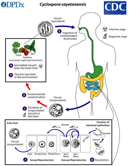
Cyclospora cayetanensis is an intestinal coccidian parasite transmitted to humans through the consumption of oocysts in fecally contaminated food and water. Infection is found worldwide and is highly endemic in tropical and subtropical regions with poor sanitation. Disease in developed countries is usually observed in travelers and in seasonal outbreaks associated with imported produce from endemic areas. Recently, summertime outbreaks in the United States have also been linked to locally grown produce. Cyclosporiasis causes a diarrheal illness which may be severe in infants, the elderly, and immunocompromised individuals. The increased adoption of highly sensitive molecular diagnostic tests, including commercially available multiplex panels for gastrointestinal pathogens, has facilitated the detection of infection and likely contributed to the increased reports of cases in developed countries. 

Cyclospora protozoan diarrhea diagnostics pathology parasite parasitic

1. Introduction

Cyclosporiasis is a foodborne and waterborne intestinal parasitic disease caused by the coccidian parasite Cyclospora cayetanensis. While other Cyclospora species have been described from non-human primates [1], C. cayetanensis is the only species known to infect humans and to date has only been isolated from humans, although isolates from captive chimpanzees and macaques in Europe have been found to have isolates genetically similar to C. cayetanensis [2].
Cyclospora cayetanensis occurs worldwide, with hot spots of endemicity including tropical and subtropical regions of Latin America (including the Caribbean), Central and Southeast Asia, the Middle East, and North Africa [3][4]. This parasite is particularly prevalent in settings with poor sanitation where the environment is contaminated with human feces from infected individuals. Cyclosporiasis exhibits varied seasonality worldwide, which may be affected by precipitation, temperature, and humidity [3].
Isolated cases in developed countries are usually from individuals returning from endemic areas [4]. As surveillance for cyclosporiasis has improved in recent years, seasonal outbreaks are becoming commonplace in many parts of the world. In the last 10 years, summertime outbreaks have been documented in Canada [5][6][7][8], Poland [9], the United Kingdom [7], and the USA [10][11][12][13][14][15][16][17]. The sources of outbreaks are usually fresh produce that is typically eaten raw, especially plants that grow low to the ground and are prone to being exposed to fecally contaminated water and soil. Common vehicles implicated in outbreaks include raspberries, blackberries, strawberries, blueberries, basil, cilantro, snow peas, snap peas, and various lettuces [3][4]. Often, the source of the outbreak is not known, because fresh produce has a short shelf life and the products are usually consumed or expired and discarded before an outbreak is realized [3].
According to the U.S. Centers for Disease Control and Prevention (CDC), there have been approximately 6000 domestically acquired cases of cyclosporiasis in the past three years [18]. Outbreaks in the US have historically been associated with produce imported from Latin America. However, the US Food and Drug Administration (FDA) detected the first evidence of C. cayetanensis in locally grown produce (cilantro) in 2018 [3]. Since then, C. cayetanensis has been increasingly detected in domestic food and surface water [18], likely due in part to improved surveillance tools. Given the ongoing annual outbreaks of cyclosporiasis in the United States, the FDA formed the Cyclospora Task Force in 2019, and this group produced the “Cyclospora Prevention, Response and Research Action Plan” to combat foodborne illness associated with imported and domestically grown produce [19].

2. Biology and Life Cycle

Cyclospora cayetanensis has a complex life cycle involving both sexual and asexual development within a single host. Infection is initiated by the ingestion of fully sporulated oocysts in fecally contaminated food or water. The oocysts excyst in the lumen of the small intestine, and sporozoites invade the epithelial cells lining the duodenum and jejunum. The sporozoites become trophozoites, which in turn become either Type I meronts (schizonts) or Type II meronts. Type I meronts contain 8–12 merozoites and perpetuate autoinfection in the host. Type II meronts each contain four merozoites, which go on to form microgametocytes (microgamonts) and macrogametocytes (macrogamonts) to initiate the sexual cycle. A microgametocyte fertilizes a macrogametocyte, resulting in the formation of a zygote. Zygotes become oocysts in the enterocytes and are shed in an unsporulated state in the feces [3][4][20]. Oocysts sporulate in the environment, at which time they become infective to other people. Factors affecting sporulation in nature are still unresolved, but the process may be influenced by humidity, soil chemistry, and exposure to ultraviolet light. Under laboratory conditions, sporulation takes approximately 7–14 days at 22 °C and 30 °C [21] (Figure 1).
Figure 1. Life cycle of Cyclospora cayetanensis. Unsporulated, non-infective oocysts are passed in the feces (1). In the environment (2), sporulation occurs after days or weeks at temperatures between 22 °C to 32 °C, resulting in the division of the sporont into two sporocysts, each containing two elongated sporozoites (3). The sporulated oocysts can contaminate fresh produce and water (4) images, which are then ingested (5). The oocysts excyst in the gastrointestinal tract, freeing the sporozoites, which invade the epithelial cells of the small intestine (6). Inside the cells, they undergo asexual multiplication into Type I and Type II meronts. Merozoites from Type I meronts perpetuate the asexual cycle, while merozoites from Type II meronts undergo sexual development into macrogametocytes and microgametocytes upon invasion of another host cell. Fertilization occurs, and the zygote develops to an oocyst, which is released from the host cell and shed in the stool (7). Figure courtesy of the CDC-DPDx.
Cyclospora cayetanensis is usually confined to the upper small intestine in immunocompetent hosts, but it can cause ectopic infection of the biliary tree and gall bladder in patients with HIV infection and AIDS [22][23][24].

3. Pathogenesis

Parasite invasion and replication within enterocytes damages the small intestinal epithelium, leading to the disruption of the brush border, loss of membrane bound digestive enzymes, and intestinal villous blunting and atrophy [25][26]. An influx of lymphocytes, plasma cells, and occasionally eosinophils occurs in the lamina propria. These changes have the overall effect of decreasing the small intestinal absorptive capacity, leading to decreased uptake of water, nutrients, and electrolytes [25].

4. Clinical Presentation

The presentation of infection varies with the age and immune status of the host, as well as the local endemicity of infection [25]. Infection is often mild or asymptomatic [4], particularly in residents of highly endemic countries. When present, symptoms include profuse watery diarrhea, abdominal cramping, nausea, fatigue, low-grade fever, anorexia and weight loss. Less commonly, mucus or blood may be found in the stool. More severe disease occurs most commonly in infants, the elderly, and profoundly immunocompromised patients such as those with HIV/AIDS [25]. Travelers from non-endemic countries are also likely to experience severe infection. Prolonged diarrhea can result in dehydration and malnutrition and may rarely result in death, particularly in infants and individuals with other infections or morbidities.
Symptom onset usually occurs after a median incubation period of seven days following the ingestion of infectious oocysts (with a range from 2 to ≥2 weeks) and may last for weeks to months without treatment [27][28]. Some patients experience a single self-limited episode, whereas others have waxing and waning symptoms [29][30].
Rarely, C. cayetanensis may infect the biliary tract and cause acalculous cholecystitis [23][24], particularly in immunocompromised hosts. Guillain-Barré syndrome, ocular inflammation, reactive arthritis, and sterile urethritis have also been reported [25].

5. Treatment

Trimethoprim/sulfamethoxazole (TMP/SMX; trade names Bactrim, Cotrim, Septra) is the treatment of choice for cyclosporiasis [31][32]. It is administered at a dose of one double-strength (DS) 160 mg/800 mg tablet given orally twice per day for 7–10 days and has been shown to provide >90% cure rates in immunocompetent patients. The efficacy of TMP/SMX for treating cyclosporiasis was first demonstrated in a placebo-controlled trial of 40 adult expatriates and tourists in Nepal [29]. The authors found that only 1 of 16 patients (6.3%) had detectable oocysts in stool after seven days of treatment with TMP/SMX, compared with 15 of 17 patients (88.2%) who received a placebo. Importantly, an improvement in symptomatology was correlated with the eradication of oocysts. Nitazoxanide or ciprofloxacin are recommended for patients that are unable to take TMP/SMX due to sulfa allergy, although treatment failure may occur [32][33].
Profoundly immunocompromised patients such as those with AIDS and transplant recipients may require a longer course of treatment and/or a higher dose of TMP/SMX. Ongoing prophylaxis is also recommended to prevent relapse [34]. A 1994 study of HIV-positive adults in Haiti found that symptomatic infection recurred in 12 of 28 patients (43%) who were monitored for more than one month after a 10 day course of TMP/SMX given orally four times per day [34]. All responded promptly to repeat treatment and were subsequently given TMP/SMX three times a week for secondary prophylaxis; of these, only one patient recurred after seven months. These authors published a follow up study in 2000 showing that patients with HIV were successfully treated with seven days of TMP/SMX and DS tablets given orally twice per day, followed by prophylaxis for 10 weeks (DS tablet given orally, three times per week). Regardless of the initial dose, these studies clearly show the importance of prophylaxis for preventing relapse. Based on these data, the 2019 guidelines from the American Society of Transplantation recommend a 10 day course of TMP/SMX (one DS tablet given orally four times per day) for solid organ transplant recipients, followed by secondary prophylaxis with TMP/SMX (one DS tablet given orally three time per week) [35]. The reduction of immunosuppression is also indicated if possible.
There is currently no vaccine for cyclosporiasis. Instead, preventative measures focus on improving sanitation (e.g., measures to prevent human feces from entering the environment and contaminating the food and water supply) and treating food to inactivate contaminating oocysts. The oocysts are highly resistant to commonly used disinfectants but are inactivated by cooking. Travelers to highly endemic areas are advised to avoid eating uncooked raw vegetables and unpeeled fruits and preferentially to choose foods that are fully cooked and served hot [35]. Similarly, patients with HIV and solid organ transplant recipients should avoid consuming untreated well and surface water to avoid gastroenteric infections [36][37].

6. Diagnosis

The diagnosis of intestinal parasites is laborious, time-consuming, and often requires specialized expertise [38]. Still, ova-and-parasite (O&P) exams and other forms of stool microscopy are routinely ordered for patients presenting with diarrhea and other intestinal manifestations, even when other diagnostic methods may be more appropriate. In developed counties, if a parasitic disease is suspected in an immunocompetent patient with diarrhea and no travel history to endemic areas for parasitic diseases, parasites such as Giardia duodenalis and Cryptosporidium spp. should be considered before ordering O&P exams [38]. In the United States and Canada, where cyclosporiasis has become a seasonal illness in the summer, C. cayetanensis should also be considered as a primary differential in any patient presenting with compatible symptoms and illness onset during the cyclosporiasis peak period (i.e., May through August). A history of consuming fresh leafy greens, berries, basil, and cilantro within 2 weeks prior to the onset of illness may raise the clinical suspicion for cyclosporiasis, although this type of nutritional history is not commonly obtained [12]. Cyclosporiasis should also be considered in patients returning or emigrating from areas endemic for the disease, in which case specialized assays such as modified acid-fast (MAF) stain, safranin stain, and UV autofluorescence (see below) should be ordered to compliment the routine O&P exams [38]. Except during seasonal outbreaks, cyclosporiasis is rarely considered by a health care provider, and C. cayetanensis may be overlooked when only routine O&P examinations are ordered. Importantly, it may be necessary to examine multiple stool specimens for C. cayetanensis to make a diagnosis of cyclosporiasis, as the number of oocysts shed in stool may be relatively few.
Unfortunately, while there are numerous rapid antigen-detection assays for G. duodenalis and Cryptosporidium spp., the diagnosis of cyclosporiasis still relies heavily on stool microscopy (Table 1). Only recently have nucleic acid amplification tests (NAATs) started to become available, and even then, options are limited and tests may be cost prohibitive. To date, there are no antibody or antigen detection assays for the routine clinical diagnosis of cyclosporiasis.
Table 1. Microscopic methods for the detection of Cyclospora cayetanensis.
Diagnostic Method Advantages Disadvantages
Stool Microscopy    
Direct wet mount Fast, inexpensive; simultaneous detection of other intestinal parasites Lack of sensitivity without concentration step; lack of defined morphologic features might make detection difficult for microscopists
Concentrated wet mount Fast, inexpensive; simultaneous detection of other intestinal parasites Lack of defined morphologic features might make detection difficult for microscopists
Differential Interference Contrast (DIC) Increased sensitivity by highlighting internal structures Not routinely available in many diagnostic labs
Ultraviolet autofluorescence More sensitive than permanent smears; simultaneous detection of other coccidian oocysts and several helminth eggs Requires specific UV filters that may not be routinely present in diagnostic labs
Lacto-phenol cotton blue Fast, inexpensive; may be advantageous in resource-poor areas where acid-fast staining is not available Non-specific; likely false positives with fungal elements
Trichrome/iron hematoxylin stain Simultaneous detection of other intestinal protozoans Oocysts do not stain with trichrome
Modified Ziehl-Neelsen (ZN) stain Increased sensitivity over traditional O&P exams Inconsistent staining of oocysts
Kinyoun’s modified acid-fast (MAF) stain Increased sensitivity over traditional O&P exams Inconsistent staining of oocysts
Modified safranin More consistent staining of oocysts over ZN and MAF Requires heating of stain
Auramine O (auramine-phenol) More sensitive than traditional O&P exams May be less sensitive than MAF, ZN; requires fluorescent microscope
Histopathology    
Hematoxylin-and-eosin (H&E), periodic acid Schiff (PAS) Identify multiple developmental stages of C. cayetanensis Not routinely ordered for C. cayetanensis; may be difficult to distinguish from Cystoisospora belli
Ziehl–Neelsen stain, Fite’s acid-fast stain Can detect oocysts in tissues Pre-oocyst stages may not stain

References

  1. Eberhard, M.L.; da Silva, A.J.; Lilley, B.G.; Pieniazek, N.J. Morphologic and molecular characterization of new Cyclospora species from Ethiopian monkeys: C. cercopitheci sp.n. C. colobi sp.n. and C. papionis sp.n. Emerg. Infect. Dis. 1999, 5, 651–658.
  2. Marangi, M.; Koehler, A.V.; Zanzani, S.A.; Manfredi, M.T.; Brianti, E.; Giangaspero, A.; Gasser, R.B. Detection of Cyclospora in captive chimpanzees and macaques by a quantitative PCR-based mutation scanning approach. Parasit. Vectors 2015, 8, 274.
  3. Almeria, S.; Cinar, H.N.; Dubey, J.P. Cyclospora cayetanensis and Cyclosporiasis: An Update. Microorganisms 2019, 7, 317.
  4. Ortega, Y.R.; Sanchez, R. Update on Cyclospora cayetanensis, a food-borne and waterborne parasite. Clin. Microbiol. Rev. 2010, 23, 218–234.
  5. CFIA-PHAC Public Health Notice-Outbreak of Cyclospora Infections under Investigation, October 11, 2017-Final Update. Available online: https://www.canada.ca/en/public-health/services/public-health-notices/2017/public-health-notice-outbreak-cyclospora-infections-under-investigation.html (accessed on 30 April 2021).
  6. Dixon, B.; Mihajlovic, B.; Couture, H.; Farber, J.M. Qualitative risk assessment: Cyclospora cayetanensis on fresh raspberries and blackberries imported into Canada. Food Prot. Trends 2016, 36, 18–32.
  7. Nichols, G.L.; Freedman, J.; Pollock, K.G.; Rumble, C.; Chalmers, R.M.; Chiodini, P.; Hawkins, G.; Alexander, C.L.; Godbole, G.; Williams, C.; et al. Cyclospora infection linked to travel to Mexico, June to September 2015. Euro Surveill 2015, 20.
  8. CFIA-PHAC Public Health Notice: Outbreak of Cyclospora Infections Linked to Salad Products and Fresh Herbs. Available online: https://www.canada.ca/en/public-health/services/public-health-notices/2020/outbreak-cyclospora-infections-salad-products.html (accessed on 30 April 2021).
  9. Bednarska, M.; Bajer, A.; Welc-Faleciak, R.; Pawelas, A. Cyclospora cayetanensis infection in transplant traveller: A case report of outbreak. Parasit. Vectors 2015, 8, 411.
  10. Abanyie, F.; Harvey, R.R.; Harris, J.R.; Wiegand, R.E.; Gaul, L.; Desvignes-Kendrick, M.; Irvin, K.; Williams, I.; Hall, R.L.; Herwaldt, B.; et al. 2013 multistate outbreaks of Cyclospora cayetanensis infections associated with fresh produce: Focus on the Texas investigations. Epidemiol. Infect. 2015, 143, 3451–3458.
  11. Casillas, S.M.; Bennett, C.; Straily, A. Notes from the Field: Multiple Cyclosporiasis Outbreaks-United States, 2018. MMWR Morb. Mortal. Wkly. Rep. 2018, 67, 1101–1102.
  12. Casillas, S.M.; Hall, R.L.; Herwaldt, B.L. Cyclosporiasis Surveillance-United States, 2011–2015. MMWR Surveill. Summ. 2019, 68, 1–16.
  13. CDC. U.S. Foodborne Outbreaks of Cyclosporiasis-2000–2017. Available online: https://www.cdc.gov/parasites/cyclosporiasis/outbreaks/foodborneoutbreaks.html (accessed on 30 April 2021).
  14. CDC. Domestically Acquired Cases of Cyclosporiasis-United States, May–August 2018. Available online: https://www.cdc.gov/parasites/cyclosporiasis/outbreaks/2018/c-082318/index.html (accessed on 30 April 2021).
  15. CDC. Multistate Outbreak of Cyclosporiasis Linked to Fresh Express Salad Mix Sold at McDonald’s Restaurants—United States, 2018: Final Update. Available online: https://www.cdc.gov/parasites/cyclosporiasis/outbreaks/2018/b-071318/index.html (accessed on 30 April 2021).
  16. CDC. Multistate Outbreak of Cyclosporiasis Linked to Del Monte Fresh Produce Vegetable Trays—United States, 2018: Final Update. Available online: https://www.cdc.gov/parasites/cyclosporiasis/outbreaks/2018/a-062018/index.html (accessed on 30 April 2021).
  17. CDC. Outbreak of Cyclospora Infections Linked to Bagged Salad Mix. Available online: https://www.cdc.gov/parasites/cyclosporiasis/outbreaks/2020/ (accessed on 30 April 2021).
  18. CDC. Cyclosporiasis-Outbreak Investigations and Updates. Available online: https://www.cdc.gov/parasites/cyclosporiasis/outbreaks/index.html (accessed on 22 July 2021).
  19. FDA. Cyclospora Prevention, Response and Research Action Plan. Available online: https://www.fda.gov/food/foodborne-pathogens/cyclospora-prevention-response-and-research-action-plan (accessed on 22 July 2021).
  20. Cama, V.A.; Mathison, B.A. Infections by Intestinal Coccidia and Giardia duodenalis. Clin. Lab. Med. 2015, 35, 423–444.
  21. Smith, H.V.; Paton, C.A.; Mitambo, M.M.; Girdwood, R.W. Sporulation of Cyclospora sp. oocysts. Appl. Environ. Microbiol. 1997, 63, 1631–1632.
  22. de Gorgolas, M.; Fortes, J.; Guerrero, M.L.F. Cyclospora cayetanensis Cholecystitis in a Patient with AIDS. Ann. Intern. Med. 2001, 134, 166.
  23. Sifuentes-Osornio, J.; Porras-Cortés, G.; Bendall, R.P.; Morales-Villarreal, F.; Reyes-Terán, G.; Ruiz-Palacios, G.M. Cyclospora cayetanensis Infection in Patients with and Without AIDS: Biliary Disease as Another Clinical Manifestation. Clin. Infect. Dis. 1995, 21, 1092–1097.
  24. Zar, F.A.; El-Bayoumi, E.; Yungbluth, M.M. Histologic Proof of Acalculous Cholecystitis Due to Cyclospora cayetanensis. Clin. Infect. Dis. 2001, 33, e140–e141.
  25. Giangaspero, A.; Gasser, R.B. Human cyclosporiasis. Lancet Infect. Dis. 2019, 19, e226–e236.
  26. Sun, T.; Ilardi, C.F.; Asnis, D.; Bresciani, A.R.; Goldenberg, S.; Roberts, B.; Teichberg, S. Light and electron microscopic identification of Cyclospora species in the small intestine. Evidence of the presence of asexual life cycle in human host. Am. J. Clin. Pathol. 1996, 105, 216–220.
  27. Fleming, C.A.; Caron, D.; Gunn, J.E.; Barry, M.A. A foodborne outbreak of Cyclospora cayetanensis at a wedding: Clinical features and risk factors for illness. Arch. Intern. Med. 1998, 158, 1121–1125.
  28. Herwaldt, B.L.; Ackers, M.L. An outbreak in 1996 of cyclosporiasis associated with imported raspberries. The Cyclospora Working Group. N. Engl. J. Med. 1997, 336, 1548–1556.
  29. Hoge, C.W.; Shlim, D.R.; Ghimire, M.; Rabold, J.G.; Pandey, P.; Walch, A.; Rajah, R.; Gaudio, P.; Echeverria, P. Placebo-controlled trial of co-trimoxazole for Cyclospora infections among travellers and foreign residents in Nepal. Lancet 1995, 345, 691–693.
  30. Huang, P.; Weber, J.T.; Sosin, D.M.; Griffin, P.M.; Long, E.G.; Murphy, J.J.; Kocka, F.; Peters, C.; Kallick, C. The first reported outbreak of diarrheal illness associated with Cyclospora in the United States. Ann. Intern. Med. 1995, 123, 409–414.
  31. CDC, Treatment for Cyclosporiasis. Available online: https://www.cdc.gov/parasites/cyclosporiasis/health_professionals/tx.html (accessed on 22 July 2021).
  32. Medical Letter. The Medical Letter: Drugs for Parasitic Infections, 3rd ed.; The Medical Letter, Inc.: New Rochelle, NY, USA, 2013.
  33. Verdier, R.I.; Fitzgerald, D.W.; Johnson, W.D., Jr.; Pape, J.W. Trimethoprim-sulfamethoxazole compared with ciprofloxacin for treatment and prophylaxis of Isospora belli and Cyclospora cayetanensis infection in HIV-infected patients. A randomized, controlled trial. Ann. Intern. Med. 2000, 132, 885–888.
  34. Pape, J.W.; Verdier, R.-I.; Boncy, M.; Boncy, J.; Johnson, W.D. Cyclospora Infection in Adults Infected with HIV: Clinical Manifestations, Treatment, and Prophylaxis. Ann. Intern. Med. 1994, 121, 654–657.
  35. CDC. Chapter 2-Preparing Interntional Travelers. In CDC Yellow Book; Oxford University Press: Oxford, UK. Available online: https://wwwnc.cdc.gov/travel/yellowbook/2020/preparing-international-travelers/food-and-water-precautions (accessed on 22 July 2021).
  36. La Hoz, R.M.; Morris, M.I.; AST Infectious Diseases Community of Practice. Intestinal parasites including Cryptosporidium, Cyclospora, Giardia, and Microsporidia, Entamoeba histolytica, Strongyloides, Schistosomiasis, and Echinococcus: Guidelines from the American Society of Transplantation Infectious Diseases Community of Practice. Clin. Transplant 2019, 33, e13618.
  37. NIH HIV.gov-Clinical Guidelines. Available online: https://clinicalinfo.hiv.gov/en/guidelines (accessed on 22 July 2021).
  38. Garcia, L.S.; Arrowood, M.; Kokoskin, E.; Paltridge, G.P.; Pillai, D.R.; Procop, G.W.; Ryan, N.; Shimizu, R.Y.; Visvesvara, G. Laboratory Diagnosis of Parasites from the Gastrointestinal Tract. Clin. Microbiol. Rev. 2018, 31, e00025-17.
More
Information
Subjects: Microbiology
Contributor MDPI registered users' name will be linked to their SciProfiles pages. To register with us, please refer to https://encyclopedia.pub/register :
View Times: 550
Revisions: 2 times (View History)
Update Date: 22 Sep 2021
1000/1000
Video Production Service