Your browser does not fully support modern features. Please upgrade for a smoother experience.
Submitted Successfully!
 +1 credit
Thank you for your contribution! You can also upload a video entry or images related to this topic.
For video creation, please contact our Academic Video Service.
| Version | Summary | Created by | Modification | Content Size | Created at | Operation |
|---|---|---|---|---|---|---|
| 1 | DAZHONG XU | + 2041 word(s) | 2041 | 2021-07-02 12:55:56 |
Video Upload Options
We provide professional Academic Video Service to translate complex research into visually appealing presentations. Would you like to try it?
Cite
If you have any further questions, please contact Encyclopedia Editorial Office.
Xu, D. Gene 33. Encyclopedia. Available online: https://encyclopedia.pub/entry/11962 (accessed on 30 June 2026).
Xu D. Gene 33. Encyclopedia. Available at: https://encyclopedia.pub/entry/11962. Accessed June 30, 2026.
Xu, Dazhong. "Gene 33" Encyclopedia, https://encyclopedia.pub/entry/11962 (accessed June 30, 2026).
Xu, D. (2021, July 12). Gene 33. In Encyclopedia. https://encyclopedia.pub/entry/11962
Xu, Dazhong. "Gene 33." Encyclopedia. Web. 12 July, 2021.
Copy Citation
Gene 33 (also named Mig6, RALT, and ERRFI1) is an adapter/scaffold protein with a calculated molecular weight of about 50 kD. It contains multiple domains known to mediate protein–protein interaction, suggesting that it has the potential to interact with many cellular partners and have multiple cellular functions. The research over the last two decades has confirmed that it indeed regulates multiple cell signaling pathways and is involved in many pathophysiological processes. Gene 33 has long been viewed as an exclusively cytosolic protein. However, recent evidence suggests that it also has nuclear and chromatin-associated functions. These new findings highlight a significantly broader functional spectrum of this protein.
gene 33
ERRFI1
adapter protein
DNA damage
EGFR
cancer
signal transduction
1. Introduction
Gene 33 (also named Mig6, RALT, and ERRFI1) was discovered as a glucocorticoid-induced transcript from the rat liver using the differential hybridization technique in 1985 [1]. The gene that encodes this transcript was initially named p33 and was later renamed gene 33 to avoid confusion with its protein product [2]. The human homologue of gene 33 was later identified from quiescent fibroblasts treated with serum and named as mitogen-inducible gene 6 (Mig6 or Mig-6), for its high inducibility by serum [3]. Subsequent studies revealed that the transcript of gene 33 can be induced by a wide variety of extracellular stimuli and is widely expressed [2][3][4][5][6][7][8][9][10][11][12]. The induction of gene 33 by multiple signaling inputs is consistent with the fact that the promotor region of gene 33 contains an array of regulatory elements [2][13]. gene 33 is considered an immediate early response gene, defined as quick induction in response to stimuli without the requirement of de novo protein synthesis [3][14]. Gene 33 appeared rather late in the evolution, existing only in vertebrates. It shares considerable homology to the C-terminal portion of activated CDC42-associated kinase 1 (ACK1). Structurally, it is more or less an ACK1 without the kinase domain and the SRC-homology 3 domain (SH3) at the N-terminus (Figure 1). It is likely that Gene 33 was descended from ACK1 during evolution to fulfill the functional needs of more advanced animals.
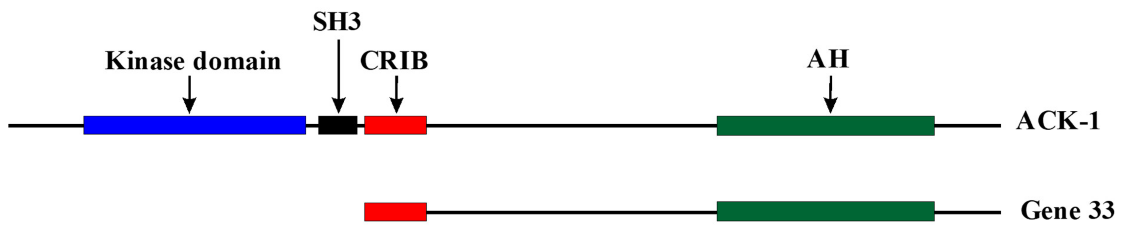 Figure 1. Comparison of linear structures of Gene 33 and ACK-1. SH3: Src homology 3 domain, CRIB: Cdc42/Rac interactive and binding domain. AH: Ack homology domain.
Although the highly inducible nature of Gene 33 by multiple stimuli, particularly insulin, drew considerable interest in this gene soon after its discovery, the function of its protein product remained elusive until early 2000, when three studies were published on the function of this protein [4][15][16]. The laboratory of John Kyriakis showed that Gene 33 can be induced in the kidney after unilateral nephrectomy and/or streptozotocin-induced diabetes [4]. Gene 33 can also be induced by a mechanical strain in a JNK-dependent fashion in rat renal mesangial cells [4]. The study also described the domain structure of the protein and the potential signaling pathway connecting Gene 33, the small GTPase Cdc42, and JNK [4]. The laboratories of Oreste Segotto and Axel Ullrich independently discovered the interaction between rat Gene 33 with ErbB2 and Mig6 with EGFR, respectively, using the yeast two-hybrid system [15][16]. The interactions were found to inhibit the signaling pathways and fibroblast transformation mediated by these receptors [15][16]. These studies also showed that Gene 33 can also be induced by the activation of these receptors, establishing that Gene 33 is a potential feedback inhibitor of the signaling pathway mediated by these receptors. The Segotto group renamed Gene 33 as receptor-associated late transducer (RALT). The official name given later by the Human Genome Organization to this gene is ErbB receptor feedback inhibitor 1 (ERRFI1). This review will refer to this protein using its original common name “Gene 33” and the gene encoding it using its official name ERRFI1 (Errfi1 for the rodent gene).
The involvement of Gene 33 in the signaling of the ErbB family receptor tyrosine kinases (RTKs) sparked intense interest in this then little-known protein and led to a series of studies that solidified its role in the ErbB receptor signaling pathway [11][17][18][19][20][21][22][23][24][25][26]. Although the regulation of ErbB receptors appears to be the most prominent function of Gene 33, its involvement in other signaling pathways has also been revealed. The biological roles of Gene 33 in various pathophysiological processes have become clearer as well. Although Gene 33 has long been regarded as an exclusively cytoplasmic protein, recent evidence showed that a fraction of it is localized in the nucleus and associated with chromatin. This nuclear/chromatin fraction of Gene 33 has been shown to modulate the DNA damage response in response to genotoxic stresses [27][28]. These new findings significantly expand the functional profile of this protein. Several excellent and focused review articles on Gene 33 have been published in the past, with main emphases on its association with the ErbB family RTKs and its role in cancer [29][30][31][32].
2. Gene 33 and Human Diseases
2.1. Gene 33 in Cancer
The involvement of Gene 33 in the signaling of receptor tyrosine kinases of the EGFR family prompted immediate interest in its potential role in cancer, as these receptors are heavily involved in various types of human cancer [33]. The function of Gene 33 as an inhibitor of these receptors suggests a potential tumor suppressive role of this protein. Other known functions of Gene 33, including the proapoptotic and the nuclear function of Gene 33, are also largely in line with this notion. Indeed, Errfi1-null mice develop neoplasms in multiple tissues [20]. In humans, ERRFI1 is located in chromosome 1p36, a locus long believed to contain multiple putative tumor suppressor genes and frequently altered in many cancers [34]. These data support a general role of Gene 33 in human cancer as a tumor suppressor.
2.2. Gene 33 in Diabetes
Errfi1 was shown initially by the laboratory of Joseph Messina to be an insulin-inducible gene in H4 hepatocarcinoma cells soon after its discovery [35]. Follow-up studies confirmed this observation [2][8][36][37][35]. As in other settings, the induction of Gene 33 by insulin is MEK-ERK dependent [37]. These findings suggest a potential role of Gene 33 in the development of diabetes. Supporting this notion, Gene 33 expression is strongly induced in a murine model for diabetic nephropathy and was proposed to mediate diabetic renal hypertrophy [4]. Furthermore, a study based on a Korean population revealed a single nucleotide polymorphism (SNP) in the third intron of the ERRFI1 gene (+808(T/G)) that is negatively associated with diabetic nephropathy [38]. This SNP is believed to negatively regulate the expression of Gene 33 [38].
Errfi1 haploinsufficiency protects mice from streptozotocin-induced diabetes [39]. This prodiabetic function of Gene 33 appears to be a result of the ability of Gene 33 to promote DNA damage-induced apoptotic death of pancreatic β cells in response to proinflammatory cytokines and ER stress [40][39]. Gene 33 may also mediate glucocorticoid-induced insulin resistance and diabetes by suppressing β cell proliferation via inhibition of EGFR signaling [41]. In contrast, mice with liver-specific knockout of Errfi1 exhibit hyperglycemia as a result of hepatic insulin resistance [42]. Deletion of Errfi1 increases EGFR signaling, mTOR activity, JNK activity, and IRS-1 phosphorylation at serine 307 [42]. On the other hand, hepatic expression of glucokinase, glucose-6-phosphatase, and phosphoenolpyruvate carboxykinase 1 is reduced after Errfi1 ablation [42]. Interestingly, another study showed that liver-specific knockout of Errfi1 in mice leads to fatty liver, fasting hyperglycemia, and hypercholesterolemia but lower body weight and higher insulin sensitivity [43]. The hypercholesterolemia in the knockout mice is apparently a result of increased EGFR activation, as inhibition of EGFR with TKI reduces hypercholesterolemia [44]. These findings highlight a rather complex role of Gene 33 in diabetes and metabolic syndrome, and possible differential roles in conditions mimicking type 1 and type 2 diabetes.
2.3. Gene 33 in Cardiovascular Diseases
ErbB2 signaling is well established to promote cardiomyocyte survival while inhibition of it leads to cardiomyocyte apoptosis and dilated heart failure [45][46][47][48][49]. Gene 33 overexpression using the adenoviral vector leads to strong apoptosis of rat neonatal myocytes as a result of inhibition of the antiapoptotic AKT and ERK activity downstream of ErbB family tyrosine kinases [17]. Gene 33 is induced in rat neonatal myocytes by hypoxia and hypoxia/reoxygenation and in turn promotes apoptosis [17]. Moreover, Gene 33 is strikingly induced in the murine cardiac tissue suffering experimentally induced myocardial infarction or ischemia and ischemia/reperfusion [17]. These findings point to a significant role of Gene 33 in the loss of cardiomyocytes during ischemia induced by myocardial infarction by promoting cardiomyocyte apoptosis.
On the other hand, constitutive cardiac-specific ectopic expression of Gene 33 in mouse heart attenuates both hypertrophic and inflammatory responses and helps preserve cardiac functions [50]. This is accomplished through inhibition of the angiotensin II and isoproterenol-stimulated vasoactive signaling pathways, which are known to transactivate EGFR [51][50]. Unlike in the case of ischemic stress, transgenic expression of Gene 33 does not cause significant cardiomyocyte apoptosis in mice, highlighting the differential role of Gene 33 in acute and chronic cardiac conditions [17][50].
Using smooth muscle-specific Errfi1 knockout mice and the cellular model, Lee et al. showed that Gene 33 inhibits EGF-dependent smooth muscle proliferation and migration that lead to neointimal hyperplasia, thereby potentially inhibiting atherosclerosis [52].
2.4. Gene 33 in Other Diseases
Gene 33 has been documented to promote intervertebral disk degeneration, in which miR-2355 is upregulated and suppresses the expression of Gene 33 [53]. Gene 33 in turn inhibits EGFR-mediated proliferation of intervertebral disc cells, particularly nucleus pulposus cells [53]. Microarray analyses detected upregulation of ERRFI1 in the central nervous system after acute infection by pseudorabies virus, suggesting a potential involvement of Gene 33 in the response of the central nervous system to viral infection [54]. Downregulation of Errfi1 was observed in the liver of mice subjected to disrupted growth hormone and insulin-like growth factor-1 signaling as well as caloric restriction, suggesting a potential involvement of Gene 33 in dwarfism and longevity [55]. Interestingly, a reduced plasma level of Gene 33 has been shown to be associated with children with autism [56].
A recent integrated analysis of public genomic data revealed a potential association between ERRFI1 with the risk of psoriasis, a chronic inflammatory skin condition [57]. An enhancer variant in the chromosome 1p36.23 region and within the enhancer of ERRFI1 is associated with increased susceptibility for psoriasis. The variant may render the ERRFI1 enhancer less able to interact with the AP-1 complex at the ERRFI1 promoter, thereby destabilizing the expression of the gene [57]. Interestingly, psoriasis has been shown to be associated with nonalcoholic fatty liver disease, type 2 diabetes, and metabolic syndrome [58]. These findings provide further support to the involvement of Gene 33 in diabetes and metabolic syndrome. Table 1 summarizes the known association of Gene 33 with human diseases.
Table 1. Gene 33 and human diseases.
| Function in Cancer | Cancer Type | Reference |
| Suppressor | Lung cancer | [20][28][59][60][61][62][63][64][65] |
| Endometrial cancer | [66][67][68][69][70] | |
| Glioma | [25][71][72][73] | |
| Thyroid cancer | [74][75][76][77] | |
| Liver cancer | [26][78] | |
| Suppressor or promotor | Breast cancer | [24][79][80][81] |
| Skin cancer | [20][82][83] | |
| Marker for liver metastasis | Colorectal cancer | [84] |
| Function in other diseases | Disease | Reference |
| Suppressor | Cholangiocarcinoma | [85] |
| promotor | Intervertebral disk degeneration | [53] |
| Suppressor or promotor | Diabetes | [4][41][40][38][39][42][43] |
| Suppressor or promoter | Cardiovascular diseases | [17][50][52] |
| Positive correlation | Dwarfism and longevity | [55] |
| Negative correlation | Psoriasis | [57] |
| Negative correlation | Autism | [56] |
3. Conclusions and Perspectives
Our understanding of Gene 33 has come a long way since its discovery more than 30 years ago. The nature of this protein as an adapter/scaffold and the relative late appearance in the evolution suggest that its main functions are likely to coordinate multiple cellular pathways in response to the increased biological complexity of higher metazoans. The functions of Gene 33 discovered to date appear to support this notion. It would not be surprising that more functions of Gene 33 will be revealed along this line of reasoning. Recent evidence on its nuclear and chromatin-associated functions in DDR and DNA repair points to a new direction in searching for additional functions of this protein. Although Gene 33 may not be one of the core regulators of these biological processes, its importance in pathophysiological processes should not be overlooked. In fact, many biological processes, chronic pathological conditions in particular, are strongly associated with seemingly weak biological alterations rather than overt biological defects with severe consequences, e.g., early mortality. Therefore, the study of proteins, such as Gene 33, is important for better understanding of these chronic conditions, which could lead to novel therapeutic strategies.
For instance, the nuclear function of Gene 33 on DDR may contribute to carcinogenesis induced by genotoxic agents with environmental or occupational relevance, such as hexavalent chromium, by affecting Cr(VI)-induced genomic instability. Gene 33 dysregulation may also facilitate spontaneous gene mutations that lead to genomic instability and carcinogenesis. In diabetes, abnormal function of nuclear Gene 33 could contribute to chronic loss of β cells through dysregulation of cell cycle checkpoint control at the G2/M phase. A similar mechanism could be involved in psoriasis, where abnormal proliferation of skin cells contributes to the symptom of the disease. The nuclear function of Gene 33 adds additional complexity to the already complex functional profile of Gene 33. Further investigation into the role of nuclear Gene 33 in chronic human diseases is clearly needed and potentially fruitful.
References
- Lee, K.L.; Isham, K.R.; Stringfellow, L.; Rothrock, R.; Kenney, F.T. Molecular cloning of cDNAs cognate to genes sensitive to hormonal control in rat liver. J. Biol. Chem. 1985, 260, 16433–16438.
- Messina, J.L. Regulation of Gene 33 expression by insulin. In Molecular Biology of Diabetes, Part II; Draznin, B., LeRooith, D., Eds.; Humana Press: Totowa, NJ, USA, 1994; pp. 263–281.
- Wick, M.; Burger, C.; Funk, M.; Muller, R. Identification of a novel mitogen-inducible gene (mig-6): Regulation during G1 progression and differentiation. Exp. Cell Res. 1995, 219, 527–535.
- Makkinje, A.; Quinn, D.A.; Chen, A.; Cadilla, C.L.; Force, T.; Bonventre, J.V.; Kyriakis, J.M. Gene 33/Mig-6, a transcriptionally inducible adapter protein that binds GTP-Cdc42 and activates SAPK/JNK. A potential marker transcript for chronic pathologic conditions, such as diabetic nephropathy. Possible role in the response to persistent stress. J. Biol. Chem. 2000, 275, 17838–17847.
- Saarikoski, S.T.; Rivera, S.P.; Hankinson, O. Mitogen-inducible gene 6 (MIG-6), adipophilin and tuftelin are inducible by hypoxia. FEBS Lett. 2002, 530, 186–190.
- Van Laar, T.; Schouten, T.; van der Eb, A.J.; Terleth, C. Induction of the SAPK activator MIG-6 by the alkylating agent methyl methanesulfonate. Mol. Carcinog. 2001, 31, 63–67.
- Cadilla, C.; Isham, K.R.; Lee, K.L.; Ch’ang, L.Y.; Johnson, A.C.; Kenney, F.T. Insulin increases transcription of rat gene 33 through cis-acting elements in 5’-flanking DNA. Gene 1992, 118, 223–229.
- Kent, T.A.; Messina, J.L.; Weinstock, R.S.; Stein, J.P. Synergistic induction of gene 33 expression by retinoic acid and insulin. Endocrinology 1994, 134, 2237–2244.
- Romero, N.; Jimenez, B.D.; Cadilla, C.L. Insulin and phorbol ester regulation of gene 33 expression in CHO cells. P. R. Health Sci. J. 1999, 18, 257–265.
- Varley, C.L.; Armitage, S.; Dickson, A.J. Activation of stress-activated protein kinases by hepatocyte isolation induces gene 33 expression. Biochem. Biophys. Res. Commun. 1999, 254, 728–733.
- Xu, D.; Makkinje, A.; Kyriakis, J.M. Gene 33 is an endogenous inhibitor of epidermal growth factor (EGF) receptor signaling and mediates dexamethasone-induced suppression of EGF function. J. Biol. Chem. 2005, 280, 2924–2933.
- Anastasi, S.; Fiorentino, L.; Fiorini, M.; Fraioli, R.; Sala, G.; Castellani, L.; Alema, S.; Alimandi, M.; Segatto, O. Feedback inhibition by RALT controls signal output by the ErbB network. Oncogene 2003, 22, 4221–4234.
- Chrapkiewicz, N.B.; Davis, C.M.; Chu, D.T.; Caldwell, C.M.; Granner, D.K. Rat gene 33: Analysis of its structure, messenger RNA and basal promoter activity. Nucleic Acids Res. 1989, 17, 6651–6667.
- Mohn, K.L.; Laz, T.M.; Melby, A.E.; Taub, R. Immediate-early gene expression differs between regenerating liver, insulin-stimulated H-35 cells, and mitogen-stimulated Balb/c 3T3 cells. Liver-specific induction patterns of gene 33, phosphoenolpyruvate carboxykinase, and the jun, fos, and egr families. J. Biol. Chem. 1990, 265, 21914–21921.
- Fiorentino, L.; Pertica, C.; Fiorini, M.; Talora, C.; Crescenzi, M.; Castellani, L.; Alema, S.; Benedetti, P.; Segatto, O. Inhibition of ErbB-2 mitogenic and transforming activity by RALT, a mitogen-induced signal transducer which binds to the ErbB-2 kinase domain. Mol. Cell. Biol. 2000, 20, 7735–7750.
- Hackel, P.O.; Gishizky, M.; Ullrich, A. Mig-6 is a negative regulator of the epidermal growth factor receptor signal. Biol. Chem. 2001, 382, 1649–1662.
- Xu, D.; Patten, R.; Force, T.; Kyriakis, J.M. Gene 33/RALT is induced in cardiomyocytes by hypoxia where it promotes cell death by suppressing PI-3-kinase and ERK survival signaling. Mol. Cell. Biol. 2006, 26, 5043–5054.
- Anastasi, S.; Baietti, M.F.; Frosi, Y.; Alema, S.; Segatto, O. The evolutionarily conserved EBR module of RALT/MIG6 mediates suppression of the EGFR catalytic activity. Oncogene 2007, 26, 7833–7846.
- Boopathy, G.T.K.; Lynn, J.L.S.; Wee, S.; Gunaratne, J.; Hong, W. Phosphorylation of Mig6 negatively regulates the ubiquitination and degradation of EGFR mutants in lung adenocarcinoma cell lines. Cell. Signal. 2018, 43, 21–31.
- Ferby, I.; Reschke, M.; Kudlacek, O.; Knyazev, P.; Pante, G.; Amann, K.; Sommergruber, W.; Kraut, N.; Ullrich, A.; Fassler, R.; et al. Mig6 is a negative regulator of EGF receptor-mediated skin morphogenesis and tumor formation. Nat. Med. 2006, 12, 568–573.
- Frosi, Y.; Anastasi, S.; Ballaro, C.; Varsano, G.; Castellani, L.; Maspero, E.; Polo, S.; Alema, S.; Segatto, O. A two-tiered mechanism of EGFR inhibition by RALT/MIG6 via kinase suppression and receptor degradation. J. Cell Biol. 2010, 189, 557–571.
- Zhang, X.; Pickin, K.A.; Bose, R.; Jura, N.; Cole, P.A.; Kuriyan, J. Inhibition of the EGF receptor by binding of MIG6 to an activating kinase domain interface. Nature 2007, 450, 741–744.
- Descot, A.; Hoffmann, R.; Shaposhnikov, D.; Reschke, M.; Ullrich, A.; Posern, G. Negative regulation of the EGFR-MAPK cascade by actin-MAL-mediated Mig6/Errfi-1 induction. Mol. Cell 2009, 35, 291–304.
- Anastasi, S.; Sala, G.; Huiping, C.; Caprini, E.; Russo, G.; Iacovelli, S.; Lucini, F.; Ingvarsson, S.; Segatto, O. Loss of RALT/MIG-6 expression in ERBB2-amplified breast carcinomas enhances ErbB-2 oncogenic potency and favors resistance to Herceptin. Oncogene 2005, 24, 4540–4548.
- Ying, H.; Zheng, H.; Scott, K.; Wiedemeyer, R.; Yan, H.; Lim, C.; Huang, J.; Dhakal, S.; Ivanova, E.; Xiao, Y.; et al. Mig-6 controls EGFR trafficking and suppresses gliomagenesis. Proc. Natl. Acad. Sci. USA 2010, 107, 6912–6917.
- Reschke, M.; Ferby, I.; Stepniak, E.; Seitzer, N.; Horst, D.; Wagner, E.F.; Ullrich, A. Mitogen-inducible gene-6 is a negative regulator of epidermal growth factor receptor signaling in hepatocytes and human hepatocellular carcinoma. Hepatology 2010, 51, 1383–1390.
- Li, C.; Park, S.; Zhang, X.; Eisenberg, L.M.; Zhao, H.; Darzynkiewicz, Z.; Xu, D. Nuclear Gene 33/Mig6 Regulates the DNA Damage Response through an ATM-dependent Mechanism. J. Biol. Chem. 2017, 292, 16746–16759.
- Park, S.; Li, C.; Zhao, H.; Darzynkiewicz, Z.; Xu, D. Gene 33/Mig6 inhibits hexavalent chromium-induced DNA damage and cell transformation in human lung epithelial cells. Oncotarget 2016, 7, 8916–8930.
- Zhang, Y.W.; Vande Woude, G.F. Mig-6, signal transduction, stress response and cancer. Cell Cycle 2007, 6, 507–513.
- Gotoh, N. Feedback inhibitors of the epidermal growth factor receptor signaling pathways. Int. J. Biochem. Cell Biol. 2009, 41, 511–515.
- Segatto, O.; Anastasi, S.; Alema, S. Regulation of epidermal growth factor receptor signalling by inducible feedback inhibitors. J. Cell Sci. 2011, 124, 1785–1793.
- Anastasi, S.; Lamberti, D.; Alema, S.; Segatto, O. Regulation of the ErbB network by the MIG6 feedback loop in physiology, tumor suppression and responses to oncogene-targeted therapeutics. Semin. Cell. Dev. Biol. 2016, 50, 115–124.
- Zhang, H.; Berezov, A.; Wang, Q.; Zhang, G.; Drebin, J.; Murali, R.; Greene, M.I. ErbB receptors: From oncogenes to targeted cancer therapies. J. Clin. Investig. 2007, 117, 2051–2058.
- Bagchi, A.; Mills, A.A. The quest for the 1p36 tumor suppressor. Cancer Res. 2008, 68, 2551–2556.
- Messina, J.L.; Hamlin, J.; Larner, J. Effects of insulin alone on the accumulation of a specific mRNA in rat hepatoma cells. J. Biol. Chem. 1985, 260, 16418–16423.
- Weinstock, R.S.; Messina, J.L. Vanadate and insulin stimulate gene 33 expression. Biochem. Biophys. Res. Commun. 1992, 189, 931–937.
- Keeton, A.B.; Xu, J.; Franklin, J.L.; Messina, J.L. Regulation of Gene33 expression by insulin requires MEK-ERK activation. Biochim. Biophys. Acta 2004, 1679, 248–255.
- Lee, I.S.; Lee, J.H.; Kim, H.J.; Lee, J.M.; Lee, S.K.; Kim, H.S.; Lee, J.M.; Park, K.S.; Ku, B.J. Novel ERBB receptor feedback inhibitor 1 (ERRFI1) + 808 T/G polymorphism confers protective effect on diabetic nephropathy in a Korean population. Dis. Markers 2013, 34, 113–124.
- Chen, Y.C.; Colvin, E.S.; Griffin, K.E.; Maier, B.F.; Fueger, P.T. Mig6 haploinsufficiency protects mice against streptozotocin-induced diabetes. Diabetologia 2014, 57, 2066–2075.
- Chen, Y.C.; Colvin, E.S.; Maier, B.F.; Mirmira, R.G.; Fueger, P.T. Mitogen-inducible gene 6 triggers apoptosis and exacerbates ER stress-induced beta-cell death. Mol. Endocrinol. 2013, 27, 162–171.
- Colvin, E.S.; Ma, H.Y.; Chen, Y.C.; Hernandez, A.M.; Fueger, P.T. Glucocorticoid-induced suppression of beta-cell proliferation is mediated by Mig6. Endocrinology 2013, 154, 1039–1046.
- Yoo, J.Y.; Kim, T.H.; Kong, S.; Lee, J.H.; Choi, W.; Kim, K.S.; Kim, H.J.; Jeong, J.W.; Ku, B.J. Role of Mig-6 in hepatic glucose metabolism. J. Diabetes 2016, 8, 86–97.
- Park, B.K.; Lee, E.A.; Kim, H.Y.; Lee, J.C.; Kim, K.S.; Jeong, W.H.; Kim, K.Y.; Ku, B.J.; Rhee, S.D. Fatty Liver and Insulin Resistance in the Liver-Specific Knockout Mice of Mitogen Inducible Gene-6. J. Diabetes Res. 2016, 2016, 1632061.
- Lee, J.C.; Park, B.K.; Choung, S.; Kim, J.M.; Joung, K.H.; Lee, J.H.; Kim, K.S.; Kim, H.J.; Jeong, J.W.; Rhee, S.D.; et al. Amelioration of hypercholesterolemia by an EGFR tyrosine kinase inhibitor in mice with liver-specific knockout of Mig-6. PLoS ONE 2014, 9, e114782.
- Crone, S.A.; Zhao, Y.Y.; Fan, L.; Gu, Y.; Minamisawa, S.; Liu, Y.; Peterson, K.L.; Chen, J.; Kahn, R.; Condorelli, G.; et al. ErbB2 is essential in the prevention of dilated cardiomyopathy. Nat. Med. 2002, 8, 459–465.
- Gordon, L.I.; Burke, M.A.; Singh, A.T.; Prachand, S.; Lieberman, E.D.; Sun, L.; Naik, T.J.; Prasad, S.V.; Ardehali, H. Blockade of the erbB2 receptor induces cardiomyocyte death through mitochondrial and reactive oxygen species-dependent pathways. J. Biol. Chem. 2009, 284, 2080–2087.
- Grazette, L.P.; Boecker, W.; Matsui, T.; Semigran, M.; Force, T.L.; Hajjar, R.J.; Rosenzweig, A. Inhibition of ErbB2 causes mitochondrial dysfunction in cardiomyocytes: Implications for herceptin-induced cardiomyopathy. J. Am. Coll. Cardiol. 2004, 44, 2231–2238.
- Ozcelik, C.; Erdmann, B.; Pilz, B.; Wettschureck, N.; Britsch, S.; Hubner, N.; Chien, K.R.; Birchmeier, C.; Garratt, A.N. Conditional mutation of the ErbB2 (HER2) receptor in cardiomyocytes leads to dilated cardiomyopathy. Proc. Natl. Acad. Sci. USA 2002, 99, 8880–8885.
- Kuramochi, Y.; Cote, G.M.; Guo, X.; Lebrasseur, N.K.; Cui, L.; Liao, R.; Sawyer, D.B. Cardiac endothelial cells regulate reactive oxygen species-induced cardiomyocyte apoptosis through neuregulin-1beta/erbB4 signaling. J. Biol. Chem. 2004, 279, 51141–51147.
- Cai, J.; Yi, F.F.; Yang, L.; Shen, D.F.; Yang, Q.; Li, A.; Ghosh, A.K.; Bian, Z.Y.; Yan, L.; Tang, Q.Z.; et al. Targeted expression of receptor-associated late transducer inhibits maladaptive hypertrophy via blocking epidermal growth factor receptor signaling. Hypertension 2009, 53, 539–548.
- Avraham, R.; Yarden, Y. Feedback regulation of EGFR signalling: Decision making by early and delayed loops. Nat. Rev. Mol. Cell Biol. 2011, 12, 104–117.
- Lee, J.H.; Choung, S.; Kim, J.M.; Lee, J.U.; Kim, K.S.; Kim, H.J.; Jeong, J.W.; Ku, B.J. Mig-6 gene knockout induces neointimal hyperplasia in the vascular smooth muscle cell. Dis. Markers 2014, 2014, 549054.
- Guo, Y.; Tian, L.; Liu, X.; He, Y.; Chang, S.; Shen, Y. ERRFI1 Inhibits Proliferation and Inflammation of Nucleus Pulposus and Is Negatively Regulated by miR-2355-5p in Intervertebral Disc Degeneration. Spine (Phila Pa 1976) 2019, 44, E873–E881.
- Paulus, C.; Sollars, P.J.; Pickard, G.E.; Enquist, L.W. Transcriptome signature of virulent and attenuated pseudorabies virus-infected rodent brain. J. Virol. 2006, 80, 1773–1786.
- Tsuchiya, T.; Dhahbi, J.M.; Cui, X.; Mote, P.L.; Bartke, A.; Spindler, S.R. Additive regulation of hepatic gene expression by dwarfism and caloric restriction. Physiol. Genom. 2004, 17, 307–315.
- Russo, A. Decreased Mitogen Inducible Gene 6 (MIG-6) Associated with Symptom Severity in Children with Autism. Biomark Insights 2014, 9, 85–89.
- Kubota, N.; Suyama, M. An integrated analysis of public genomic data unveils a possible functional mechanism of psoriasis risk via a long-range ERRFI1 enhancer. BMC Med. Genom. 2020, 13, 8.
- Gelfand, J.M. Psoriasis, Type 2 Diabetes Mellitus, and Obesity: Weighing the Evidence. JAMA Dermatol. 2016, 152, 753–754.
- Maity, T.K.; Venugopalan, A.; Linnoila, I.; Cultraro, C.M.; Giannakou, A.; Nemati, R.; Zhang, X.; Webster, J.D.; Ritt, D.; Ghosal, S.; et al. Loss of MIG6 Accelerates Initiation and Progression of Mutant Epidermal Growth Factor Receptor-Driven Lung Adenocarcinoma. Cancer Discov. 2015, 5, 534–549.
- Park, S.; Zhang, X.; Li, C.; Yin, C.; Li, J.; Fallon, J.T.; Huang, W.; Xu, D. Single-cell RNA sequencing reveals an altered gene expression pattern as a result of CRISPR/cas9-mediated deletion of Gene 33/Mig6 and chronic exposure to hexavalent chromium in human lung epithelial cells. Toxicol. Appl. Pharmacol. 2017, 330, 30–39.
- Li, Z.; Dong, Q.; Wang, Y.; Qu, L.; Qiu, X.; Wang, E. Downregulation of Mig-6 in nonsmall-cell lung cancer is associated with EGFR signaling. Mol. Carcinog. 2011, 51, 522–534.
- Zhang, Y.W.; Staal, B.; Su, Y.; Swiatek, P.; Zhao, P.; Cao, B.; Resau, J.; Sigler, R.; Bronson, R.; Vande Woude, G.F. Evidence that MIG-6 is a tumor-suppressor gene. Oncogene 2007, 26, 269–276.
- Nagashima, T.; Ushikoshi-Nakayama, R.; Suenaga, A.; Ide, K.; Yumoto, N.; Naruo, Y.; Takahashi, K.; Saeki, Y.; Taiji, M.; Tanaka, H.; et al. Mutation of epidermal growth factor receptor is associated with MIG6 expression. FEBS J. 2009, 276, 5239–5251.
- Liu, J.; Cho, S.N.; Wu, S.P.; Jin, N.; Moghaddam, S.J.; Gilbert, J.L.; Wistuba, I.; DeMayo, F.J. Mig-6 deficiency cooperates with oncogenic Kras to promote mouse lung tumorigenesis. Lung Cancer 2017, 112, 47–56.
- Liu, J.; Cho, S.N.; Akkanti, B.; Jin, N.; Mao, J.; Long, W.; Chen, T.; Zhang, Y.; Tang, X.; Wistub, I.I.; et al. ErbB2 Pathway Activation upon Smad4 Loss Promotes Lung Tumor Growth and Metastasis. Cell Rep. 2015, 10, 1599–1613.
- Ando, H.; Miyamoto, T.; Kashima, H.; Higuchi, S.; Ida, K.; Mvunta, D.H.; Shiozawa, T. Panobinostat Enhances Growth Suppressive Effects of Progestin on Endometrial Carcinoma by Increasing Progesterone Receptor and Mitogen-Inducible Gene-6. Horm. Cancer 2017, 8, 257–267.
- Xu, W.; Zhu, S.; Zhou, Y.; Jin, Y.; Dai, H.; Wang, X. Upregulation of mitogen-inducible gene 6 triggers antitumor effect and attenuates progesterone resistance in endometrial carcinoma cells. Cancer Gene Ther. 2015, 22, 536–541.
- Kim, T.H.; Franco, H.L.; Jung, S.Y.; Qin, J.; Broaddus, R.R.; Lydon, J.P.; Jeong, J.W. The synergistic effect of Mig-6 and Pten ablation on endometrial cancer development and progression. Oncogene 2010, 29, 3770–3780.
- Nagy, P.; Kiss, A.; Schnur, J.; Thorgeirsson, S.S. Dexamethasone inhibits the proliferation of hepatocytes and oval cells but not bile duct cells in rat liver. Hepatology 1998, 28, 423–429.
- Kim, T.H.; Lee, D.K.; Cho, S.N.; Orvis, G.D.; Behringer, R.R.; Lydon, J.P.; Ku, B.J.; McCampbell, A.S.; Broaddus, R.R.; Jeong, J.W. Critical tumor suppressor function mediated by epithelial Mig-6 in endometrial cancer. Cancer Res. 2013, 73, 5090–5099.
- Kim, J.; Zhang, Y.; Skalski, M.; Hayes, J.; Kefas, B.; Schiff, D.; Purow, B.; Parsons, S.; Lawler, S.; Abounader, R. microRNA-148a is a prognostic oncomiR that targets MIG6 and BIM to regulate EGFR and apoptosis in glioblastoma. Cancer Res. 2014, 74, 1541–1553.
- Duncan, C.G.; Killela, P.J.; Payne, C.A.; Lampson, B.; Chen, W.C.; Liu, J.; Solomon, D.; Waldman, T.; Towers, A.J.; Gregory, S.G.; et al. Integrated genomic analyses identify ERRFI1 and TACC3 as glioblastoma-targeted genes. Oncotarget 2010, 1, 265–277.
- Wong, A.J.; Ruppert, J.M.; Bigner, S.H.; Grzeschik, C.H.; Humphrey, P.A.; Bigner, D.S.; Vogelstein, B. Structural alterations of the epidermal growth factor receptor gene in human gliomas. Proc. Natl. Acad. Sci. USA 1992, 89, 2965–2969.
- Lin, C.I.; Du, J.; Shen, W.T.; Whang, E.E.; Donner, D.B.; Griff, N.; He, F.; Moore, F.D., Jr.; Clark, O.H.; Ruan, D.T. Mitogen-inducible gene-6 is a multifunctional adaptor protein with tumor suppressor-like activity in papillary thyroid cancer. J. Clin. Endocrinol. Metab. 2011, 96, E554–E565.
- Milewska, M.; Romano, D.; Herrero, A.; Guerriero, M.L.; Birtwistle, M.; Quehenberger, F.; Hatzl, S.; Kholodenko, B.N.; Segatto, O.; Kolch, W.; et al. Mitogen-Inducible Gene-6 Mediates Feedback Inhibition from Mutated BRAF towards the Epidermal Growth Factor Receptor and Thereby Limits Malignant Transformation. PLoS ONE 2015, 10, e0129859.
- Lin, C.I.; Barletta, J.A.; Nehs, M.A.; Morris, Z.S.; Donner, D.B.; Whang, E.E.; Jeong, J.W.; Kimura, S.; Moore, F.D., Jr.; Ruan, D.T. Thyroid-specific knockout of the tumor suppressor mitogen-inducible gene 6 activates epidermal growth factor receptor signaling pathways and suppresses nuclear factor-kappaB activity. Surgery 2011, 150, 1295–1302.
- Ruan, D.T.; Warren, R.S.; Moalem, J.; Chung, K.W.; Griffin, A.C.; Shen, W.; Duh, Q.Y.; Nakakura, E.; Donner, D.B.; Khanafshar, E.; et al. Mitogen-inducible gene-6 expression correlates with survival and is an independent predictor of recurrence in BRAF(V600E) positive papillary thyroid cancers. Surgery 2008, 144, 908–913; discussion 913–914.
- Okada, H.; Honda, M.; Campbell, J.S.; Takegoshi, K.; Sakai, Y.; Yamashita, T.; Shirasaki, T.; Takabatake, R.; Nakamura, M.; Tanaka, T.; et al. Inhibition of microRNA-214 ameliorates hepatic fibrosis and tumor incidence in platelet-derived growth factor C transgenic mice. Cancer Sci. 2015, 106, 1143–1152.
- Xu, J.; Keeton, A.B.; Wu, L.; Franklin, J.L.; Cao, X.; Messina, J.L. Gene 33 inhibits apoptosis of breast cancer cells and increases poly(ADP-ribose) polymerase expression. Breast Cancer Res. Treat. 2005, 91, 207–215.
- Amatschek, S.; Koenig, U.; Auer, H.; Steinlein, P.; Pacher, M.; Gruenfelder, A.; Dekan, G.; Vogl, S.; Kubista, E.; Heider, K.H.; et al. Tissue-wide expression profiling using cDNA subtraction and microarrays to identify tumor-specific genes. Cancer Res. 2004, 64, 844–856.
- Wendt, M.K.; Williams, W.K.; Pascuzzi, P.E.; Balanis, N.G.; Schiemann, B.J.; Carlin, C.R.; Schiemann, W.P. The antitumorigenic function of EGFR in metastatic breast cancer is regulated by expression of Mig6. Neoplasia 2015, 17, 124–133.
- Vu, H.L.; Rosenbaum, S.; Capparelli, C.; Purwin, T.J.; Davies, M.A.; Berger, A.C.; Aplin, A.E. MIG6 Is MEK Regulated and Affects EGF-Induced Migration in Mutant NRAS Melanoma. J. Investig. Dermatol. 2016, 136, 453–463.
- Jager, K.; Larribere, L.; Wu, H.; Weiss, C.; Gebhardt, C.; Utikal, J. Expression of Neural Crest Markers GLDC and ERRFI1 is Correlated with Melanoma Prognosis. Cancers 2019, 11, 76.
- Donner, D.B.; Ruan, D.T.; Toriguchi, K.; Bergsland, E.K.; Nakakura, E.K.; Lin, M.H.; Antonia, R.J.; Warren, R.S. Mitogen Inducible Gene-6 Is a Prognostic Marker for Patients with Colorectal Liver Metastases. Transl. Oncol. 2019, 12, 550–560.
- Borad, M.J.; Champion, M.D.; Egan, J.B.; Liang, W.S.; Fonseca, R.; Bryce, A.H.; McCullough, A.E.; Barrett, M.T.; Hunt, K.; Patel, M.D.; et al. Integrated genomic characterization reveals novel, therapeutically relevant drug targets in FGFR and EGFR pathways in sporadic intrahepatic cholangiocarcinoma. PLoS Genet. 2014, 10, e1004135.
More
Information
Subjects:
Cell Biology
Contributor
MDPI registered users' name will be linked to their SciProfiles pages. To register with us, please refer to https://encyclopedia.pub/register
:
View Times:
752
Revision:
1 time
(View History)
Update Date:
12 Jul 2021
Table of Contents


Notice
You are not a member of the advisory board for this topic. If you want to update advisory board member profile, please contact office@encyclopedia.pub.
OK
Confirm
Only members of the Encyclopedia advisory board for this topic are allowed to note entries. Would you like to become an advisory board member of the Encyclopedia?
Yes
No
${ textCharacter }/${ maxCharacter }
Submit
Cancel
Back
Comments
${ item }
|
${ item.createdUser.fullName }
${ item.createdAt }
${ item.vote }
${ item.reply }
Delete
${ reply.createdUser.fullName }
${ reply.createdAt }
${ reply.vote }
Delete
There is no reply to this comment~
${ item.replyTextCharacter }/${ item.replyMaxCharacter }
Submit
Cancel
More
No more~
There is no comment~
${ textCharacter }/${ maxCharacter }
Submit
Cancel
${ selectedItem.replyTextCharacter }/${ selectedItem.replyMaxCharacter }
Submit
Cancel
Confirm
Are you sure to Delete?
Yes
No