Submitted Successfully!
To reward your contribution, here is a gift for you: A free trial for our video production service.
Thank you for your contribution! You can also upload a video entry or images related to this topic.
Version Summary Created by Modification Content Size Created at Operation
1 + 2745 word(s) 2745 2021-09-13 08:59:22 |
2 format correct Meta information modification 2745 2021-09-24 03:15:43 |

Video Upload Options

Do you have a full video?

Confirm

Are you sure to Delete?
Cite
If you have any further questions, please contact Encyclopedia Editorial Office.
Ryu, B. Seaweeds and Cardiovascular Disease. Encyclopedia. Available online: https://encyclopedia.pub/entry/14443 (accessed on 26 April 2024).
Ryu B. Seaweeds and Cardiovascular Disease. Encyclopedia. Available at: https://encyclopedia.pub/entry/14443. Accessed April 26, 2024.
Ryu, Bomi. "Seaweeds and Cardiovascular Disease" Encyclopedia, https://encyclopedia.pub/entry/14443 (accessed April 26, 2024).
Ryu, B. (2021, September 23). Seaweeds and Cardiovascular Disease. In Encyclopedia. https://encyclopedia.pub/entry/14443
Ryu, Bomi. "Seaweeds and Cardiovascular Disease." Encyclopedia. Web. 23 September, 2021.
Seaweeds and Cardiovascular Disease
Edit

Cardiovascular disease (CVD), which involves the onset and exacerbation of various conditions including dyslipidemia, activation of the renin–angiotensin system, vascular endothelial cell damage, and oxidative stress, is a leading cause of high mortality rates and accounts for one-third of deaths worldwide. Accordingly, as dietary changes in daily life are thought to greatly reduce the prevalence of CVD, numerous studies have been conducted to examine the potential use of foods and their bioactive components for preventing and treating CVD. In particular, seaweeds contain unique bioactive metabolites that are not found in terrestrial plants because of the harsh environment in which they survive, leading to in vitro and in vivo studies of their prevention and treatment effects. 

cardiovascular disease dyslipidemia hypertension vascular endothelial cell seaweed natural product

1. Introduction

According to the National Health and Nutrition Examination Survey, from 2015 to 2018, the prevalence of cardiovascular disease (CVD) including coronary heart disease (CHD), heart failure (HF), and hypertension in adults over 20 years of age is 49.2% (126.9 million people in 2018), whereas the CVD prevalence excluding hypertension (CHD, HF, and stroke only) is 9.3% (26.1 million in 2018) [1]. In another study, the overall prevalence of lipitension, hypertension alone, and hypercholesterolemia alone was found to be 30%, 47%, and 18%, respectively [2]. CVD is the leading cause of global mortality and a major contributor to disability, representing 17.8 million deaths, which accounts for 32% of all global deaths according to the statistics update in 2019 from the World Health Organization [3]. A large proportion of CVD cases is directly related to dietary risks, high systolic blood pressure (BP), high body mass index, high total cholesterol level, high fasting plasma glucose level, tobacco smoking, and low levels of physical activity [3].
Particularly, several medical studies across diverse hospitals and patient populations have revealed that patients with coronavirus disease 2019 (COVID-19) and underlying CVD are at an increased risk for developing severe symptoms, poor prognosis, and high mortality rates [4][5][6]. Although the relationship between COVID-19 and CVD remains unclear, approximately 30–35% of COVID-related deaths are known to be associated with underlying CVD, supporting the close relationship between these conditions [5][7]. Severe acute respiratory syndrome coronavirus 2 (SARS-CoV-2), the etiologic agent of COVID-19, targets the angiotensin-converting enzyme 2 (ACE2) receptor on the host cell receptor by recognizing the viral spike protein; after entering the cells, the virus can cause infections in the heart, vascular tissues, and circulating cells [8][9]. As such, there is growing concern that CVD appears to increase the risk of serious symptoms of COVID-19, particularly in older patients and those with impaired immune system function.
CVD is a class of diseases that occur in the heart and blood vessels (veins and arteries), including heart disease, other vascular diseases, and cerebrovascular diseases [10]. Heart diseases include ischemic heart disease due to the progression of arteriosclerosis. Hypertension, heart failure, arrhythmia, cardiomyopathy, and endocarditis are forms of ischemic heart disease [11]. Vascular diseases include stroke and peripheral vascular diseases [10][11]. Hypertension and dyslipidemia, as the two main risk factors for CVD, typically cause arteriosclerosis by blocking or narrowing the coronary arteries that supply blood to the heart. In particular, plaque buildup leads to artery narrowing, making it more difficult for blood to pass through, which can block blood flow when clots form [12]. In addition, people with high blood pressure are more likely to develop coronary artery disease because high blood pressure exerts an added force against the artery walls. Over time, excess pressure can damage the arteries, making them more vulnerable to narrowing and plaque buildup associated with atherosclerosis. As the narrowed arteries reduce oxygen supply, the hardened surface of the arteries promotes the formation of small blood clots, potentially leading to a heart attack or stroke. Various epidemiological studies have shown that the prevalence of coexisting hypertension and dyslipidemia is 15–31% [12][13]. The coexistence of these two risk factors has more than an additive adverse impact on the vascular endothelium, increasing the risk of atherosclerosis and leading to CVD [14].
Dietary changes in daily life are a major approach used to reduce the prevalence of chronic diseases, such as CVD [14][15]. Accordingly, various studies have begun to reveal that foods and their physiologically active components can affect CVD [14][16]. In this context, marine seaweeds have vast biodiversity because they are exposed to a wide range of environmental factors that differ from those of terrestrial plants, leading to the pro-duction of secondary metabolites with various characteristics and applicability. Various in vitro, in vivo, and clinical studies have reported on the efficacy of seaweeds and their natural products for reducing the risk of CVD [17][18]. For example, several studies have revealed an association between dietary intake of seaweed and increased life expectancy or reduced incidence of certain diseases, such as CVD [19].

2. Marine Natural Product on Hyperlipidemia

With changes in lifestyle and improvements in living standards, consumption of a high-fat diet has become common, gradually increasing the prevalence of hyperlipidemia. Hyperlipidemia is typically caused by increases in serum total cholesterol (TC), triglyceride (TG), and low-density lipoprotein cholesterol (LDL-C) levels and decreased levels of high-density lipoprotein cholesterol (HDL-C). This condition is reported to be closely correlated with atherosclerosis and is a common cause of CVD [8][20]. Adults in their 40s and 50s with hyperlipidemia have an increased risk of coronary heart disease, including those with a low cardiovascular risk [21]. In addition, it has been reported that long-term use of lipid-lowering agents improves the survival of patients with coronary heart disease to improve patient prognosis [22].
As controlling blood lipid levels is important for preventing and improving CVD, many studies are being conducted to identify active components with lipid-lowering activities. Specifically, the prevalence of obesity-related diseases in people who consume marine products was shown to be low, suggesting that marine products and their active components have lipid-lowering effects [17][23]. Based on this information, various marine products, including seaweed with lipid-lowering effects, have been evaluated to promote the development and utilization of related bioactive components. Table 1 shows the lipid-lowering effects of seaweed extracts observed in in vivo models.
Table 1. Lipid-lowering effect of seaweed and its components in in vitro and in vivo models.
Seaweeds Experimental Models Effects (% or mmol/L) Ref.
Himanthalia elongate, B Hypercholesterolaemic wistar rats
: 21% in diets for four weeks
↓: TG by 28%
↑: HDL-C by 20%
[24]
Gigartina pistillata, R Hypercholesterolaemic wistar rats
: 23% in diets for four weeks
↓: TG by 30%, TC by 18%, LDL-C by 16% [24]
Derbesia tenuissima, G High-Fat Fed Rats
: 5% in diets for eight weeks
↓: TG by 38% and TC by 17% [25]
Gracilaria changii, R High-cholesterol/high-fat Sprague Dawley rats
: 5% or 10% in diets for eight weeks
5%
↓: TC by 39.19%, LDL-C by 36.36%, TG by 25.45%
10%
↓: TC by 40.34%, LDL-C by 35.95%, TG by 30.91%
[26]
Ecklonia cava, B STZ-diabetic mice
: 5% in diets for four weeks
↓: TG by 72%, TC by 53%, and LDL-C by 78% [27]
Ecklonia stolonifera, B 3T3-L1 preadipocyte cells
: Phloroglucinol, Eckol, Dieckol, Dioxinodehydroeckol, Phlorofucofuroeckol A,
12.5 to 100 µM, eight days
↓: lipid accumulation.
↓: level of adipocyte marker genes
[28]
Rhizoclonium implexum, G A: Adult Albino rats (Sprauge-Dawley)
T: Triton-induced hyperlipidaemic rats
H: High-fat diet-induced hyperlipidaemic rats
: 10 mg/200 g/day for 12 days, OA
A: ↓: TC by 14.4%, TG by 26.4%, LDL-C by 25.5%
↑: HDL-C by 3.1%
[29]
Dictyota Indica, B A:↓: TC by 13.5%, TG by 24.6%, LDL-C by 25.4%
↑: HDL-C by 3.1%
Padina pavonia, B A: ↓: TC by 26.5%, TG by 37%, LDL-C by 54.3%
↑: HDL-C by 23.5%
Stoechospermum marginatum, B A: ↓: TC by 21.7%, TG by 40.2%, LDL-C by 30%
↑: HDL-C by 6.2%
Stokeyia indica, B A: ↓: TC by 22.6%, TG by 17.2%, LDL-C by 40.9%
↑: HDL-C by 0.7%
Jolyna laminarioides, B A: ↓: TC by 10%, TG by 49%, LDL-C by 28.7%
↑: HDL-C by 23.5%
T: ↓: TC by 41.2%, TG by 25.2%, LDL-C by 92.4%
↑: HDL-C by 60.6%
H: ↓: TC by 19.8%, TG by 31.6%, LDL-C by 34.5%
↑: HDL-C by 33.1%
Sargassum binderi, B A: ↓: TC by 20.5%, TG by 4.2%, LDL-C by 28.0%, HDL-C by 17.4%
T: ↓: TC by 37.6%, TG by 52.2%, LDL-C by 51.1%
↑: HDL-C by 8.6%
H: ↓: TC by 2.5%, TG by 33%, LDL-C by 2.9%
↑: HDL-C by 30%
Melanothamnus afaqhusainii, R A: ↓: TC by 10.3%, TG by 36.1%, LDL-C by 17.5%
↑: HDL-C by 5%
T: ↓: TC by 35.2%, TG by 43.2%, LDL-C by 71.4%
↑: HDL-C by 57.3%
H: ↓: TC by 14.2%, TG by 25.1%, LDL-C by 5.4%
↑: HDL-C by 16.8%
Fucoidan from Sargassum henslowianum (B) High-fat diet albino mice of BALB/c strain
: 100 mg/kg/day for four weeks, OA
↓: TC by 21.09%, TG by 6.35%, LDL-C by 18.74% [30]
Carrageenans Ischemic Heart Disease (IHD) patients
: 250 mg/day for 20 days, OA
↓: TC by 16.5%, LDL-C by 33.5% [31]
Kappaphycus alvarezii, R High-cholesterol diet Male Sprague–Dawley rats
: 300 mg/kg/day for eight weeks, OA
↓: TC by 1.91±0.62%, TG by 0.65±0.05, LDL-C by 1.65±0.08 (mmol/L)
↑: HDL-C by 1.74±0.08 (mmol/L)
[32]
Sargassum polycystum, B ↓: TC by 1.91±0.62%, TG by 0.65±0.05, LDL-C by 1.65±0.08 (mmol/L)
↑: HDL-C by 1.74±0.08 (mmol/L)
Ulva fasciata, G High-cholesterol diet rats
: 175 mg/kg/day for four weeks, OA
↓: TC by 46.43%, TG by 69.03%, LDL-C by 81.04%
↑: HDL-C by 668.31%
[33]
Ulva lactuca, G Hypercholesterolemic diet rats
: 250 mg/kg/day for four weeks, OA
↑: HDL-C by 180% [34]
Monostroma nitidum, G lipid-loaded hepatocytes (HepG2 cell line)
: 200 µg/mL for one day
↓: Cellular cholesterol by 36%, TG by 31%, [35]
Fucoidan Hyperlipidemic diet mice
: 10 to 50 mg/kg/day for four weeks, OA
↓: TC, TG and LDL-C
↑: HDL-C
[36]
Fucoxanthin Hyperlipidemic diet mice
: 21% in diets for six weeks, OA
↓: Liver TG synthesis, adipocyte fatty acid synthesis, and cholesterol-regulating enzyme activity
↑: Plasma HDL-C
↑: Fecal TG level
[37]
B: brown seaweed; R: red seaweed; G: green seaweed; STZ: streptozotocin; OA: oral administration.
Supplementing the diets of hypercholesterolemic Wistar rats with 21% or 23% of Himanthalia elongata or Gigartina pistillata, which is equivalent to 8% dietary fiber for four weeks, improved the serum lipid profile as compared to in hypercholesterolemic Wistar rats without dietary intervention [24]. The Himanthalia diet significantly reduced the TG content by 28% and increased the HDL-C content by 20%. The diet containing Gigartina improved the lipid profile by decreasing TG, TC, or LDL-C levels by 30%, 18%, and 16%, respectively. Villanueva et al. also found that seaweed intake improved the lipid profile [24]. Kumar et al. reported that intake of Derbesia tenuissima for eight weeks decreased plasma TG and TC levels by 38% and 17%, respectively, in rats fed a high-fat diet because of the insoluble fiber content (23.4%) [25]. Chan et al. also found that Gracilaria changii, which has a high dietary fiber content of 61.29%, significantly improved the lipid profile of high-cholesterol/high-fat Sprague Dawley rats [26]. Rats fed a HF diet supplemented with 5% or 10% G. changii exhibited significantly reduced plasma TC, LDL-C, and TG contents. In addition, changes in the lipid profile were observed even in rats given normal feed supplemented with G. changii during the experimental period, but the authors explained that the lipid changes were caused by the normal growth process of the experimental model and were not related to the feed supplement. However, a change in the lipid profile of a normal animal model following seaweed intake was reported by Kim et al. [27] and Ruqqia et al. [29]. Kim et al. observed that Ecklonia cava had lipid-lowering effects in both normal mice and streptozotocin-diabetic mice, demonstrating the potential of this supplement to prevent the progression of coronary heart disease. Jung et al. [28] further evaluated the properties of phlorotannins from Ecklonia stolonifera in vitro. In addition, various seaweeds, including Rhizoclonium implexumDictyota indicaPadina pavoniaStoechospermum marginatumStokeyia indicaJolyna laminarioidesSargassum binderi, and Melanothamnus afaqhusainii showed lipid-lowering effects by reducing TC, TC and LDL-C and increasing HDL-C in normal rats according to Ruqqia et al. [28]. The authors emphasized the medical importance of seaweed, as consumption of seaweed not only inhibited the progression of CVD, but also regulated the accumulation of lipids in daily life and may play an important role in improving the survival of humans. Based on the lipid-lowering effect in normal rats, Ruqqia et al. further investigated J. laminarioidesS. binderi, and M. afaqhusainii for their antihyperlipidemic effects in Triton-induced hyperlipidemic rats and in high-fat diet-induced hyperlipidemic rats. They found that the brown seaweeds J. laminarioides and S. binderi significantly decreased TG levels by 31.6% and 33% in high-fat diet-induced hyperlipidemic rats. Jimenez-Escrig and Sanchez-Muniz reported that alginic acid and alginic acid isolated from brown algae play important roles in lowering blood cholesterol levels in rats by decreasing intestinal cholesterol absorption [38]. Patil et al. noted that sulfated polysaccharides in brown algae delay the intestinal absorption of cholesterol or promote cholesterol excretion [39]. Cuong et al. produced fucoidan, a sulfated polysaccharide from the brown seaweed S. henslowianum, and found that it lowered cholesterol, TG, and LDL-C levels when administered orally at 100 mg/kgP/day to obese rats [30]. The red seaweed M. afaqhusainii, which contains 0.46 ± 0.01% sterols, also exerted lipid-lowering effects. In the 1970s, Bhakuni and Silva reported that cholesterol is the most commonly occurring sterol in red seaweed and can reduce blood cholesterol levels [40]. In addition, Ruqqia et al. found that the non-toxic sterols of red algae can lower blood cholesterol and fat accumulation in the heart and liver [29]. In a clinical study, carrageenans from red seaweed significantly decreased cholesterol levels (16.5%) and LDL-C levels (33.5%), leading to a reduced atherosclerotic index [31]. Dousip et al. compared the cholesterol-lowering properties of the red seaweed Kappaphycus alvarezii and brown seaweed Sargassum polycystum [32]Kappaphycus alvarezii contains 42.09 ± 0.97% carrageenan and S. polycystum contains 8.98 ± 0.33% alginate. Sargassum polycystum consumption significantly decreased the plasma cholesterol level by 37.52% over an eight-week treatment period compared to K. alvarezii. Jiménez-Escrig and Sánchez-Muniz reported that the antihyperlipidemic activity of alginate in brown algae is affected by the degree of polymerization [38]. Accordingly, Dousip et al. explained that the cholesterol-lowering effect was lowered as the alginate of S. polycystum contained a high-molecular weight alginate polymer [32]. In addition, the beneficial effects of polysaccharides and ulvans in green seaweed extracted from Ulva fasciataUlva lactuca, and Monostroma nitidum were suggested to improve lipid profiles [33][34][35]. Marine-derived active components such as fucoidan and fucoxanthin have also been evaluated and shown to have beneficial effects on lipid profiles in in vivo models [36][37].

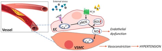
3. Marine Natural Products Affect Endothelial Dysfunction

Atherosclerosis, mainly caused by hypertension and dyslipidemia, begins with dysfunction of vascular endothelial cells and develops into CVD via plaque accumulation and related lesion formation in the blood vessels [12].
Vascular endothelial dysfunction is caused by (1) decreased eNOS activation by reduced intracellular Ca2+ level in the endothelium, (2) decreased bioavailability of nitric oxide produced from eNOS, (3) increased production of endothelial-derived vasoconstrictor factors, and (4) increased levels of oxidative stress and inflammation-inducing cytokines [41] (Figure 1). Table 2 shows the effects of various seaweed components on endothelial dysfunction in in vitro and in vivo models.
Figure 1. Crosstalk between endothelium (EC) and vascular smooth muscle cells (VSMCs) in hypertension.
Table 2. Effects of seaweed components on endothelial dysfunction.
Component Experimental Model Effects Ref
Astaxanthin ISO-induced myocardial infarction and cardiac hypertrophy model in rats
: 25 mg/kg/day for two weeks, OA
↓: ROS generation in heart tissue
↓: Oxidative damage
↑: Antioxidant enzyme activity
[42]
STZ-induced diabetes in male rats
: 10 mg/kg/d, OA
↓: Blunted endothelium-dependent vasodilator responses to Ach
↓: Aorta-induced oxidative stress and LOX-1 levels
↑: eNOS levels
[43]
Dieckol High glucose stimulation in cultured vascular endothelial cells.
: 10 or 50 μg/mL
↓: ROS production
↓: iNOS, COX-2, and NF-κB levels
[44]
Eckol and its derivates Cultured vascular endothelial cells/mice
: 50∼200 μg/mL
Protects the vascular barrier [45]
DPHC from Ishige okamurae Cultured vascular endothelial (EA.hy926) cells)/Tg(flk:EGFP) Transgenic Zebrafish
: 100 μM/0.6 μM
↑: Ach receptor and VEGF receptor 2
↑: NO production
↑: Ca2+ release
↑: Endothelium vasodilation
[46]
Sulfated polysaccharides from Padina tetrastromatica ISO induced myocardial infarction in rats
: 50 mg/kg/day for 12 days, OA
↓: hyperlipidemia
↓: Endothelial dysfunction
↓: Inflammatory reactions
[47]
ISO: isoproterenol; OA: oral administration; ROS: reactive oxygen species; STZ: streptozotocin; Ach: acetylcholine; LOX-1: lectin-like oxidized low-density lipoprotein receptor-1; eNOS: endothelial nitric oxide synthase; iNOS: inducible nitric oxide synthase; COX-2: cyclooxygenase-2; NF-κB: nuclear Factor kappa B; DPHC: diphlorethohydroxycarmalol; VEGF: vascular endothelial growth factor; NO: nitric oxide.
Alam et al. reported that the natural carotenoid astaxanthin extracted from microalgae Haematococcus pluvialis can penetrate the endothelial cell membrane and significantly inhibit ROS, thereby inhibiting oxidative stress in ISO-induced myocardial infarction and cardiac hypertrophy in rats, suggesting its cardioprotective action [42]. Zhao et al. found that astaxanthin protects against endothelial dysfunction of the aorta in diabetic rats and predicted the molecular mechanism involved in their effects [43]. They suggested that astaxanthin can attenuate blunted endothelium-dependent vasodilator responses to acetylcholine, upregulate endothelial nitric oxide synthase expression, and decrease excessive oxidative stress and endothelial dysfunction. Lee et al. isolated dieckol from the brown seaweed E. cava and found that it protected human umbilical vein endothelial cells damaged by high glucose via its antioxidant properties [44]. In addition, the positive effects of eckol and its derivatives, including dieckol from the brown seaweed Ecklonia bicyclis, were investigated in both human umbilical vein endothelial cells and mice [45]. They suggested that the abundance of hydroxyl groups of eckol and its derivatives contribute to their vascular barrier protective functions. Another phlorotannin, diphlorethohydroxycarmalol (DPHC) isolated from Ishige okamurae, was observed to have vasodilatory effects by increasing nitric oxide production and Ca2+ release in endothelial cells via stimulating the Ach receptor and VEGF-receptor 2 [46]. The author further demonstrated the vasodilatory ability of DPHC in Tg(flk:EGFP) transgenic zebrafish. In addition, another crucial components in brown seaweed, the sulfated polysaccharides extracted from Padina tetrastromatica, were investigated for their effect on ISO-induced myocardial infarction in a rat model [47]. ISO-induced hyperlipidemia, endothelial dysfunction, and inflammatory reactions were significantly reduced by the sulfated polysaccharides. Specifically, the authors emphasized that sulfated polysaccharides can be used as a new functional food ingredient for CVD, as they showed therapeutic ability similar to that of aspirin, a reference drug.

References

  1. Virani, S.S.; Alonso, A.; Aparicio, H.J.; Benjamin, E.J.; Bittencourt, M.S.; Callaway, C.W.; Carson, A.P.; Chamberlain, A.M.; Cheng, S.; Delling, F.N. Heart disease and stroke statistics—2021 update: A report from the American Heart Association. Circulation 2021, 143, e254–e743.
  2. Wong, N.D.; Lopez, V.; Tang, S.; Williams, G.R. Prevalence, treatment, and control of combined hypertension and hypercholesterolemia in the United States. Am. J. Cardiol. 2006, 98, 204–208.
  3. Zou, Z.; Cini, K.; Dong, B.; Ma, Y.; Ma, J.; Burgner, D.P.; Patton, G.C. Time trends in cardiovascular disease mortality across the BRICS: An age-period-cohort analysis of key nations with emerging economies using the global burden of disease study 2017. Circulation 2020, 141, 790–799.
  4. Grasselli, G.; Zangrillo, A.; Zanella, A.; Antonelli, M.; Cabrini, L.; Castelli, A.; Cereda, D.; Coluccello, A.; Foti, G.; Fumagalli, R. Baseline characteristics and outcomes of 1591 patients infected with SARS-CoV-2 admitted to ICUs of the Lombardy Region, Italy. JAMA 2020, 323, 1574–1581.
  5. Guo, T.; Fan, Y.; Chen, M.; Wu, X.; Zhang, L.; He, T.; Wang, H.; Wan, J.; Wang, X.; Lu, Z. Cardiovascular implications of fatal outcomes of patients with coronavirus disease 2019 (COVID-19). JAMA Cardiol. 2020, 5, 811–818.
  6. Figueroa, J.F.; Wadhera, R.K.; Lee, D.; Yeh, R.W.; Sommers, B.D. Community-level factors associated with racial and ethnic disparities in COVID-19 rates in Massachusetts: Study examines community-level factors associated with racial and ethnic disparities in COVID-19 rates in Massachusetts. Health Aff. 2020, 39, 1984–1992.
  7. Rate, C.-F.; Onder, G.; Rezza, G.; Brusaferro, S. Characteristics of patients dying in relation to COVID-19 in Italy. JAMA 2020, 23, 1775–1776.
  8. Bonow, R.O.; O’Gara, P.T.; Yancy, C.W. Cardiology and COVID-19. JAMA 2020, 324, 1131–1132.
  9. Colling, M.E.; Kanthi, Y. COVID–19-associated coagulopathy: An exploration of mechanisms. Vasc. Med. 2020, 25, 471–478.
  10. Moran, A.E.; Roth, G.A.; Narula, J.; Mensah, G.A. 1990–2010 global cardiovascular disease atlas. Glob. Heart 2014, 9, 3–16.
  11. Lopez, E.O.; Ballard, B.D.; Jan, A. Cardiovascular Disease. StatPearls, 2020. Available online: https://www.ncbi.nlm.nih.gov/books/NBK535419/ (accessed on 5 September 2021).
  12. Eaton, C.B.; Feldman, H.A.; Assaf, A.R.; McPhillips, J.B.; Hume, A.L.; Lasater, T.M.; Levinson, P.; Carleton, R.A. Prevalence of hypertension, dyslipidemia, and dyslipidemic hypertension. J. Fam. Pract. 1994, 38, 17–24.
  13. Chobanian, A.V.; Bakris, G.L.; Black, H.R.; Cushman, W.C.; Green, L.A.; Izzo, J.L., Jr.; Jones, D.W.; Materson, B.J.; Oparil, S.; Wright, J.T., Jr. The seventh report of the joint national committee on prevention, detection, evaluation, and treatment of high blood pressure: The JNC 7 report. JAMA 2003, 289, 2560–2571.
  14. Yamagata, K. Prevention of cardiovascular disease through modulation of endothelial cell function by dietary seaweed intake. Phytomed. Plus 2021, 2, 100026.
  15. Murray, M.; Dordevic, A.L.; Ryan, L.; Bonham, M.P. An emerging trend in functional foods for the prevention of cardiovascular disease and diabetes: Marine algal polyphenols. Crit. Rev. Food Sci. Nutr. 2018, 58, 1342–1358.
  16. Parmenter, B.H.; Croft, K.D.; Hodgson, J.M.; Dalgaard, F.; Bondonno, C.P.; Lewis, J.R.; Cassidy, A.; Scalbert, A.; Bondonno, N.P. An overview and update on the epidemiology of flavonoid intake and cardiovascular disease risk. Food Funct. 2020, 11, 6777–6806.
  17. Cardoso, S.M.; Pereira, O.R.; Seca, A.M.; Pinto, D.C.; Silva, A. Seaweeds as preventive agents for cardiovascular diseases: From nutrients to functional foods. Mar. Drugs 2015, 13, 6838–6865.
  18. Sabirin, F.; Soo, K.K.; Ziau, H.S.; Kuen, L.S. Antihypertensive effects of edible brown seaweeds in rats. Int. J. Adv. Appl. Sci. 2016, 3, 103–109.
  19. Brown, E.M.; Allsopp, P.J.; Magee, P.J.; Gill, C.I.; Nitecki, S.; Strain, C.R.; McSorley, E.M. Seaweed and human health. Nutr. Rev. 2014, 72, 205–216.
  20. Kim, Y.-G.; Cho, Y.-R.; Park, G.-M.; Won, K.-B.; Ann, S.H.; Yang, D.H.; Kang, J.-W.; Lim, T.-H.; Kim, H.-K.; Choe, J. High-density lipoprotein cholesterol and the risk of obstructive coronary artery disease beyond low-density lipoprotein cholesterol in non-diabetic individuals. Eur. J. Prevent. Cardiol. 2020, 27, 706–714.
  21. Navar-Boggan, A.M.; Peterson, E.D.; D’Agostino Sr, R.B.; Neely, B.; Sniderman, A.D.; Pencina, M.J. Hyperlipidemia in early adulthood increases long-term risk of coronary heart disease. Circulation 2015, 131, 451–458.
  22. Scandinavian Simvastatin Survival Study Group. Randomised trial of cholesterol lowering in 4444 patients with coronary heart disease: The Scandinavian Simvastatin Survival Study (4S). Lancet 1994, 344, 1383–1389.
  23. Jin, Q.; Yu, H.; Li, P. The evaluation and utilization of marine-derived bioactive compounds with anti-obesity effect. Curr. Med. Chem. 2018, 25, 861–878.
  24. Villanueva, M.-J.; Morcillo, M.; Tenorio, M.-D.; Mateos-Aparicio, I.; Andrés, V.; Redondo-Cuenca, A. Health-promoting effects in the gut and influence on lipid metabolism of Himanthalia elongata and Gigartina pistillata in hypercholesterolaemic Wistar rats. Eur. Food Res. Technol. 2014, 238, 409–416.
  25. Kumar, S.A.; Magnusson, M.; Ward, L.C.; Paul, N.A.; Brown, L. Seaweed supplements normalise metabolic, cardiovascular and liver responses in high-carbohydrate, high-fat fed rats. Mar. Drugs 2015, 13, 788–805.
  26. Chan, P.T.; Matanjun, P.; Yasir, S.M.; Tan, T.S. Antioxidant and hypolipidaemic properties of red seaweed, Gracilaria changii. J. Appl. Phycol. 2014, 26, 987–997.
  27. Kim, M.J.; Kim, H.K. Insulinotrophic and hypolipidemic effects of Ecklonia cava in streptozotocin-induced diabetic mice. Asian Pac. J. Trop. Med. 2012, 5, 374–379.
  28. Jung, H.A.; Jung, H.J.; Jeong, H.Y.; Kwon, H.J.; Ali, M.Y.; Choi, J.S. Phlorotannins isolated from the edible brown alga Ecklonia stolonifera exert anti-adipogenic activity on 3T3-L1 adipocytes by downregulating C/EBPα and PPARγ. Fitoterapia 2014, 92, 260–269.
  29. Ruqqia, K.; Sultana, V.; Ara, J.; Ehteshamul-Haque, S.; Athar, M. Hypolipidaemic potential of seaweeds in normal, triton-induced and high-fat diet-induced hyperlipidaemic rats. J. Appl. Phycol. 2015, 27, 571–579.
  30. Cuong, H.D.; Thuy, T.T.; Huong, T.T.; Ly, B.M.; Van, T.T. Structure and hypolipidaemic activity of fucoidan extracted from brown seaweed Sargassum henslowianum. Nat. Prod. Res. 2015, 29, 411–415.
  31. Sokolova, E.V.; Bogdanovich, L.N.; Ivanova, T.B.; Byankina, A.O.; Kryzhanovskiy, S.P.; Yermak, I.M. Effect of carrageenan food supplement on patients with cardiovascular disease results in normalization of lipid profile and moderate modulation of immunity system markers. PharmaNutrition 2014, 2, 33–37.
  32. Dousip, A.; Matanjun, P.; Sulaiman, M.R.; Tan, T.S.; Ooi, Y.B.H.; Lim, T.P. Effect of seaweed mixture intake on plasma lipid and antioxidant profile of hyperholesterolaemic rats. J. Appl. Phycol. 2014, 26, 999–1008.
  33. Borai, I.H.; Ezz, M.K.; Rizk, M.Z.; Matloub, A.; Aly, H.; El, A.; Farrag, R.; Fouad, G.I. Hypolipidemic and anti-atherogenic effect of sulphated polysaccharides from the green alga Ulva fasciata. Int. J. Pharm. Sci. Rev. Res. 2015, 31, 1–12.
  34. Hassan, S.; El-Twab, S.A.; Hetta, M.; Mahmoud, B. Improvement of lipid profile and antioxidant of hypercholesterolemic albino rats by polysaccharides extracted from the green alga Ulva lactuca Linnaeus. Saudi J. Biol. Sci. 2011, 18, 333–340.
  35. Hoang, M.H.; Kim, J.-Y.; Lee, J.H.; You, S.; Lee, S.-J. Antioxidative, hypolipidemic, and anti-inflammatory activities of sulfated polysaccharides from Monostroma nitidum. Food Sci. Biotechnol. 2015, 24, 199–205.
  36. Park, J.; Yeom, M.; Hahm, D.-H. Fucoidan improves serum lipid levels and atherosclerosis through hepatic SREBP-2-mediated regulation. J. Pharmacol. Sci. 2016, 131, 84–92.
  37. Woo, M.-N.; Jeon, S.-M.; Kim, H.-J.; Lee, M.-K.; Shin, S.-K.; Shin, Y.C.; Park, Y.-B.; Choi, M.-S. Fucoxanthin supplementation improves plasma and hepatic lipid metabolism and blood glucose concentration in high-fat fed C57BL/6N mice. Chem.-Biol. Interact. 2010, 186, 316–322.
  38. Jiménez-Escrig, A.; Sánchez-Muniz, F.J. Dietary fibre from edible seaweeds: Chemical structure, physicochemical properties and effects on cholesterol metabolism. Nutr. Res. 2000, 20, 585–598.
  39. Patil, N.P.; Le, V.; Sligar, A.D.; Mei, L.; Chavarria, D.; Yang, E.Y.; Baker, A.B. Algal polysaccharides as therapeutic agents for atherosclerosis. Front. Cardiovasc. Med. 2018, 5, 153.
  40. Bhakuni, D.S.; Silva, M. Biodynamic substances from marine flora. Bot. Mar. 1974, 17, 40–51.
  41. Gimbrone, M.A., Jr.; Garcia-Cardena, G. Endothelial cell dysfunction and the pathobiology of atherosclerosis. Circ. Res. 2016, 118, 620–636.
  42. Alam, M.N.; Hossain, M.M.; Rahman, M.M.; Subhan, N.; Mamun, M.A.A.; Ulla, A.; Reza, H.M.; Alam, M.A. Astaxanthin prevented oxidative stress in heart and kidneys of isoproterenol-administered aged rats. J. Diet. Suppl. 2018, 15, 42–54.
  43. Zhao, Z.-W.; Cai, W.; Lin, Y.-L.; Lin, Q.-F.; Jiang, Q.; Lin, Z.; Chen, L.-L. Ameliorative effect of astaxanthin on endothelial dysfunction in streptozotocin-induced diabetes in male rats. Arzneimittelforschung 2011, 61, 239–246.
  44. Lee, S.-H.; Han, J.-S.; Heo, S.-J.; Hwang, J.-Y.; Jeon, Y.-J. Protective effects of dieckol isolated from Ecklonia cava against high glucose-induced oxidative stress in human umbilical vein endothelial cells. Toxicol. In Vitro 2010, 24, 375–381.
  45. Kim, T.H.; Lee, T.; Ku, S.-K.; Bae, J.-S. Vascular barrier protective effects of eckol and its derivatives. Bioorg. Med. Chem. Lett. 2012, 22, 3710–3712.
  46. Lu, Y.A.; Jiang, Y.; Yang, H.W.; Hwang, J.; Jeon, Y.J.; Ryu, B. Diphlorethohydroxycarmalol Isolated from Ishige okamurae Exerts Vasodilatory Effects via Calcium Signaling and PI3K/Akt/eNOS Pathway. Int. J. Mol. Sci. 2021, 22, 1610.
  47. Lekshmi, V.S.; Kurup, G.M. Sulfated polysaccharides from the edible marine algae Padina tetrastromatica protects heart by ameliorating hyperlipidemia, endothelial dysfunction and inflammation in isoproterenol induced experimental myocardial infarction. J. Funct. Foods 2019, 54, 22–31.
More
Information
Contributor MDPI registered users' name will be linked to their SciProfiles pages. To register with us, please refer to https://encyclopedia.pub/register :
View Times: 407
Revisions: 2 times (View History)
Update Date: 24 Sep 2021
1000/1000