Submitted Successfully!
To reward your contribution, here is a gift for you: A free trial for our video production service.
Thank you for your contribution! You can also upload a video entry or images related to this topic.
Version Summary Created by Modification Content Size Created at Operation
1 + 4080 word(s) 4080 2021-04-26 10:43:59 |
2 format correct Meta information modification 4080 2021-05-06 05:25:47 | |
3 move out from EC Meta information modification 4080 2021-09-22 04:10:28 |

Video Upload Options

Do you have a full video?

Confirm

Are you sure to Delete?
Cite
If you have any further questions, please contact Encyclopedia Editorial Office.
Arii, J. Nuclear Egress of HSV-1. Encyclopedia. Available online: https://encyclopedia.pub/entry/9320 (accessed on 26 April 2024).
Arii J. Nuclear Egress of HSV-1. Encyclopedia. Available at: https://encyclopedia.pub/entry/9320. Accessed April 26, 2024.
Arii, Jun. "Nuclear Egress of HSV-1" Encyclopedia, https://encyclopedia.pub/entry/9320 (accessed April 26, 2024).
Arii, J. (2021, May 06). Nuclear Egress of HSV-1. In Encyclopedia. https://encyclopedia.pub/entry/9320
Arii, Jun. "Nuclear Egress of HSV-1." Encyclopedia. Web. 06 May, 2021.
Nuclear Egress of HSV-1
Edit

Herpes simplex virus 1 (HSV-1) replicates its genome and packages it into capsids within the nucleus. HSV-1 has evolved a complex mechanism of nuclear egress whereby nascent capsids bud on the inner nuclear membrane to form perinuclear virions that subsequently fuse with the outer nuclear membrane, releasing capsids into the cytosol. The viral-encoded nuclear egress complex (NEC) plays a crucial role in this vesicle-mediated nucleocytoplasmic transport. Nevertheless, similar system mediates the movement of other cellular macromolecular complexes in normal cells. Therefore, HSV-1 may utilize viral proteins to hijack the cellular machinery in order to facilitate capsid transport. 

herpesviruses nuclear egress primary envelopment de-envelopment

1. Introduction

The nuclear membrane (NM) consists of an inner nuclear membrane (INM) and an outer nuclear membrane (ONM), which separate nuclear and cytoplasmic activities in the eukaryotic cell (Figure 1A). The segregation of the nucleoplasm from the cytoplasm separates translation from transcription and maintains genome integrity. The INM and ONM are biochemically distinct [1]. While the ONM is continuous and functionally interacts with the endoplasmic reticulum (ER), the INM contains its own set of integral membrane proteins [2]. The ONM and the INM are separated by the perinuclear space and connected at annular junctions, where nuclear pore complexes (NPCs) are found. NPCs are large macromolecular assemblies that form transport channels and regulate trafficking between the cytoplasm and the nucleoplasm [3]. It had long been assumed that the NPC was the only route out of the nucleus except during the early phases of mitosis when the nuclear envelope breaks down.

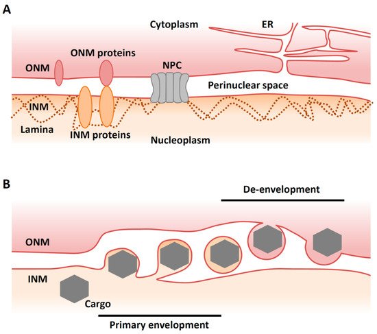
Figure 1. Overview of nuclear egress. (A) Illustration of a ‘normal’ nuclear envelope. INM, inner nuclear membrane; ONM, outer nuclear membrane; NPC, nuclear pore complex; ER, endoplasmic reticulum. (B) Illustration of vesicle-mediated nucleocytoplasmic transport. A cargo (e.g., viral capsid) in the nucleus buds through the INM to form a vesicle in the perinuclear space that then fuses with the ONM to release the complex into the cytoplasm (de-envelopment).

Recently, however, an alternative and NPC-independent pathway for nuclear export has been described in cells infected with viruses. Vesicle-mediated nucleocytoplasmic transport is a mechanism for the nuclear export of macromolecular complexes [4]. In this system, a macromolecular complex in the nucleus buds through the INM to form a vesicle in the perinuclear space (‘primary envelopment’). This vesicle then fuses with the ONM to release the complex into the cytoplasm in a process called ‘de-envelopment’ [4] (Figure 1B). This type of transport is observed in herpesvirus-infected mammalian cells and is required for the nuclear export of viral capsids that assemble in the nucleus [5]. Nucleocytoplasmic transport (or ‘nuclear egress’) of capsids is essential for the life cycle of all herpesviruses, as final maturation of their virions occurs in the cytoplasm. Although vesicle-mediated nucleocytoplasmic transport is not common in uninfected mammalian cells, it has been reported that Drosophila cellular large ribonucleoprotein complexes (RNPs) utilize this nuclear export mechanism in the absence of viral infection [6].

2. Overview of Nuclear Egress of Herpesviruses

Herpesviruses are enveloped double-stranded DNA viruses that establish lifelong latent infections in their natural hosts [7]. The Herpesviridae family is subdivided into the Alphaherpesvirinae, Betaherpesvirinae, and Gammaherpesvirinae subfamilies, based on their molecular and biological properties [7]. HSV-1 is the prototype of the alphaherpesvirus subfamily, which is comprised of the most common pathogenic agents in humans, and causes a variety of conditions such as mucocutaneous disease, keratitis, skin disease and encephalitis [8]. Herpesviruses share a common virion structure and similar proliferation strategies. A mature virion consists of an icosahedral capsid with a linear double-stranded DNA genome, a proteinaceous layer called tegument, and a host-membrane derived envelope spiked with viral glycoproteins.

Herpesviruses replicate their genomes and package them into capsids within the host cell nucleus. These capsids must traverse from the nucleus to the cytoplasm through a process called nuclear egress. In brief, the nascent nucleocapsids bud at the INM to form primary virions in the perinuclear space, and then the envelopes of primary virions fuse with the ONM, releasing the nucleocapsids into the cytoplasm for further maturation (Figure 1B). In the cytoplasm, nucleocapsids bud into vesicles derived from trans-Golgi networks or endosomes in a process called ‘secondary envelopment’. Finally, enveloped virions are released from the cells through an exocytotic pathway [5].

This envelopment/de-envelopment model of herpesvirus egress is now generally accepted and appears to be the dominant mechanism employed by this virus. However, other routes have been proposed. For example, capsids may be transported concomitantly with nuclear envelope breakdown in certain situations [9]. It has also been reported that capsids may egress from the nucleus through defective nuclear pores [10][11].

3. Primary Envelopment

3.1. Lamina Dissociation

Nuclear shape and mechanical stability are maintained by the nuclear lamina, which is localized beneath the INM. This structure is primarily composed of lamins, which are members of the intermediate filament family of cytoskeletal proteins (Figure 1A). Lamins interact with a large number of proteins at the INM and in the nucleoplasm, thereby influencing structural stability of the nucleus, DNA replication, transcription, and chromatin remodeling [12].

Before HSV-1 capsids access the INM, the nuclear lamina meshwork has to be dissolved. Between prophase and metaphase of mitosis, the lamina disassembles before reassembling during interphase [1]. This lamina disassembly is tightly regulated by phosphorylation [1]. During herpesvirus infection, the lamina is disrupted locally [13][14][15][16][17] (Figure 2). It appears that the NEC component UL34 recruits protein kinase C (PKC) isoforms that phosphorylate lamin B during infection [13][18]. In addition, viral kinases are also required for lamina dissolution. Viral kinase Us3, which is conserved in alphaherpesviruses, phosphorylates the lamin A/C protein (LMNA) [19][20][21]. HSV-1 encodes the other kinase UL13, which is conserved in Herpesviridae. UL13 homologues found in Betaherpesvirinae and Gammaherpesvirinae have predominant roles in lamin dissociation [22][23][24]. However, this function of UL13 homologues is not conserved in HSV-1 UL13 [22].

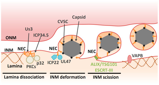
Figure 2. Multiple proteins are involved in primary envelopment of HSV-1 capsids. NEC and kinases (e.g., PKCs and Us3) induce lamina dissociation to allow capsid recruitment to the INM. ICP34.5 and p32 contribute to the recruitment of PKCs. The NEC is responsible for capsid recruitment and mediates budding by assembling into a hexagonal lattice. CVSC decorates vertices of DNA-filled nucleocapsid and promotes nuclear egress through interaction with the NEC. Viral proteins (e.g., UL47 and ICP22) and cellular protein VAPB regulate primary envelopment through as-yet unidentified mechanisms. The NEC recruits ESCRT-III via ALIX to pinch off primary virions from the INM.

Furthermore, the NEC binds directly to LMNA to modulate its conformation [15] and perturbs the interaction of lamins with emerin, a protein that links the lamina to the INM [25][26]. Taken together, the NEC directly and indirectly disrupts the fibrillar network of the nuclear lamina to ensure correct docking of capsids to the INM. Viral proteins also regulate cellular components beyond the NEC in order to facilitate capsid egress. For example, the HSV-1 neurovirulence factor ICP34.5 also contributes to lamin dissociation through recruitment of PKC to the NM via the cellular protein p32 [27][28] (Figure 2).

New data have underscored the importance of the lamin meshwork as a barrier to viral replication. Lamin degradation is required for HSV-1 nuclear egress and growth in dendritic cells [29]. Tripartite Motif Containing (TRIM) 43 is a factor that restricts a broad range of herpesviruses and is upregulated during viral infection [30]. Interestingly, TRIM43 mediates degradation of the centrosomal protein, pericentrin, which subsequently leads to alterations of the nuclear lamina. This alteration suppresses transcriptionally active viral chromatin states and viral replication.

In general, lamins contribute to the regulation of chromatin organization by tethering peripheral heterochromatin and chromatin remodeling complexes to the nuclear envelope [12]. Evidence of LMNA’s role in chromatin organization is provided by the finding that mutations in the human LMNA gene lead to premature aging and progressive loss of heterochromatin [31]. The same is true for the HSV-1 genome, as epigenetic regulation of viral DNA is modulated by its binding to LMNA [32][33][34]. These observations suggest that viral gene expression and nuclear egress must proceed in a coordinated manner.

3.2. Deformation of the INM

In addition to the disintegration of lamins, the NEC is responsible for recruitment of capsids to the INM [35][36][37][38]. Capsid vertex-specific component (CVSC), which consists of the UL17 and UL25 proteins, decorates vertices of DNA-filled nucleocapsids [39][40][41][42]. CVSC is proposed to promote nuclear egress of DNA-filled nucleocapsids through interaction with the NEC [43][44][45][46][47]. Once the capsid is recruited to the INM, primary envelopment occurs. This process involves membrane deformation around the capsid, followed by a ‘pinching off’ of the nascent bud; the resulting vesicle is formed in the perinuclear space and is referred to as the ‘primary virion’ (Figure 2). The NEC plays a direct role in nucleocapsid budding at the INM. Transient coexpression of NEC from pseudorabiesvirus (PRV, a porcine alphaherpesvirus) or Kaposi’s sarcoma-associated herpesvirus (KSHV, a human gammaherpesvirus) induces formation of perinuclear vesicles [48][49][50]. Moreover, recombinant NEC from HSV-1 or PRV can promote vesiculation of synthetic lipid membranes in the absence of any other viral or cellular proteins [51][52]. These findings suggest that the NEC has an intrinsic ability to deform membranes. Disruption of the membrane-coupled hexagonal NEC lattice by mutation of the inter- or intrahexagonal contact site impairs vesiculation both in vitro and in infected cells [4][53][37][51][54]. Thus, it is likely that formation of the NEC lattice drives vesiculation [4][53][51].

Biochemical analysis of the NEC also shows the requirement for a specific lipid composition for vesicle formation in vitro. Although the role of lipid in infected cells has not been analyzed, a high concentration of phosphatidic acid (PA) is required for in vitro vesicle formation [51]. Recently, it has been reported that PA-rich membranes at nuclear envelope herniation sites recruit cellular membrane remodeling machinery to the NMs in budding yeast [55]. Cell membranes mainly consist of lipids and thus lipid composition must affect the viral entry and the process by which capsid vesicles form during the budding processes. Protein kinase D (PKD) regulates vesicle transport through phosphorylation of phosphatidylinositol 4-kinase β (PI4KIIIKβ), which converts phosphoinositide (PI) to phosphoinositol 4-phosphate (PI4P). Knockdown of PKD or its modulators impairs both primary envelopment and virion release [56], suggesting that lipid composition plays a role during primary envelopment. Phosphatidylethanolamine (PE) a phospholipid that plays significant roles in cellular processes, such as vesicle transport and membrane fusion. Inhibition of the PE synthesis pathway impairs envelopment of HSV-1 capsids at the cytoplasm, but has no effect on nuclear egress [57]. The exact composition of specific lipids and the requirement for these during primary envelopment in infected cells should be determined during future studies.

3.3. Factors Involved in INM Deformation

Although the NEC has ability to vesiculate membranes in transfected cells and in vitro [4][53][51], empty perinuclear vesicles are rarely observed during infection. Therefore, in infected cells, the intrinsic budding potential of the NEC has to be regulated tightly before capsids arrive to the INM. In other words, NEC activity alone may not be sufficient for primary envelopment in infected cells where the environment is more complex.

Indeed, several other viral factors are reported to contribute nuclear egress in infected cells (Table 1). Tegument is a layer comprising thousands of densely packaged proteins that are localized between the virion envelope and capsid. The assembly of tegument on capsids occurs predominantly in the cytoplasm following nuclear egress. However, accumulating evidence suggests that capsids acquire some of the tegument proteins in the nucleus. In particular, tegument proteins UL36 (VP1/2), UL37, UL41 (vhs), UL47 (VP13/14) UL48 (VP16), UL49 (VP22), Us3, ICP0 and ICP4 have been reported to localize in the nucleus or associate with capsids [58][59][60][61][62]. While UL36 is not essential for nuclear egress of HSV-1 or PRV, a nuclear-specific isoform comprising the C-terminal region of UL36 is recruited to PRV capsids in the nucleus and may enhance their nuclear egress [63][64]. Cryo-EM analyses reveal that UL36 forms part of the HSV-1 CVSC, which was originally considered to be a complex of the UL17 and UL25 proteins [65][66]. Contributions of other HSV-1 proteins on primary envelopment have been described. UL47, the major tegument protein of HSV-1, interacts with the NEC and promotes primary envelopment [67]. The immediate early protein ICP22, which is conserved in the subfamily Alphaherpesvirinae, interacts with the NEC and regulates NEC localization and HSV-1 primary envelopment [68] (Figure 2).

Table 1. Viral factors involved in nuclear egress of HSV capsid.

Protein Conservation Function
NEC (UL31/UL34) Herpesviridae Vesicle formation via hexagonal lattice [51].
Incorporation of capsid [35][36].
Dissociation of lamins [15].
Recruitment of ESCRT-III [69].
De-envelopment [70].
UL13 Herpesviridae Dissociation of lamins [71].
(Weak in Alphaherpesvirinae).
Us3 Alphaherpesvirinae Dissociation of lamins [19].
Promote de-envelopment [72].
Phosphorylate UL34 [73].
Phosphorylate gB [74][75].
Phosphorylate UL31 [76].
ICP34.5 HSV-1 and HSV-2 Dissociation of lamins via PKC [28].
CVSC (UL17/UL25) Herpesviridae Link between NEC and capsid [43][44].
UL47 Alphaherpesvirinae Promote primary envelopment [67].
(Not observed in PRV [77][78]).
ICP22 Alphaherpesvirinae Promote primary envelopment [68].
gB and gH/gL Herpesviridae Redundantly promote de-envelopment [79].
(Not observed in PRV [80]).
UL51 Herpesviridae Promote de-envelopment [81].
(Not observed in PRV [82]).

In addition to the viral factors described above, host cell factors also contribute to vesicle formation at the INM during primary envelopment through as-yet undefined mechanisms. Vesicle-associated membrane protein-associated protein B (VAPB), which mediates cytoplasmic vesicle transport at the ER, facilitates HSV-1 nuclear egress [83]. Viral and cellular factors involved in HSV-1 primary envelopment are listed in Table 1 and Table 2, respectively.

Table 2. Cellular factors involved in nuclear egress of HSV capsids.

Protein Interactor Function Drosophila RNP
Lamins NEC, Us3, UL13 Prevent nuclear egress [15]. Yes [6][84]
PKC family NEC Dissociate lamins [18]. Yes [6]
p32 ICP34.5, UL47 Recruit PKC [28].
Promote de-envelopment [85].
-
PKD - Promote nuclear egress indirectly [56]. -
VAPB - Promote nuclear egress [83]. -
ESCRT-III NEC Mediate scission of INM [69]. Yes [69]
ALIX NEC Recruit ESCRT-III to INM [69]. -
TSG101 - Nuclear egress [86].  
CD98hc
β1 integrin
NEC, gB, gH Promote de-envelopment [87].
(Modulate fusion activity?).
-
Torsins - Promote de-envelopment indirectly [88]. Yes [89]

3.4. Scission at the INM during Primary Envelopment

Although NECs have the intrinsic capability to remodel membranes, they are unable to mediate primary envelopment by themselves in infected cells, as described above. It appears that host endosomal sorting complexes required for transport (ESCRT)-III machinery is responsible for the scission step during HSV-1 primary envelopment [69]. Viral budding can be divided into two stages: membrane deformation (the membrane is wrapped around the assembling virion) and membrane scission (the bud neck is severed) (Figure 2). A remarkable variety of enveloped viruses appear to use host ESCRT-III machinery at the scission stage of budding [90]. The ESCRT-III proteins were initially discovered as factors that are required for the biogenesis of multivesicular bodies (MVBs). It is now clear that the functions of the ESCRT-III proteins extend far beyond their role in MVB formation. Indeed, ESCRT complexes also regulate cytokinesis, the biogenesis of microvesicles and exosomes, plasma membrane repair, neuron pruning, the quality control of NPC, nuclear envelope reformation, and autophagy [91]. ESCRT-III proteins exist as soluble monomers in solution. When activated by membrane-associated upstream factors such as ESCRT-I/II, ALG-2-interacting protein X (ALIX) or charged multivesicular body protein (CHMP)7, ESCRT-III assembles into filamentous structures on membranes. These ESCRT-III assemblies are thought to be the main drivers of membrane remodeling and scission. The hexameric AAA ATPase Vps4 drives remodeling and disassembly of ESCRT-III filaments, and inactivation of Vps4 severely impairs ESCRT-III-mediated membrane remodeling [91].

Herpesvirus replication appears to rely on the ESCRT-III machinery [92][93][94][95]. At first, HSV-1 nuclear egress was thought to be independent of the ESCRT-III machinery in HEK293 cells as it is insensitive to a dominant negative mutation of Vps4 [92]. However, depletion of an ESCRT-III protein or its adaptor, ALIX in HeLa cells, reveals the role of ESCRT-III machinery during nuclear egress of HSV-1 [69]. In these cells, viral budding is arrested at the lollipop stage and nearly complete immature virions remained tethered to the INM. These nearly complete virions were morphologically similar to the virions of other viruses that are arrested during budding in the absence of ESCRT factors [90]. Furthermore, ESCRT-III is recruited by the NEC to the INM during the nuclear egress of HSV-1 capsids [69]. ESCRT-II, ALIX and CHMP7 have been identified as upstream factors of ESCRT-III that recruit ESCRT-III proteins and initiate their assembly [91]. In these contexts, CHMP7, but not ESCRT-II or ALIX, is required for NM reformation [96][97]. However, ALIX is required for the scission in the INM during nuclear egress [69] (Figure 2). In agreement with this observation, ALIX is required for recruitment of ESCRT-III to the NM during infection of cells with Epstein–Barr virus (EBV, a human gammaherpesvirus) [98][99]. In addition, a compound that binds to ESCRT-I protein TSG101 inhibits primary envelopment of HSV-1 in Vero cells [86], supporting the idea that ESCRT-III machinery is required for nuclear egress of HSV-1 capsids. The other ESCRT-III adaptor CHMP7 has a predominant role in reformation of the nuclear membrane in various situations [96][97]. Thus, it is unlikely that ESCRT-III is recruited to repair HSV-1-induced defects in the nuclear envelope. Taken together, these data suggest that, while NEC has ability to promote vesiculation in vitro, it recruits ESCRT-III machinery to complete scission in infected cells. Similarly, Ebola virus VP40 alone can carry out membrane budding in an in vitro system [100], although Ebola virus budding is highly dependent on ESCRT-III machinery [90].

4. De-Envelopment

4.1. Overview of De-Envelopment

De-envelopment is a process in which perinuclear virions fuse with the ONM to release naked capsids into the cytosol (Figure 1B). Membrane fusion and disassembly of the NEC are required in this process. The molecular mechanism of de-envelopment is not well understood. Nonetheless, several viral and cellular proteins have been reported as regulators of HSV-1 de-envelopment (Table 1 and Table 2).

In particular, the Us3 protein kinase, which is conserved among Alphaherpesvirinae, has a prominent role in the de-envelopment step. Inactivation of Us3 enzymatic activity leads to an accumulation of primary enveloped HSV-1 virions in large invaginations of the INM [72][101][102], indicating that phosphorylation may regulate the de-envelopment process, even though it is not essential. The NEC lattices within the perinuclear virions are stable structures that must be disassembled during de-envelopment. Phosphorylation of the NEC by Us3 may loosen NEC lattices in perinuclear virions to promote de-envelopment [70][76] (Figure 3). In agreement with this hypothesis, Us3 is present in perinuclear virions [72]. As is the case with Us3 kinase inactive mutants, mutations in other viral tegument protein UL51 of HSV-1 also lead to accumulation of primary envelope virions in the perinuclear space [81][103][104], suggesting that the de-envelopment process is modulated by various viral proteins.

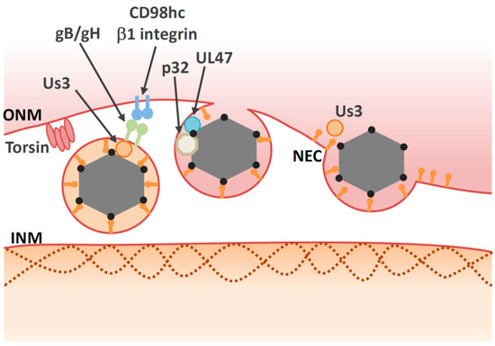
Figure 3. Multiple proteins are involved in the de-envelopment step. Some viral proteins, such as gB/gH and Us3 may be involved in the de-envelopment step, but their roles are unclear. Us3 may induce disassembly of the NEC lattice. Torsin and p32 promote de-envelopment through unidentified mechanisms. Viral protein UL47 interacts with cellular protein p32. The CD98hc/β1 integrin complex may influence fusion activity of the primary virions.

Similar to membrane fusion during virus entry, virion glycoproteins may play roles in mediating fusion between the envelope of the primary virions and the ONM. Primary virions accumulate in the perinuclear space during infection with recombinant HSV-1s harboring null or point mutations in both gB and gH [74][79][105]. To date, it is unclear how either gB or gH mediate fusion during de-envelopment, although both are required for fusion during viral entry [106] and Us3 phosphorylates gB [74][75]. In the case of the closely related PRV protein, gB or gH are not present in the perinuclear vesicles or the NMs and they do not have any role on nuclear egress [80]. Thus, gB or gH/gL do not appear to function as conserved components of the fusion machinery during de-envelopment step.

Deletion of either UL31 or UL34 completely halts perinuclear virion formation, which occurs before de-envelopment. Thus, it is conceivable that UL31 and/or UL34 also play an essential role in the de-envelopment step as well as in primary envelopment. UL34 is a transmembrane protein, the C-terminus of which is exposed to the perinuclear space. However, the C-terminal domain (including the transmembrane region) can be replaced by corresponding regions of other viral or cellular proteins without loss of function [107][108]. These observations indicate that the NEC does not directly play a role in the fusion process and that other components of the cellular fusion machinery are involved in de-envelopment. Thus, viral proteins that are involved in the de-envelopment process described above (Table 1) might also modulate cellular fusion machinery at the ONM.

4.2. Cellular Factors Involved in De-Envelopment

In addition to viral factors, cellular factors also regulate the de-envelopment process (Figure 3 and Table 2). Cellular factors including p32, CD98 heavy chain (CD98hc) and β1 integrin are recruited to the NM in HSV-1-infected cells [87][85]. Knockdown of these proteins leads to aberrant accumulation of enveloped virions in the invagination structures derived from the INM; this is phenocopied by inactivation of Us3 protein kinase activity. These observations suggest that the cellular factors mentioned above regulate the de-envelopment step. Accumulation of the multifunctional scaffold protein, p32, at the nuclear rim in infected cells depends on UL47 [85], suggesting that UL47 has an optimal role in the de-envelopment step in addition to its role in primary envelopment [67]. Plasma membrane protein CD98hc may regulate membrane fusion, since anti-CD98hc monoclonal antibodies enhance or inhibit cell–cell fusion, mediated by fusion proteins [109][110][111][112]. This CD98hc function in membrane fusion involves its binding partner, β1 integrin [111], suggesting that the CD98hc/β1 integrin interaction regulates fusion between perinuclear virions and the ONM. In the absence of UL34, CD98hc is not redistributed to the NM, but is dispersed throughout the cytoplasm [113]. At the ultrastructural level, HSV-1 infection causes ER compression around the nuclear envelope, whereas the UL34-null mutation causes cytoplasmic dispersion of the ER [113]. These observations suggest that HSV-1 infection redistributes ER around the nuclear envelope through unidentified mechanisms, thereby enabling accumulation of ER-associated de-envelopment factors, such as CD98hc, gB, and gH in the region where de-envelopment occurs.

Several lines of evidence indicate that torsins might be involved in HSV-1 nuclear egress. Torsins are members of the AAA+ ATPase superfamily, which is comprised of enzymes that mediate ATP-dependent conformational remodeling of target proteins or protein complexes [114]. Among torsin orthologues in humans, Torsin A (TorA) is the best studied due to its association with a disease designated as early onset dystonia; this association underlies the alternate name for this protein, which is dystonia 1 protein (DYT1) [114]. Torsins are located within the lumen of the ER and NM [115]. In contrast to other AAA+ ATPases, torsins lack a key catalytic residue and need the INM protein lamina-associated polypeptide 1 (LAP1) or the ER protein lumenal domain like LAP1 (LULL1) as a cofactor for full activation [116][117]. Deletion or mutation of TorA resulted in the accumulation of vesicles in the perinuclear space [115]; these vesicles resembled those produced in cells expressing NEC [48]. Ectopic expression of wild-type TorA reduced HSV-1 production and led to accumulation of virus-like structures in the perinuclear space and the lumen of the ER [88], indicating that torsins play a role in de-envelopment. Similarly, perinuclear virions accumulate in TorA-mutant murine cells infected with HSV-1 [118]. Although torsins are involved in nuclear egress, the precise molecular mechanisms by which they regulate this process remain unclear. Another study showed that knockout of torsin itself or the INM-specific torsin cofactor, LAP1, produced only subtle defects in viral replication [119]. In this study, the authors also showed that knockout of LULL1, a torsin cofactor in the ER, reduces HSV-1 growth by one order of magnitude in the absence of nuclear egress defects. Taken together, torsins may be involved in HSV-1 nuclear egress indirectly, as they appear to regulate the nuclear envelope [120][121] and lipid metabolism [122], and both these biological processes have an impact on nuclear egress. Although torsins have been extensively investigated in biochemical studies and animal models, their precise biological functions remain elusive. Further study of their roles in the nuclear egress of herpesvirus might help elucidate these functions.

Neither the de-envelopment step nor viral replication are completely dependent on any of the individual cellular factors described above (Table 2). This suggests a degree of redundancy among these factors, which presents a challenge when attempting to characterize the precise contribution of each. Future biochemical analyses or fusion assays designed to reconstitute the de-envelopment step are desirable in this regard, as they will help to elucidate molecular mechanisms. Alternatively, it is possible that the major cellular players in vesicle-mediated nuclear egress have not been identified yet. Notably, there have been no reports to date of an a ONM-resident factor that is involved in fusion.

In general, an entry receptor is required for viral glycoprotein-driven fusion. The entry receptor restricts susceptible cells and determines the tropism of a particular virus. A receptor-like molecule that increases affinity for the perinuclear virion would therefore restrict the ‘perinuclear tropism’, thereby preventing back-fusion to the INM. If viral fusion protein gB mediates fusion between the perinuclear virion and ONM [74][79][105], it might also associate with membrane proteins in the ONM, as gB receptors are important during viral entry [123][124][125]. As described above, however, gB is not essential for the de-envelopment step. Thus, both a ‘ligand’ in the perinuclear virion and a ‘receptor’ in the ONM must be identified to prove this hypothesis.

References

  1. Hetzer, M.W. The nuclear envelope. Cold Spring Harb. Perspect. Biol. 2010, 2, a000539.
  2. Crisp, M.; Burke, B. The nuclear envelope as an integrator of nuclear and cytoplasmic architecture. FEBS Lett. 2008, 582, 2023–2032.
  3. Paci, G.; Caria, J.; Lemke, E.A. Cargo transport through the nuclear pore complex at a glance. J. Cell Sci. 2021, 134.
  4. Hagen, C.; Dent, K.C.; Zeev-Ben-Mordehai, T.; Grange, M.; Bosse, J.B.; Whittle, C.; Klupp, B.G.; Siebert, C.A.; Vasishtan, D.; Bauerlein, F.J.; et al. Structural Basis of Vesicle Formation at the Inner Nuclear Membrane. Cell 2015, 163, 1692–1701.
  5. Johnson, D.C.; Baines, J.D. Herpesviruses remodel host membranes for virus egress. Nat. Rev. Microbiol. 2011, 9, 382–394.
  6. Speese, S.D.; Ashley, J.; Jokhi, V.; Nunnari, J.; Barria, R.; Li, Y.; Ataman, B.; Koon, A.; Chang, Y.T.; Li, Q.; et al. Nuclear envelope budding enables large ribonucleoprotein particle export during synaptic Wnt signaling. Cell 2012, 149, 832–846.
  7. Pellet, P.E.; Roizman, B. Herpesviridae. In Fields Virology, 6th ed.; Knipe, D.M., Howley, P.M., Cohen, J.I., Griffin, D.E., Lamb, R.A., Martin, M.A., Racaniello, V.R., Roizman, B., Eds.; Lippincott-Williams &Wilkins: Philadelphia, PA, USA, 2013; pp. 1802–1822.
  8. Roizman, B.; Knipe, D.M.; Whitley, R.J. Herpes simplex viruses. In Fields Virology, 6th ed.; Knipe, D.M., Howley, P.M., Cohen, J.I., Griffin, D.E., Lamb, R.A., Martin, M.A., Racaniello, V.R., Roizman, B., Eds.; Lippincott-Williams &Wilkins: Philadelphia, PA, USA, 2013; pp. 1823–1897.
  9. Schulz, K.S.; Klupp, B.G.; Granzow, H.; Passvogel, L.; Mettenleiter, T.C. Herpesvirus nuclear egress: Pseudorabies Virus can simultaneously induce nuclear envelope breakdown and exit the nucleus via the envelopment-deenvelopment-pathway. Virus Res. 2015, 209, 76–86.
  10. Leuzinger, H.; Ziegler, U.; Schraner, E.M.; Fraefel, C.; Glauser, D.L.; Heid, I.; Ackermann, M.; Mueller, M.; Wild, P. Herpes simplex virus 1 envelopment follows two diverse pathways. J. Virol. 2005, 79, 13047–13059.
  11. Wild, P.; Senn, C.; Manera, C.L.; Sutter, E.; Schraner, E.M.; Tobler, K.; Ackermann, M.; Ziegler, U.; Lucas, M.S.; Kaech, A. Exploring the nuclear envelope of herpes simplex virus 1-infected cells by high-resolution microscopy. J. Virol. 2009, 83, 408–419.
  12. Gruenbaum, Y.; Margalit, A.; Goldman, R.D.; Shumaker, D.K.; Wilson, K.L. The nuclear lamina comes of age. Nat. Rev. Mol. Cell Biol. 2005, 6, 21–31.
  13. Muranyi, W.; Haas, J.; Wagner, M.; Krohne, G.; Koszinowski, U.H. Cytomegalovirus recruitment of cellular kinases to dissolve the nuclear lamina. Science 2002, 297, 854–857.
  14. Scott, E.S.; O’Hare, P. Fate of the inner nuclear membrane protein lamin B receptor and nuclear lamins in herpes simplex virus type 1 infection. J. Virol. 2001, 75, 8818–8830.
  15. Reynolds, A.E.; Liang, L.; Baines, J.D. Conformational changes in the nuclear lamina induced by herpes simplex virus type 1 require genes U(L)31 and U(L)34. J. Virol. 2004, 78, 5564–5575.
  16. Simpson-Holley, M.; Baines, J.; Roller, R.; Knipe, D.M. Herpes simplex virus 1 U(L)31 and U(L)34 gene products promote the late maturation of viral replication compartments to the nuclear periphery. J. Virol. 2004, 78, 5591–5600.
  17. Simpson-Holley, M.; Colgrove, R.C.; Nalepa, G.; Harper, J.W.; Knipe, D.M. Identification and functional evaluation of cellular and viral factors involved in the alteration of nuclear architecture during herpes simplex virus 1 infection. J. Virol. 2005, 79, 12840–12851.
  18. Park, R.; Baines, J.D. Herpes simplex virus type 1 infection induces activation and recruitment of protein kinase C to the nuclear membrane and increased phosphorylation of lamin B. J. Virol. 2006, 80, 494–504.
  19. Bjerke, S.L.; Roller, R.J. Roles for herpes simplex virus type 1 UL34 and US3 proteins in disrupting the nuclear lamina during herpes simplex virus type 1 egress. Virology 2006, 347, 261–276.
  20. Mou, F.; Forest, T.; Baines, J.D. US3 of herpes simplex virus type 1 encodes a promiscuous protein kinase that phosphorylates and alters localization of lamin A/C in infected cells. J. Virol. 2007, 81, 6459–6470.
  21. Mou, F.; Wills, E.G.; Park, R.; Baines, J.D. Effects of lamin A/C, lamin B1, and viral US3 kinase activity on viral infectivity, virion egress, and the targeting of herpes simplex virus U(L)34-encoded protein to the inner nuclear membrane. J. Virol. 2008, 82, 8094–8104.
  22. Kuny, C.V.; Chinchilla, K.; Culbertson, M.R.; Kalejta, R.F. Cyclin-dependent kinase-like function is shared by the beta- and gamma- subset of the conserved herpesvirus protein kinases. PLoS Pathog. 2010, 6, e1001092.
  23. Lee, C.P.; Huang, Y.H.; Lin, S.F.; Chang, Y.; Chang, Y.H.; Takada, K.; Chen, M.R. Epstein-Barr virus BGLF4 kinase induces disassembly of the nuclear lamina to facilitate virion production. J. Virol. 2008, 82, 11913–11926.
  24. Hamirally, S.; Kamil, J.P.; Ndassa-Colday, Y.M.; Lin, A.J.; Jahng, W.J.; Baek, M.C.; Noton, S.; Silva, L.A.; Simpson-Holley, M.; Knipe, D.M.; et al. Viral mimicry of Cdc2/cyclin-dependent kinase 1 mediates disruption of nuclear lamina during human cytomegalovirus nuclear egress. PLoS Pathog. 2009, 5, e1000275.
  25. Leach, N.; Bjerke, S.L.; Christensen, D.K.; Bouchard, J.M.; Mou, F.; Park, R.; Baines, J.; Haraguchi, T.; Roller, R.J. Emerin is hyperphosphorylated and redistributed in herpes simplex virus type 1-infected cells in a manner dependent on both UL34 and US3. J. Virol. 2007, 81, 10792–10803.
  26. Morris, J.B.; Hofemeister, H.; O’Hare, P. Herpes simplex virus infection induces phosphorylation and delocalization of emerin, a key inner nuclear membrane protein. J. Virol. 2007, 81, 4429–4437.
  27. Wu, S.; Pan, S.; Zhang, L.; Baines, J.; Roller, R.; Ames, J.; Yang, M.; Wang, J.; Chen, D.; Liu, Y.; et al. Herpes Simplex Virus 1 Induces Phosphorylation and Reorganization of Lamin A/C through the gamma134.5 Protein That Facilitates Nuclear Egress. J. Virol. 2016, 90, 10414–10422.
  28. Wang, Y.; Yang, Y.; Wu, S.; Pan, S.; Zhou, C.; Ma, Y.; Ru, Y.; Dong, S.; He, B.; Zhang, C.; et al. p32 is a novel target for viral protein ICP34.5 of herpes simplex virus type 1 and facilitates viral nuclear egress. J. Biol. Chem. 2014, 289, 35795–35805.
  29. Turan, A.; Grosche, L.; Krawczyk, A.; Muhl-Zurbes, P.; Drassner, C.; Duthorn, A.; Kummer, M.; Hasenberg, M.; Voortmann, S.; Jastrow, H.; et al. Autophagic degradation of lamins facilitates the nuclear egress of herpes simplex virus type 1. J. Cell Biol. 2019, 218, 508–523.
  30. Full, F.; van Gent, M.; Sparrer, K.M.J.; Chiang, C.; Zurenski, M.A.; Scherer, M.; Brockmeyer, N.H.; Heinzerling, L.; Sturzl, M.; Korn, K.; et al. Centrosomal protein TRIM43 restricts herpesvirus infection by regulating nuclear lamina integrity. Nat. Microbiol. 2019, 4, 164–176.
  31. Schreiber, K.H.; Kennedy, B.K. When lamins go bad: Nuclear structure and disease. Cell 2013, 152, 1365–1375.
  32. Silva, L.; Cliffe, A.; Chang, L.; Knipe, D.M. Role for A-type lamins in herpesviral DNA targeting and heterochromatin modulation. PLoS Pathog. 2008, 4, e1000071.
  33. Silva, L.; Oh, H.S.; Chang, L.; Yan, Z.; Triezenberg, S.J.; Knipe, D.M. Roles of the nuclear lamina in stable nuclear association and assembly of a herpesviral transactivator complex on viral immediate-early genes. mBio 2012, 3.
  34. Oh, H.S.; Traktman, P.; Knipe, D.M. Barrier-to-Autointegration Factor 1 (BAF/BANF1) Promotes Association of the SETD1A Histone Methyltransferase with Herpes Simplex Virus Immediate-Early Gene Promoters. mBio 2015, 6, e00345-15.
  35. Chang, Y.E.; Van Sant, C.; Krug, P.W.; Sears, A.E.; Roizman, B. The null mutant of the U(L)31 gene of herpes simplex virus 1: Construction and phenotype in infected cells. J. Virol. 1997, 71, 8307–8315.
  36. Roller, R.J.; Zhou, Y.; Schnetzer, R.; Ferguson, J.; DeSalvo, D. Herpes simplex virus type 1 U(L)34 gene product is required for viral envelopment. J. Virol. 2000, 74, 117–129.
  37. Roller, R.J.; Bjerke, S.L.; Haugo, A.C.; Hanson, S. Analysis of a charge cluster mutation of herpes simplex virus type 1 UL34 and its extragenic suppressor suggests a novel interaction between pUL34 and pUL31 that is necessary for membrane curvature around capsids. J. Virol. 2010, 84, 3921–3934.
  38. Funk, C.; Ott, M.; Raschbichler, V.; Nagel, C.H.; Binz, A.; Sodeik, B.; Bauerfeind, R.; Bailer, S.M. The Herpes Simplex Virus Protein pUL31 Escorts Nucleocapsids to Sites of Nuclear Egress, a Process Coordinated by Its N-Terminal Domain. PLoS Pathog. 2015, 11, e1004957.
  39. Trus, B.L.; Newcomb, W.W.; Cheng, N.; Cardone, G.; Marekov, L.; Homa, F.L.; Brown, J.C.; Steven, A.C. Allosteric signaling and a nuclear exit strategy: Binding of UL25/UL17 heterodimers to DNA-Filled HSV-1 capsids. Mol. Cell 2007, 26, 479–489.
  40. Sheaffer, A.K.; Newcomb, W.W.; Gao, M.; Yu, D.; Weller, S.K.; Brown, J.C.; Tenney, D.J. Herpes simplex virus DNA cleavage and packaging proteins associate with the procapsid prior to its maturation. J. Virol. 2001, 75, 687–698.
  41. Thurlow, J.K.; Rixon, F.J.; Murphy, M.; Targett-Adams, P.; Hughes, M.; Preston, V.G. The herpes simplex virus type 1 DNA packaging protein UL17 is a virion protein that is present in both the capsid and the tegument compartments. J. Virol. 2005, 79, 150–158.
  42. Newcomb, W.W.; Homa, F.L.; Brown, J.C. Herpes simplex virus capsid structure: DNA packaging protein UL25 is located on the external surface of the capsid near the vertices. J. Virol. 2006, 80, 6286–6294.
  43. Yang, K.; Baines, J.D. Selection of HSV capsids for envelopment involves interaction between capsid surface components pUL31, pUL17, and pUL25. Proc. Natl. Acad. Sci. USA 2011, 108, 14276–14281.
  44. Yang, K.; Wills, E.; Lim, H.Y.; Zhou, Z.H.; Baines, J.D. Association of herpes simplex virus pUL31 with capsid vertices and components of the capsid vertex-specific complex. J. Virol. 2014, 88, 3815–3825.
  45. Takeshima, K.; Arii, J.; Maruzuru, Y.; Koyanagi, N.; Kato, A.; Kawaguchi, Y. Identification of the Capsid Binding Site in the Herpes Simplex Virus 1 Nuclear Egress Complex and Its Role in Viral Primary Envelopment and Replication. J. Virol. 2019, 93.
  46. Newcomb, W.W.; Fontana, J.; Winkler, D.C.; Cheng, N.; Heymann, J.B.; Steven, A.C. The Primary Enveloped Virion of Herpes Simplex Virus 1: Its Role in Nuclear Egress. mBio 2017, 8.
  47. Draganova, E.B.; Zhang, J.; Zhou, Z.H.; Heldwein, E.E. Structural basis for capsid recruitment and coat formation during HSV-1 nuclear egress. Elife 2020, 9.
  48. Klupp, B.G.; Granzow, H.; Fuchs, W.; Keil, G.M.; Finke, S.; Mettenleiter, T.C. Vesicle formation from the nuclear membrane is induced by coexpression of two conserved herpesvirus proteins. Proc. Natl. Acad. Sci. USA 2007, 104, 7241–7246.
  49. Luitweiler, E.M.; Henson, B.W.; Pryce, E.N.; Patel, V.; Coombs, G.; McCaffery, J.M.; Desai, P.J. Interactions of the Kaposi’s Sarcoma-associated herpesvirus nuclear egress complex: ORF69 is a potent factor for remodeling cellular membranes. J. Virol. 2013, 87, 3915–3929.
  50. Desai, P.J.; Pryce, E.N.; Henson, B.W.; Luitweiler, E.M.; Cothran, J. Reconstitution of the Kaposi’s sarcoma-associated herpesvirus nuclear egress complex and formation of nuclear membrane vesicles by coexpression of ORF67 and ORF69 gene products. J. Virol. 2012, 86, 594–598.
  51. Bigalke, J.M.; Heuser, T.; Nicastro, D.; Heldwein, E.E. Membrane deformation and scission by the HSV-1 nuclear egress complex. Nat. Commun 2014, 5, 4131.
  52. Lorenz, M.; Vollmer, B.; Unsay, J.D.; Klupp, B.G.; Garcia-Saez, A.J.; Mettenleiter, T.C.; Antonin, W. A single herpesvirus protein can mediate vesicle formation in the nuclear envelope. J. Biol. Chem. 2015, 290, 6962–6974.
  53. Bigalke, J.M.; Heldwein, E.E. Structural basis of membrane budding by the nuclear egress complex of herpesviruses. EMBO J. 2015, 34, 2921–2936.
  54. Arii, J.; Takeshima, K.; Maruzuru, Y.; Koyanagi, N.; Kato, A.; Kawaguchi, Y. Roles of the Interhexamer Contact Site for Hexagonal Lattice Formation of the Herpes Simplex Virus 1 Nuclear Egress Complex in Viral Primary Envelopment and Replication. J. Virol. 2019, 93.
  55. Thaller, D.J.; Tong, D.; Marklew, C.J.; Ader, N.R.; Mannino, P.J.; Borah, S.; King, M.C.; Ciani, B.; Lusk, C.P. Direct binding of ESCRT protein Chm7 to phosphatidic acid-rich membranes at nuclear envelope herniations. J. Cell Biol. 2021, 220.
  56. Roussel, E.; Lippe, R. Cellular Protein Kinase D Modulators Play a Role during Multiple Steps of Herpes Simplex Virus 1 Egress. J. Virol. 2018, 92.
  57. Arii, J.; Fukui, A.; Shimanaka, Y.; Kono, N.; Arai, H.; Maruzuru, Y.; Koyanagi, N.; Kato, A.; Mori, Y.; Kawaguchi, Y. Role of Phosphatidylethanolamine Biosynthesis in Herpes Simplex Virus 1-Infected Cells in Progeny Virus Morphogenesis in the Cytoplasm and in Viral Pathogenicity In Vivo. J. Virol. 2020, 94.
  58. Naldinho-Souto, R.; Browne, H.; Minson, T. Herpes simplex virus tegument protein VP16 is a component of primary enveloped virions. J. Virol. 2006, 80, 2582–2584.
  59. Bucks, M.A.; O’Regan, K.J.; Murphy, M.A.; Wills, J.W.; Courtney, R.J. Herpes simplex virus type 1 tegument proteins VP1/2 and UL37 are associated with intranuclear capsids. Virology 2007, 361, 316–324.
  60. Donnelly, M.; Elliott, G. Fluorescent tagging of herpes simplex virus tegument protein VP13/14 in virus infection. J. Virol. 2001, 75, 2575–2583.
  61. Pomeranz, L.E.; Blaho, J.A. Modified VP22 localizes to the cell nucleus during synchronized herpes simplex virus type 1 infection. J. Virol. 1999, 73, 6769–6781.
  62. Henaff, D.; Remillard-Labrosse, G.; Loret, S.; Lippe, R. Analysis of the early steps of herpes simplex virus 1 capsid tegumentation. J. Virol. 2013, 87, 4895–4906.
  63. Luxton, G.W.; Lee, J.I.; Haverlock-Moyns, S.; Schober, J.M.; Smith, G.A. The pseudorabies virus VP1/2 tegument protein is required for intracellular capsid transport. J. Virol. 2006, 80, 201–209.
  64. Leelawong, M.; Lee, J.I.; Smith, G.A. Nuclear egress of pseudorabies virus capsids is enhanced by a subspecies of the large tegument protein that is lost upon cytoplasmic maturation. J. Virol. 2012, 86, 6303–6314.
  65. Huet, A.; Makhov, A.M.; Huffman, J.B.; Vos, M.; Homa, F.L.; Conway, J.F. Extensive subunit contacts underpin herpesvirus capsid stability and interior-to-exterior allostery. Nat. Struct. Mol. Biol. 2016, 23, 531–539.
  66. Cardone, G.; Newcomb, W.W.; Cheng, N.; Wingfield, P.T.; Trus, B.L.; Brown, J.C.; Steven, A.C. The UL36 tegument protein of herpes simplex virus 1 has a composite binding site at the capsid vertices. J. Virol. 2012, 86, 4058–4064.
  67. Liu, Z.; Kato, A.; Shindo, K.; Noda, T.; Sagara, H.; Kawaoka, Y.; Arii, J.; Kawaguchi, Y. Herpes simplex virus 1 UL47 interacts with viral nuclear egress factors UL31, UL34, and Us3 and regulates viral nuclear egress. J. Virol. 2014, 88, 4657–4667.
  68. Maruzuru, Y.; Shindo, K.; Liu, Z.; Oyama, M.; Kozuka-Hata, H.; Arii, J.; Kato, A.; Kawaguchi, Y. Role of herpes simplex virus 1 immediate early protein ICP22 in viral nuclear egress. J. Virol. 2014, 88, 7445–7454.
  69. Arii, J.; Watanabe, M.; Maeda, F.; Tokai-Nishizumi, N.; Chihara, T.; Miura, M.; Maruzuru, Y.; Koyanagi, N.; Kato, A.; Kawaguchi, Y. ESCRT-III mediates budding across the inner nuclear membrane and regulates its integrity. Nat. Commun. 2018, 9, 3379.
  70. Mou, F.; Wills, E.; Baines, J.D. Phosphorylation of the U(L)31 protein of herpes simplex virus 1 by the U(S)3-encoded kinase regulates localization of the nuclear envelopment complex and egress of nucleocapsids. J. Virol. 2009, 83, 5181–5191.
  71. Cano-Monreal, G.L.; Wylie, K.M.; Cao, F.; Tavis, J.E.; Morrison, L.A. Herpes simplex virus 2 UL13 protein kinase disrupts nuclear lamins. Virology 2009, 392, 137–147.
  72. Reynolds, A.E.; Wills, E.G.; Roller, R.J.; Ryckman, B.J.; Baines, J.D. Ultrastructural localization of the herpes simplex virus type 1 UL31, UL34, and US3 proteins suggests specific roles in primary envelopment and egress of nucleocapsids. J. Virol. 2002, 76, 8939–8952.
  73. Purves, F.C.; Spector, D.; Roizman, B. The herpes simplex virus 1 protein kinase encoded by the US3 gene mediates posttranslational modification of the phosphoprotein encoded by the UL34 gene. J. Virol. 1991, 65, 5757–5764.
  74. Wisner, T.W.; Wright, C.C.; Kato, A.; Kawaguchi, Y.; Mou, F.; Baines, J.D.; Roller, R.J.; Johnson, D.C. Herpesvirus gB-induced fusion between the virion envelope and outer nuclear membrane during virus egress is regulated by the viral US3 kinase. J. Virol. 2009, 83, 3115–3126.
  75. Kato, A.; Arii, J.; Shiratori, I.; Akashi, H.; Arase, H.; Kawaguchi, Y. Herpes simplex virus 1 protein kinase Us3 phosphorylates viral envelope glycoprotein B and regulates its expression on the cell surface. J. Virol. 2009, 83, 250–261.
  76. Kato, A.; Yamamoto, M.; Ohno, T.; Kodaira, H.; Nishiyama, Y.; Kawaguchi, Y. Identification of proteins phosphorylated directly by the Us3 protein kinase encoded by herpes simplex virus 1. J. Virol. 2005, 79, 9325–9331.
  77. Kopp, M.; Klupp, B.G.; Granzow, H.; Fuchs, W.; Mettenleiter, T.C. Identification and characterization of the pseudorabies virus tegument proteins UL46 and UL47: Role for UL47 in virion morphogenesis in the cytoplasm. J. Virol. 2002, 76, 8820–8833.
  78. Fuchs, W.; Granzow, H.; Mettenleiter, T.C. A pseudorabies virus recombinant simultaneously lacking the major tegument proteins encoded by the UL46, UL47, UL48, and UL49 genes is viable in cultured cells. J. Virol. 2003, 77, 12891–12900.
  79. Farnsworth, A.; Wisner, T.W.; Webb, M.; Roller, R.; Cohen, G.; Eisenberg, R.; Johnson, D.C. Herpes simplex virus glycoproteins gB and gH function in fusion between the virion envelope and the outer nuclear membrane. Proc. Natl. Acad. Sci. USA 2007, 104, 10187–10192.
  80. Klupp, B.; Altenschmidt, J.; Granzow, H.; Fuchs, W.; Mettenleiter, T.C. Glycoproteins required for entry are not necessary for egress of pseudorabies virus. J. Virol. 2008, 82, 6299–6309.
  81. Nozawa, N.; Daikoku, T.; Koshizuka, T.; Yamauchi, Y.; Yoshikawa, T.; Nishiyama, Y. Subcellular localization of herpes simplex virus type 1 UL51 protein and role of palmitoylation in Golgi apparatus targeting. J. Virol. 2003, 77, 3204–3216.
  82. Klupp, B.G.; Granzow, H.; Klopfleisch, R.; Fuchs, W.; Kopp, M.; Lenk, M.; Mettenleiter, T.C. Functional analysis of the pseudorabies virus UL51 protein. J. Virol. 2005, 79, 3831–3840.
  83. Saiz-Ros, N.; Czapiewski, R.; Epifano, I.; Stevenson, A.; Swanson, S.K.; Dixon, C.R.; Zamora, D.B.; McElwee, M.; Vijayakrishnan, S.; Richardson, C.A.; et al. Host Vesicle Fusion Protein VAPB Contributes to the Nuclear Egress Stage of Herpes Simplex Virus Type-1 (HSV-1) Replication. Cells 2019, 8, 120.
  84. Li, Y.; Hassinger, L.; Thomson, T.; Ding, B.; Ashley, J.; Hassinger, W.; Budnik, V. Lamin Mutations Accelerate Aging via Defective Export of Mitochondrial mRNAs through Nuclear Envelope Budding. Curr. Biol. 2016, 26, 2052–2059.
  85. Liu, Z.; Kato, A.; Oyama, M.; Kozuka-Hata, H.; Arii, J.; Kawaguchi, Y. Role of Host Cell p32 in Herpes Simplex Virus 1 De-Envelopment during Viral Nuclear Egress. J. Virol. 2015, 89, 8982–8998.
  86. Leis, J.; Luan, C.H.; Audia, J.E.; Dunne, S.F.; Heath, C.M. Ilaprazole and other novel prazole-based compounds that bind Tsg101 inhibit viral budding of HSV-1/2 and HIV from cells. J. Virol. 2021.
  87. Hirohata, Y.; Arii, J.; Liu, Z.; Shindo, K.; Oyama, M.; Kozuka-Hata, H.; Sagara, H.; Kato, A.; Kawaguchi, Y. Herpes Simplex Virus 1 Recruits CD98 Heavy Chain and beta1 Integrin to the Nuclear Membrane for Viral De-Envelopment. J. Virol. 2015, 89, 7799–7812.
  88. Maric, M.; Shao, J.; Ryan, R.J.; Wong, C.S.; Gonzalez-Alegre, P.; Roller, R.J. A functional role for TorsinA in herpes simplex virus 1 nuclear egress. J. Virol. 2011, 85, 9667–9679.
  89. Jokhi, V.; Ashley, J.; Nunnari, J.; Noma, A.; Ito, N.; Wakabayashi-Ito, N.; Moore, M.J.; Budnik, V. Torsin mediates primary envelopment of large ribonucleoprotein granules at the nuclear envelope. Cell Rep. 2013, 3, 988–995.
  90. Votteler, J.; Sundquist, W.I. Virus budding and the ESCRT pathway. Cell Host Microbe 2013, 14, 232–241.
  91. Vietri, M.; Radulovic, M.; Stenmark, H. The many functions of ESCRTs. Nat. Rev. Mol. Cell Biol. 2020, 21, 25–42.
  92. Crump, C.M.; Yates, C.; Minson, T. Herpes simplex virus type 1 cytoplasmic envelopment requires functional Vps4. J. Virol. 2007, 81, 7380–7387.
  93. Tandon, R.; AuCoin, D.P.; Mocarski, E.S. Human cytomegalovirus exploits ESCRT machinery in the process of virion maturation. J. Virol. 2009, 83, 10797–10807.
  94. Kharkwal, H.; Smith, C.G.; Wilson, D.W. Blocking ESCRT-mediated envelopment inhibits microtubule-dependent trafficking of alphaherpesviruses in vitro. J. Virol. 2014, 88, 14467–14478.
  95. Pawliczek, T.; Crump, C.M. Herpes simplex virus type 1 production requires a functional ESCRT-III complex but is independent of TSG101 and ALIX expression. J. Virol. 2009, 83, 11254–11264.
  96. Vietri, M.; Schink, K.O.; Campsteijn, C.; Wegner, C.S.; Schultz, S.W.; Christ, L.; Thoresen, S.B.; Brech, A.; Raiborg, C.; Stenmark, H. Spastin and ESCRT-III coordinate mitotic spindle disassembly and nuclear envelope sealing. Nature 2015, 522, 231–235.
  97. Denais, C.M.; Gilbert, R.M.; Isermann, P.; McGregor, A.L.; te Lindert, M.; Weigelin, B.; Davidson, P.M.; Friedl, P.; Wolf, K.; Lammerding, J. Nuclear envelope rupture and repair during cancer cell migration. Science 2016, 352, 353–358.
  98. Lee, C.P.; Liu, P.T.; Kung, H.N.; Su, M.T.; Chua, H.H.; Chang, Y.H.; Chang, C.W.; Tsai, C.H.; Liu, F.T.; Chen, M.R. The ESCRT machinery is recruited by the viral BFRF1 protein to the nucleus-associated membrane for the maturation of Epstein-Barr Virus. PLoS Pathog. 2012, 8, e1002904.
  99. Lee, C.P.; Liu, G.T.; Kung, H.N.; Liu, P.T.; Liao, Y.T.; Chow, L.P.; Chang, L.S.; Chang, Y.H.; Chang, C.W.; Shu, W.C.; et al. The Ubiquitin Ligase Itch and Ubiquitination Regulate BFRF1-Mediated Nuclear Envelope Modification for Epstein-Barr Virus Maturation. J. Virol. 2016, 90, 8994–9007.
  100. Soni, S.P.; Stahelin, R.V. The Ebola virus matrix protein VP40 selectively induces vesiculation from phosphatidylserine-enriched membranes. J. Biol. Chem. 2014, 289, 33590–33597.
  101. Klupp, B.G.; Granzow, H.; Mettenleiter, T.C. Effect of the pseudorabies virus US3 protein on nuclear membrane localization of the UL34 protein and virus egress from the nucleus. J. Gen. Virol 2001, 82, 2363–2371.
  102. Ryckman, B.J.; Roller, R.J. Herpes simplex virus type 1 primary envelopment: UL34 protein modification and the US3-UL34 catalytic relationship. J. Virol. 2004, 78, 399–412.
  103. Kato, A.; Oda, S.; Watanabe, M.; Oyama, M.; Kozuka-Hata, H.; Koyanagi, N.; Maruzuru, Y.; Arii, J.; Kawaguchi, Y. Roles of the Phosphorylation of Herpes Simplex Virus 1 UL51 at a Specific Site in Viral Replication and Pathogenicity. J. Virol. 2018, 92.
  104. Riva, L.; Thiry, M.; Lebrun, M.; L’Homme, L.; Piette, J.; Sadzot-Delvaux, C. Deletion of the ORF9p acidic cluster impairs the nuclear egress of varicella-zoster virus capsids. J. Virol. 2015, 89, 2436–2441.
  105. Wright, C.C.; Wisner, T.W.; Hannah, B.P.; Eisenberg, R.J.; Cohen, G.H.; Johnson, D.C. Fusion between perinuclear virions and the outer nuclear membrane requires the fusogenic activity of herpes simplex virus gB. J. Virol. 2009, 83, 11847–11856.
  106. Arii, J.; Kawaguchi, Y. The Role of HSV Glycoproteins in Mediating Cell Entry. Adv. Exp. Med. Biol. 2018, 1045, 3–21.
  107. Ott, M.; Tascher, G.; Hassdenteufel, S.; Zimmermann, R.; Haas, J.; Bailer, S.M. Functional characterization of the essential tail anchor of the herpes simplex virus type 1 nuclear egress protein pUL34. J. Gen. Virol. 2011, 92, 2734–2745.
  108. Schuster, F.; Klupp, B.G.; Granzow, H.; Mettenleiter, T.C. Structural determinants for nuclear envelope localization and function of pseudorabies virus pUL34. J. Virol. 2012, 86, 2079–2088.
  109. Ohgimoto, S.; Tabata, N.; Suga, S.; Nishio, M.; Ohta, H.; Tsurudome, M.; Komada, H.; Kawano, M.; Watanabe, N.; Ito, Y. Molecular characterization of fusion regulatory protein-1 (FRP-1) that induces multinucleated giant cell formation of monocytes and HIV gp160-mediated cell fusion. FRP-1 and 4F2/CD98 are identical molecules. J. Immunol. 1995, 155, 3585–3592.
  110. Okamoto, K.; Tsurudome, M.; Ohgimoto, S.; Kawano, M.; Nishio, M.; Komada, H.; Ito, M.; Sakakura, Y.; Ito, Y. An anti-fusion regulatory protein-1 monoclonal antibody suppresses human parainfluenza virus type 2-induced cell fusion. J. Gen. Virol. 1997, 78, 83–89.
  111. Ohta, H.; Tsurudome, M.; Matsumura, H.; Koga, Y.; Morikawa, S.; Kawano, M.; Kusugawa, S.; Komada, H.; Nishio, M.; Ito, Y. Molecular and biological characterization of fusion regulatory proteins (FRPs): Anti-FRP mAbs induced HIV-mediated cell fusion via an integrin system. EMBO J. 1994, 13, 2044–2055.
  112. Ito, Y.; Komada, H.; Kusagawa, S.; Tsurudome, M.; Matsumura, H.; Kawano, M.; Ohta, H.; Nishio, M. Fusion regulation proteins on the cell surface: Isolation and characterization of monoclonal antibodies which enhance giant polykaryocyte formation in Newcastle disease virus-infected cell lines of human origin. J. Virol. 1992, 66, 5999–6007.
  113. Maeda, F.; Arii, J.; Hirohata, Y.; Maruzuru, Y.; Koyanagi, N.; Kato, A.; Kawaguchi, Y. Herpes Simplex Virus 1 UL34 Protein Regulates the Global Architecture of the Endoplasmic Reticulum in Infected Cells. J. Virol. 2017, 91.
  114. Rampello, A.J.; Prophet, S.M.; Schlieker, C. The Role of Torsin AAA+ Proteins in Preserving Nuclear Envelope Integrity and Safeguarding Against Disease. Biomolecules 2020, 10, 468.
  115. Laudermilch, E.; Schlieker, C. Torsin ATPases: Structural insights and functional perspectives. Curr. Opin. Cell Biol. 2016, 40, 1–7.
  116. Brown, R.S.; Zhao, C.; Chase, A.R.; Wang, J.; Schlieker, C. The mechanism of Torsin ATPase activation. Proc. Natl. Acad. Sci. USA 2014, 111, E4822–E4831.
  117. Sosa, B.A.; Demircioglu, F.E.; Chen, J.Z.; Ingram, J.; Ploegh, H.L.; Schwartz, T.U. How lamina-associated polypeptide 1 (LAP1) activates Torsin. Elife 2014, 3, e03239.
  118. Gyorgy, B.; Cruz, L.; Yellen, D.; Aufiero, M.; Alland, I.; Zhang, X.; Ericsson, M.; Fraefel, C.; Li, Y.C.; Takeda, S.; et al. Mutant torsinA in the heterozygous DYT1 state compromises HSV propagation in infected neurons and fibroblasts. Sci. Rep. 2018, 8, 2324.
  119. Turner, E.M.; Brown, R.S.; Laudermilch, E.; Tsai, P.L.; Schlieker, C. The Torsin Activator LULL1 Is Required for Efficient Growth of Herpes Simplex Virus 1. J. Virol. 2015, 89, 8444–8452.
  120. Goodchild, R.E.; Kim, C.E.; Dauer, W.T. Loss of the dystonia-associated protein torsinA selectively disrupts the neuronal nuclear envelope. Neuron 2005, 48, 923–932.
  121. Liang, C.C.; Tanabe, L.M.; Jou, S.; Chi, F.; Dauer, W.T. TorsinA hypofunction causes abnormal twisting movements and sensorimotor circuit neurodegeneration. J. Clin. Investig. 2014, 124, 3080–3092.
  122. Grillet, M.; Dominguez Gonzalez, B.; Sicart, A.; Pottler, M.; Cascalho, A.; Billion, K.; Hernandez Diaz, S.; Swerts, J.; Naismith, T.V.; Gounko, N.V.; et al. Torsins Are Essential Regulators of Cellular Lipid Metabolism. Dev. Cell 2016, 38, 235–247.
  123. Arii, J.; Goto, H.; Suenaga, T.; Oyama, M.; Kozuka-Hata, H.; Imai, T.; Minowa, A.; Akashi, H.; Arase, H.; Kawaoka, Y.; et al. Non-muscle myosin IIA is a functional entry receptor for herpes simplex virus-1. Nature 2010, 467, 859–862.
  124. Arii, J.; Hirohata, Y.; Kato, A.; Kawaguchi, Y. Nonmuscle myosin heavy chain IIb mediates herpes simplex virus 1 entry. J. Virol. 2015, 89, 1879–1888.
  125. Satoh, T.; Arii, J.; Suenaga, T.; Wang, J.; Kogure, A.; Uehori, J.; Arase, N.; Shiratori, I.; Tanaka, S.; Kawaguchi, Y.; et al. PILRalpha is a herpes simplex virus-1 entry coreceptor that associates with glycoprotein B. Cell 2008, 132, 935–944.
More
Information
Subjects: Virology
Contributor MDPI registered users' name will be linked to their SciProfiles pages. To register with us, please refer to https://encyclopedia.pub/register :
View Times: 552
Revisions: 3 times (View History)
Update Date: 22 Sep 2021
1000/1000