Viral Hepatitis
Viruses such as hepatitis B and C utilise proteins of the tight junction complex as co-receptors for entry into cells [27]. Therefore, compromised membrane integrity is of specific interest and should be monitored in pathologies such as hepatitis C, a leading cause of hepatocellular carcinoma [25].
One challenge to studying hepatitis B and C infection and antiviral therapeutics in vitro is the inability to transfect most cell lines with viruses. Two commonly used hepatic cell lines, Huh-7 and HepaRGs, have both been used extensively in the study of hepatitis B and C [28–34]. Huh-7 cells, established in 1982 from a human hepatocellular carcinoma [35], are known to produce the human plasma proteins albumin, transferrin, alpha fetoprotein and fibronectin [36]. Their ease of transfectability and their high susceptibility to hepatitis C as they promote the replication of the virus have made the development of drugs possible [30,35,37].
To the best of our knowledge, no literature has been produced to date showing the use of IBCA in the study of hepatitis infection. However, Pennington and Van de Walle39 used the ECIS Zq to show proof of the concept that viral infection can be studied using IBCAs. Their study centred around the feline herpes virus type 1 (FHV-1) in kidney cells and detected dose-dependent changes due to virus-induced cell death [38].
Further work with Huh-7 or HepaRG cells using IBCA may aid future in vitro studies of viral pathogens and possible therapeutic interventions within hepatology.
Drug Induced Liver Injury
There is a need for non-invasive label-free real-time read outs in drug development, as labels often mask or interfere with the molecule of interest. There are some techniques for monitoring cultures label-free, but combining this with a real-time platform can also identify the time kinetics of the toxic effect and could aid in defining dosing regimens.
Here we will discuss the advantages of IBCA alongside existing assays in a variety of cell lines.
Failure to detect early hepatotoxicity is a major cause of drug attrition. A study by Gerets et al., [39] used the IBCA xCELLigence™ system together with other techniques to better characterise current hepatic in vitro toxicity models using HepG2, HepaRG and PHH. The xCELLigence™ Real-Time Cell Analysis (RTCA) system has been applied in studies of cell adhesion, spreading and invasion [12], barrier function [13], cytotoxicity [14], cell signalling and co-culture studies [15,16] and more.
Gerets et al., [39] investigated the expression levels of cytochrome P450 (CYP) activity indicative of toxicity using qRTPCR and data generated by the xCELLigenceTM system for any predictive correlation with hepatotoxicity. Using this system, they reported that HepG2 cells responded poorly to CYP450 inducers, which correlated with gene expression and CYP enzyme activity [39]. CYP was inducible under all circumstances in HepaRG and PHH; again, gene expression and CYP activity correlated [39]. Using standard assays and comparing with LC50 cytotoxicity curves generated from xCELLigenceTM data, they showed that all cell lines were sensitive to hepatotoxins, though none at a high enough level to use in predictive experiments [39]. It is important to note here that a loss of impedance does not necessarily mean a loss of cell viability [3,40,41], but detects a change in the membrane.
In a further study from this group [42], the effect of 5-day exposure to 50 compounds on the viability of HepG2, HepaRG, PHH and primary rat hepatocytes was investigated by comparing the cell index (CI) generated from xCELLigence monitoring with viability measurements based on proteases present in culture using the Promega CellTiter-Fluor assay. The viability data from the two platforms showed a correlation for HepG2 and HepaRGs, but not for PHH or primary rat hepatocytes. Other studies highlight the limitations of poor correlation between in vitro rat and human hepatocyte models [43].
There is currently a lack of comparison of impedance vs. classical toxicity assays.
Atienzar et al., [42] also investigated the application of xCELLigenceTM specific real-time cell analysis to monitor the effect of calcium modulators, anti-mitotics and DNA damaging agents. They determined that it may be possible to identify specific impedance-based signatures dependent on the mechanism of action, although further work needs to be done. These studies showed an important correlation that directly relates impedance measurements to biological viability and activity.
Impedance-based assays combined with MTT, total ATP or other classical viability and metabolic assays may be important for elucidating the mechanism of drug action at a cellular level. These studies highlight the benefits of using a sensitive real-time model for DILI and the generation of predictive values, such as LC50, alongside traditional viability assays, predictability studies and gene expression data. It also highlights the benefit of seeing in real time when damage or change occurs in the cell. This information is useful in identifying the time and mechanism of the toxic effect, planning dosing regimens, and also in planning future animal experimentation. Ultimately, knowing how and when to expect an effect could reduce the number of animals needed in drug toxicity testing.
Toxicity Assays Using HepaRG Cells
The HepaRG™ cell line is a human line that has emerged in recent years as a suitable alternative to other immortalized cell lines for toxicology studies, with function comparable to PHH.[2] The HepaRG™ cell line was isolated from a female around age 30 with grade I differentiated hepatocellular carcinoma concurrent to hepatitis C [44]. These cells are unique in that they are a bipotent line, which can be differentiated to generate an intrinsic co-culture of hepatocytes and cholangiocyte-like cells, giving a better representation of the liver parenchyma.
Spectral karyotyping of HepaRGs™ revealed a stable and unique genotype which is consistently maintained through several passages. The cultures remained stable for 4 weeks and beyond, and retained characteristics of differentiation, making HepaRG™ cells a viable model for drug toxicity testing. In contrast to other hepatic cell lines, fully differentiated HepaRGs™ provide a unique co-culture of cholagiocyte-like and hepatocyte cells, making them a useful model for drug transporter studies and for use in cholestatic models [40,43,45,46].
Published work from our laboratory, measuring total impedance using ECIS technology, has shown a dose-dependent response in fully differentiated HepaRGTM cells following exposure to the model hepatotoxin paracetamol (APAP) [2] (Figure 4). The deconvolution of data revealed a mechanism of dose dependant APAP toxicity not previously described [2]. This is relevant to the clinical presentation of paracetamol toxicity and may inform future investigation into specific molecular targets in the development of therapeutics.
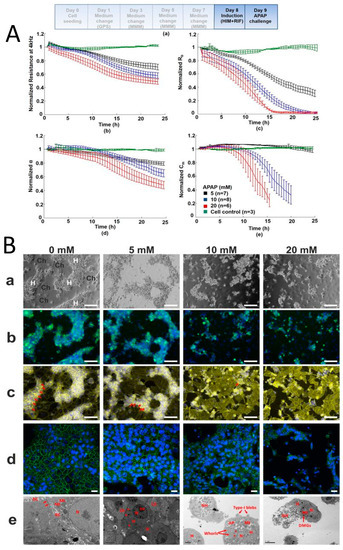
Figure 4. Direct hepatotoxicty signature of HepaRG cells treated with paracetamol; correlation between cellular impedance and immunocytostaining (A) HepaRG cells treated for 24 h with paracetamol: untreated cell control; 5 mM; 10 mM; and 20 mM. Paracetamol caused a dose-dependent decline in normalized resistance—a global indicator of cellular status. Graphs show (top left) deconvolution of total impedance, (top right) tight junctions, (bottom left) basolateral adhesion and (bottom right) membrane capacitance (B) Correlation to IBCA of confocal microscopy and TEM showing HepaRG cells’ progressive loss of tight junctions with increasing paracetamol concentration. (a) phase contrast microscopy where H refer to hepatocytes and Ch to cholangiocytes (b) transferrin and Hoescht staining showing loss of function due to destruction of tight junctions (c) F-actin staining (yellow) shows breakdown of cytoskeleton with increasing paracetamol concentration. Red arrows show tight junction associated F-actin bands (d) confocal microscopy showing loss of tight junction protein ZO-1 (green) (blue nuclear staining with Hoescht) (e) representative TEM confirming loss of tight junctions and cells showing a necrotic/apoptotic appearance. Red arrows represent mitochondria and tight junctions at 0mM, an electron dense perimeter around hepatocytes at 5 mM, type I blebbing at 10mM and dense mitochondrial granules at 20 mM (Reprinted from Gamal et al., 2017) [2].
Toxicity Assays Using Pluripotent Stem Cells
The use of human embryonic stems cells (hESC) and human induced hepatocyte-like cells derived from pluripotent stem cells (hIPS) differentiation to hepatocyte-like cells is very attractive in hepatology research. ECIS offers the advantages of providing hepatocytes from non-diseased tissue, reproducibility between experiments and IPS cells offer the opportunity to study specific disease states and genotypes known to be important in some adverse responses to drugs. Although they have already proven useful in a number of studies, further work is needed to optimise differentiation protocols to achieve mature hepatocyte phenotype and function. hESCs and hIPS could potentially generate tailored models of human liver disease and population-relevant models for drug toxicity testing [47–49]. They may also be of particular benefit in the assessment of drugs that cause idiosyncratic toxicity due to genetic polymorphisms.
Zhou et al., [50] showed that impedance data acquired using ECIS to monitor real-time growth kinetics and the differentiation of pluripotent stem cells to hepatic progenitors and hepatocyte-like cells followed established signatures of cell differentiation assessed by microscopy and were measurable in real time by ECIS. The readings from the independent wells were uniform and remained in close proximity throughout the differentiation process [50]. This result is important, as it demonstrates the reproducibility of differentiation between replicates.
They also used ECIS and Promega CellTiterGlo total ATP assay in parallel to assess the toxicity of compounds BMS4 and BMS5 on hESCs [50]. Using the ECIS Z theta system at a single frequency with one electrode per well showed that there was good correlation of results from the ECIS and CellTiterGlo assays. This demonstrates that through using standard methods, e.g., microscopy, gene expression, functional assays, to support impedance data, it is possible to establish an impedance signal for a particular cell culture under a particular set of conditions.
This study highlights two very important aspects of IBCAs; namely, their ability to show real-time differentiation and cellular growth kinetics between replicates within an experiment, and substantiating the viability of cells in toxicity testing assays [50].
It also demonstrates where the electrode configuration and applied frequency can be chosen according to the study under investigation. Using one electrode per well detects changes within a specific location of cell culture, averaging the signal only from cells growing on that electrode, useful for detecting micromotion of cells or in wound healing assays. However, assays using wells with interdigitated multi-electrode plates give a representation of the average electrical signal across various locations within the well, which is better suited to toxicological studies.
This entry is adapted from the peer-reviewed paper 10.3390/jcm9010050