1. Introduction
A polyether ionophore, SAL, can target CSCs by facilitating the movement of polar alkali metals through hydrophobic membranes [
36]. It has also been found that SAL is able to minimize the fraction of cancer cells that have stem cell-like features by inhibiting the Wnt pathway [
37]. Haruyasu et al. [
38] identified the novel polyether ionophore antibiotic SAL (molecular formula C
42H
70O
11), followed by the Miyazaki group [
39]. This molecule is a polyether antibiotic identified from the culture broth of
Streptomyces albus (strain no. 80614) by Miyazaki and colleagues [
37] at Kaken Chemicals Co., Ltd.’s research division in Tokyo, Japan, during a screening effort for novel antibiotics. SAL was produced by tank fermentation, filtering of culture broth, column chromatography on alumina or silica gel, and crystallization. SAL was isolated as a colorless prism of sodium salt using this method [
40]. SAL can also be extracted from a source of chicken feed, as described by Borgstrom et al. [
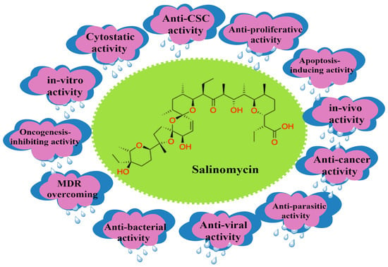
41], in which the granular feed was suspended and extracted in organic solvents, followed by purification through column chromatography. The variety of already reported pharmacological activities of SAL are depicted in
Figure 2.
Figure 2. SAL’s versatility as a naturally occurring polyether ionophore antibiotic.
2. SAL’s Mechanism of Action against CSCs
Although SAL’s ability to affect CSCs is still unclear, studies have shown that SAL’s biological effects on CSCs depend on the cell type. Based on the literature, the mechanisms of action of SAL on CSCs include:
2.1. Apoptosis of CSCs
SAL causes cell death in a different way depending on the cancer being targeted. SAL has been shown to affect the mitochondria, leading to caspase-3 cleaving poly-ADP ribose polymerase (PARP), resulting in apoptosis. SAL has shown the ability to affect prostate cancer (PC-3) cell lines through the production of reactive oxygen species (ROS), leading to programmed cell death. On the other hand, in myeloid leukemia, breast and osteosarcoma cells are also susceptible to apoptosis, while some other cell lines may not [
42]. More research is being conducted on the exact reason why some cell lines are affected by SAL, leading to apoptosis, while others do not experience apoptotic cell death.
2.2. Interference of ATP-Binding Cassette (ABC) Transporters
It has been shown that SAL has the ability to overcome the ABC transporters in most cancer cell lines, which affect cancer cells drug efflux capabilities, as exhibited in acute myeloid leukemia (AML) cancers [
42]. Based on the structure of SAL, it is a K
+ ionophore and embeds itself in the cytoplasmic or mitochondrial cell membrane. Due to the fact that ABC transporters expel substrates from the cytosol and SAL is not present in that part of the cell, it is unlikely that SAL will be affected by the transporters, and it has been shown to be a potential blocker. More research is being performed to explore the potential use of SAL as an ABC transporter inhibitor.
2.3. Inhibition of Oxidative Phosphorylation and Glycolysis
It has been demonstrated that cancers depend on aerobic glycolysis rather than oxidative phosphorylation; the neoplastic transition of human mesenchymal stem cells goes through this mechanism. It was also determined that glioma cancer cells depend on oxidative phosphorylation, and inhibition could affect these cells. In light of this knowledge, an increase in oxidative phosphorylation in certain stem cells can lead to a malignant transformation in these cells. These cells depend on this activity, and because SAL is known to impede oxidative phosphorylation in the mitochondria, blocking this route could be a useful function of this chemical as a chemotherapeutic agent against tumors [
42]. Based on this knowledge, SAL has been shown to have the effect of inhibiting the increase in oxidative phosphorylation and can contribute to the elimination of these cells.
In addition to the effects of oxidative phosphorylation inhibition by SAL, it also has an effect on the glycolysis of different cancer cells. This was seen when SAL in combination with glucose analogs (2-DG, 2-FDG) increased the toxicity of SAL towards cancer cells and showed that cancer cells are dependent on glycolysis for ATP production [
43]. In the same study, dichloroacetate (DCA), an inhibitor of pyruvate dehydrogenase kinase, which results in the activation of mitochondrial pyruvate dehydrogenase complex, which aids in converting pyruvate formichloroactate to acetyl-CoA molecules that enter the TCA cycle, was used in combination with SAL, resulting in an increase in cell death, and it can be concluded that the inhibition of oxidative phosphorylation further causes cell death induced by SAL. Glucose starvation-mediated inhibition of salinomycin-induced autophagy amplifies cancer-specific cell death [
21,
43].
2.4. Polyether Ionophore Effects on the Mitochondria
SAL is a potassium ionophore that affects the transmembrane potential and increases potassium efflux from the mitochondria and cytoplasm. It has been documented that there is an increased expression of potassium channels in AML and neuroblastoma cancerous cells, although not observed in their non-tumorigenic counterparts, hinting that potassium channels have a role in these cancers. A decrease in the intracellular concentration due to the efflux of potassium leads to a cytotoxic effect on these cells [
42,
44]. It has been found that SAL can have an effect on the mitochondria, in which enhanced Na
+ inflow leads to the suppression of breast CSCs, as discovered by Mai et al. [
21]. Miyazaki et al. [
39] were the first to explore the dual action of SAL on mitochondrial ion transport and respiration. From a rat’s liver, they discovered that 0.4 μM of SAL may release preloaded potassium from the mitochondria. The pre-treatment may limit potassium intake because SAL inhibits oxidative phosphorylation in mitochondria without selectivity and reverses cell swelling caused by potassium absorption. Furthermore, SAL inhibited mitochondrial storage of potassium more efficiently than sodium, even though with the addition of potassium uptake stimulants, it inhibited respiration at medium and low concentrations [
45]. This research demonstrates that cation transport by SAL might be conditional and influenced by ion gradients.
2.5. Induction of Autophagy, ROS, and DNA Damage
For cancer cells, autophagy is the mechanism for protecting and inhibiting cell viability. In one way, it can impede ROS production and protect against apoptotic cell death; in another way, SAL enhances the expression of microtubule-associated proteins, implying autophagy induction. It has been noticed by a number of groups that SAL itself, or in conjunction with sensitizing drugs or radiation, displayed a moderate to strong increase in DNA damage, which caused G2 cell cycle arrest and a decrease in p21 protein levels in cancer cells. Zhao et al. [
45] discovered DNA damage caused by SAL and G1 arrest in glioblastoma cells (GBM) through ROS, implying that DNA damage caused by SAL therapy could be a side effect. As a result, it is postulated that a putative autophagic mechanism for SAL in relation to radiation-induced DNA damage and subsequent tumor recurrence could be utilized as a potential avenue for treating different cancers [
46,
47].
2.6. Endoplasmic Reticulum Stress
Li et al. [
48] demonstrated that SAL causes autophagic activity; however, it is a double-edged sword in different cell types. Autophagic activity could be a defensive measure for SAL-affected cells. If the factors that inhibit autophagy are not present, the apoptotic rate increases in SAL-treated cells. Strand et al. have worked hard to create different experimental procedures, such as SAL fluorescent conjugate, to reveal SAL’s anti-CSC impact. This group also discovered that SAL causes the release of ER calcium ions and promotes their stress in breast cancer cells [
37,
47]. This was accomplished by the dispersion of a fluorescent SAL conjugate (SAL-NBD) in the ER and lipid droplets (LDs) following treatment with this conjugate. The disruption of Ca
2+ induces ER stress as cellular Ca
2+ homeostasis is closely maintained to manage Ca
2+ levels in both forms (free and bound) in all the sections of a cell [
49]. Protective factors, such as the unfolded protein response (UPR) and the mobilization of pathways to reestablish ER equilibrium, are activated. Strand et al. [
46] discovered that SAL’s fluorescent conjugate activated the UPR proteins GPR78 and ATF6. In breast cancer cells, removing CHOP hampered SAL-NBD’s capacity to downregulate β-catenin, and in addition, this compound boosts the enzymatic activity of protein kinase-C (PKC), a Wnt pathway antagonist, by 30%. This shows indirectly the potential calcium ionophore capabilities that SAL possesses, as it could have the potential to regulate calcium levels as well as Ca
2+-influenced cellular processes.
2.7. Inhibition of the Wnt Signaling Cascade
Lu et al. [
50] focused on the Wnt signaling pathway when assessing SAL’s anticancer activity, and it was found in vitro that SAL reduced Wnt1 and β-catenin while having a mild impact on Fizzled class receptor 5 (Fzd5), in addition to the fact that SAL induces an effect of lipoprotein receptor-related protein-6 (LRP6), which is essential for Wnt signaling. One example of this effect can be seen with chronic lymphocytic leukemia (CLL) cells, in which they discovered that these cells are more sensitive to SAL and had a 100-fold greater amount of apoptosis as compared to healthy donors’ peripheral blood mononuclear cells (PBMC) [
51], as well as SAL decreasing the expression of Wnt signaling, thus effecting the TCF4E complex [
52]. This finding was observed by Qi et al. [
53], who found that SAL decreased the amount of protein kinase-A (PKA) by the phosphorylation of β-catenin in NB cells. It was also found that this pathway is vital for preservation, cloning, and aspects of cancer cells, and most importantly, it aids the resistance to radiation and anticancer drugs for many cancers. SAL, however, demonstrated an ability to suppress chronic lymphocytic leukemia cells by lowering the expression of LRP6 and downregulating the Wnt target genes LEF1, cyclin D1, and fibronectin, resulting in cell death.
2.8. Sequestration of Iron in the Lysosome
In the cell, iron (Fe
2+) is closely controlled in preserving homeostasis, and based on this finding, Rodriguez et al. [
54] investigated and discovered dramatic changes in iron in CD-24 low breast cancer cells. Hence, SAL was tested, and at 0.5 μM iron accumulation in the lysosome, a reduction in iron keeper ferritin expression and elevated iron regulatory protein-2 (IRP2) were observed. Based on this finding, a novel mechanism of action of SAL affecting breast CSCs is iron accumulation in the lysosome [
1,
44,
53], and an increased amount of iron in the lysosome produces ROS, which leads to apoptosis [
53].
2.9. Intracellular Binding Targets
Based on what others have shown about SAL’s ability to suppress CSCs among different cancers, which include breast cancer, neuroblastoma (NB), GBM, medulloblastoma, pancreatic, colon, prostate, melanoma, and lung cancers, particularly in NB cells, CD-34 and CD-133 play a key role in cell proliferation and tumor formation, respectively. It was discovered that NCL is associated with binding to the CD-34 gene promoter region, which initiates the transcription of the respective gene. SAL significantly decreased the cell population of CD-34
+ and CD-133
+ NB cells through its ability at the mRNA level to prevent the binding of NCL to the CD-34 promoter region, which significantly reduced the CD-34 gene expression and affected the cell proliferation and tumor formation of NB cells [
55].
Qi et al. [
53,
55] just recently found and explored nucleolin (NCL), a protein that has many functions and is necessary for cell proliferation. Based on studies, it has the ability to bind nucleic acids (RNA and DNA) and numerous other proteins. For example, after iron chelator therapy, NCL controls matrix metallopeptidase 9 (MMP9) mRNA translation by binding to its promoter regions [
56]. NCL also controls CD-133 and CD-34 expression in hematopoietic stem/progenitor cells (HSPC) [
57,
58]. Surprisingly, Qi and colleagues discovered by combining the techniques of Drug Affinity Responsive Target Stability (DARTS) and co-immunoprecipitation (co-IP) [
59] that for SAL, NCL is a functional cellular binding target. Qi et al. showed that SAL significantly suppresses NB development with an IC
50 that is substantially lower than that seen with the majority of presently-utilized NB chemotherapeutic agents. This presents a new mechanism of SAL in the treatment of cancer. Overall, based on these findings regarding SAL’s mechanism of action, it demonstrates how versatile SAL is and how its multiple mechanisms of action can be used to treat cancer.
2.10. Differentiation of CSCs and SAL Bioactivity
There are different extracellular biomarkers, such as CD-44, that were found in solid and hematological cancers and aid in the proliferation, self-renewal, and metastasis of cancers. CD-133 is another biomarker that was identified as present in tumors of breast, liver, stomach, and colon cancers. The presence of this marker gives rise to the notion that these caners have a high rate of tumor and spheroid formation. CD-33 has been known to be a prototypical marker for CSCs; for example, it is the most common marker for AML stem cells. It is also expressed in high quantities in CML and hematopoietic stem cells [
60].
In addition to the extracellular biomarkers (CD-44 and CD-133), intracellular biomarkers also play a key role in tumorgenesis. These intracellular biomarkers, known as pluripotent transcription factors (e.g., Nanog, Oct4, and Sox2), are present in embryonic stem cells [
61]. It has been found that overexpression of these transcription factors, which are present in various carcinomas and breast and prostate cancers, causes tumorgenesis and malignant progression. The ability of these transcription factors to induce tumorgenicity is demonstrated by Oct4’s ability to transform normal mammary epithelial cells into mammosphores, which increases tumorgenicity in vivo. The expression of Sox2 and Nanog in MCF7 cells can aid in cell proliferation and the formation of mammosphores, as knockdown of either transcription factor showed inhibition of cell proliferation and mammosphore formation.
In the TNBC cell line MDA-MB-231, the administration of SAL decreased tumor growth by reducing CD-44 levels rather than inducing apoptosis. Also observed from this study was that a downregulation of Nanog, Oct4, and Sox2 leads to the suppression of mammosphore formation [
62].
SOX2 is another transcription factor that is found in embryonic stem cells and CSCs, and an increase in tumorgenesis and chemoresistance was observed when this transcription factor was expressed. This could be a potential target for the treatment of cancers at the CSC level, as seen in Sox2-positive glioblastoma cancer stem cells (GCs) treated with SAL, and it was observed that SAL decreased the expression of SOX2 at the transcriptional and translational levels. This indicated a decrease in SOX2 expression, which resulted in a decline in the tumorgenesis of GBM [
63].
SAL has the ability to promote differentiation of cancer cells early on in development, and it can reprogram epithelial growth that undergoes epithelial mesenchymal transition (EMT) and leads to the upregulation and expression of certain genes, such as mammary epithelial differentiation [
42,
64]. Based on these findings, SAL can eliminate CSCs through multiple mechanisms of action known from the literature (
Table 1). Future research is needed to find out if SAL has other relevant mechanisms of action for targeting CSCs.
Table 1. SAL’s effects on different cancer cell types.
This entry is adapted from the peer-reviewed paper 10.3390/ijtm3020016