miRNAs’ Cross-Involvement in Skin Allergies: History
Please note this is an old version of this entry, which may differ significantly from the current revision.
Contributor: , , , , , ,

Skin inflammation is a common underlying feature of atopic dermatitis, allergic contact dermatitis and chronic spontaneous urticaria. miRNAs are involved in the pathogenesis and regulation of atopic dermatitis and can reveal an atopic predisposition or indicate disease severity. In chronic spontaneous urticaria, different miRNAs which are over-expressed during urticaria exacerbations not only play a role in the possible response to therapy or remission, but also serve as a marker of chronic autoimmune urticaria and indicate associations with other autoimmune diseases. In allergic contact dermatitis, miRNAs are upregulated in inflammatory lesions and expressed during the sensitization phase of allergic response. Several miRNAs have been identified as potential biomarkers of these chronic skin conditions, but they are also possible therapeutic targets.

  • miRNA
  • atopic dermatitis
  • chronic spontaneous urticaria
  • allergic contact dermatitis
  • antagomirs
  • cytokines
  • inflammation
  • skin
  • pathogenesis
  • treatment

1. Introduction

Atopic dermatitis, allergic contact dermatitis and chronic spontaneous urticaria are common inflammatory conditions involving the skin. This inflammation may result from unique and, yet, shared pathogenic mechanisms, which may account for their coexistence and association in atopic patients.

1.1. Pathogenesis of Atopic Dermatitis

Atopic dermatitis (AD) is the most common inflammatory skin disorder in the developed world, with a lifetime prevalence of 15–20% [1]. It is often associated with other atopic conditions, such as asthma, allergic rhinitis and chronic rhinosinusitis with nasal polyps [2]. AD is a chronic, relapsing and inflammatory skin disease characterized by intense itching and excoriations with erythematous, xerotic, fissured and lichenificated skin, as well as an increased risk of skin infections [3][4]. The pathogenesis of AD has not been completely elucidated. Genetic heritage and its interactions with the environment contribute to epidermal skin barrier disruption, altered inflammatory response and skin microbiome dysbiosis [5][6], and oxidative stress also plays an important pathogenetic role [7]. Recent studies have pointed to the key role of epigenetic changes in AD development, mainly mediated by DNA methylation, histone acetylation and the action of specific non-coding RNAs, including micro-RNAs (miRNAs), small interfering RNAs, long non-coding RNAs and Pivi-interacting RNAs [8]. Skin barrier abnormalities include decreased filaggrin, loricrin, involucrin, ceramides, antimicrobial peptides and disorders of tight junctions [9][10]. In particular, filaggrin, a structural protein responsible for keratinization, moisturization and antimicrobial peptide functions, is mutated in 15–50% of patients with AD [11][12]. Skin barrier disruption stimulates keratinocytes and dendritic cells to produce activation-regulated chemokine (TARC), thymic stromal lymphopoietin (TSLP) and cytokines, including IL-1β, IL-25 and IL-33, which trigger type-2 inflammation, activating both innate and adaptative immunity [13][14]. Type-2 innate lymphoid cells and Th2 cells produce IL-4, IL-5 and IL-13 [13], key mediators of type-2 response. They promote Th-naïve lymphocytes developing Th2 phenotypes, B lymphocyte isotypes switching from IgM to IgE [15], eosinophil recruitment and activation [16], itch exacerbation [17] and skin remodeling toward fibrosis in the chronic disease phase [1].

1.2. Pathogenesis of Allergic Contact Dermatitis

Allergic contact dermatitis (ACD) is a common inflammatory skin disorder characterized by pruritus, erythema, vesicles and scaling of the skin [18]. ACD is common, with some studies demonstrating prevalence rates as high as 20% in the general population, and it accounts for the vast majority of occupational skin disorders in the Western world [18][19]. ACD involves type IV-mediated hypersensitivity to a specific allergen, resulting in an inflammatory response with exposure. The first phase is sensitization, when a person is first exposed to a hapten, which is defined as a low-molecular-weight antigen that, when bound to a larger carrier, can elicit an immune response.
After hapten has been recognized and phagocytized by dermal dendritic cells and Langerhans cells, the hapten–peptide complexes migrate to regional lymph nodes and induce the proliferation and circulation of hapten-specific T cells (Th1, Th2, Th17 and T regulatory cells) and the creation of effector and memory T cells. Clinical symptoms of ACD are produced in the elicitation phase, when re-exposure to the allergen, recognized by the sensitized hapten-specific T cells, causes an inflammatory cascade of cytokines and cellular infiltrates [20]. Since the reaction to the allergen is not always immediate, with a possible delay of up to 72 h, it may be difficult to identify the culprit agents. Thorough questioning of the patient’s occupation, hobbies and any changes in personal products or clothing is helpful [21], and certain distributions, such as on the eyelid, lateral face, central face, neck or hands suggest the consideration of ACD. Patch testing is the only practical, scientific and objective method to confirm the diagnosis of ACD.

1.3. Pathogenesis of Chronic Spontaneous Urticaria

Chronic spontaneous urticaria (CSU) is defined as the spontaneous appearance of itchy red wheals and/or angioedema for more than six weeks without any apparent cause [22][23], and it affects about 1.8% of the adult population [24]. CSU is mainly caused by the activation of cutaneous mast cells (MCs), leading to the release of histamine and other mediators, such as platelet-activating factor (PAF) and cytokines, which results in vasodilatation, plasma extravasation and sensory nerve activation, as well as cell recruitment in the urticarial lesions (mainly T cells, eosinophils and basophils). Wheals are characterized by edema of the upper and mid-dermis, whereas similar changes may occur in the lower dermis, resulting in angioedema [22]. The pathophysiology of CSU is not well understood, but there are two main pathogenetic mechanisms underlying the disease: dysregulation of intracellular signaling pathways within mast cells and basophils, which leads to defects in the trafficking or function of these cells, and development of autoantibodies to FcεRIα or IgE on both mast cells and basophils [25]. Basophil numbers appear to be reduced in at least 50% of patients [26], and they seem to be hyporeactive [27]. Furthermore, their development is dependent on the interaction of stem cell factor (SCF) with the c-kit, as well as TGF-β produced by Tregs. In CSU, circulating Tregs (CD4 + CD25 + FOXP3+) are reduced and/or defective compared with normal cells [28], and the reduced frequency of Tregs is consistent with an autoimmune hypothesis for CSU [29][30]. These events lead to mast cell degranulation and the predisposition to pathological mast cell activation if inappropriately upregulated. However, autoimmune theory is the most widely accepted hypothesis for explaining the pathogenesis of CSU, as it is thought that up to 45% of cases of CSU have autoimmune etiology [25]. Specifically, CSU is associated with the development of circulating IgG antibodies against IgE and the high-affinity IgE receptor FcεR1; approximately 40% of these patients have circulating antibodies against one of the two targets [31], with a higher frequency of positivity in CSU patients with a positive autologous serum skin test (ASST) [32]. It seems likely that different pathomechanisms are interlinked in CSU rather than independent cascades [33].

1.4. MicroRNAs

miRNAs represent an abundant class of small non-coding RNAs which regulate gene expression post-transcriptionally [34]. miRNAs are short (20–22 nucleotides long), endogenous and single-stranded RNAs, whose genes [35], highly conserved throughout evolution across species [36], are located within the introns and exons of protein-coding genes [37].
The first miRNAs were found in Caenorhabditis elegans in early 1990s [34]; afterward, the first human miRNA was discovered in the 2000s [38]. Currently, 2588 mature miRNAs have been identified for humans in miRbase [39], and it is estimated that they modulate more than 60% of all human protein-coding genes by sequence-specific base pairing [35]. In fact, it is widely recognized that one miRNA may regulate many genes as its targets, while one gene may be targeted by many miRNAs. [40]
Primary transcripts of miRNAs are generated by RNA polymerase II and sequentially processed by ribonuclease III class enzymes [37] (Drosha) to produce an miRNA precursor of 70 nucleotides (the pre-miRNA) [41]. After nuclear processing, the pre-miRNA is transferred to the cytosol, where Dicer, another ribonuclease III class enzyme, cleaves it into mature double-stranded miRNA with 19–24 nucleotides. One strand of this mature miRNA duplex is then incorporated into the RNA-induced silencing complex (RISC) [42] and serves as a guide for the recognition of target mRNAs [43]. The other strand is unrolled from the guide strand and degraded, but, in some cases, both strands of the miRNA duplex are functional [44]. The miRNA–RNA-induced silencing complexes interact with mRNA targets by sequence-specific base pairing, generally within the 3′-untranslated region of the target mRNAs [45]. Two different mechanisms of RISC-mediated gene regulation exist, and they depend on the level of miRNA–mRNA complementarity: at sites with extensive complementarity, the miRNA can mediate mRNA cleavage; however, sites with a lower degree of complementarity may lead to translational repression or mRNA destabilization [46].
Circular RNAs (CircRNAs) are single-stranded, covalently closed RNA molecules that are ubiquitous across species ranging from viruses to mammals [47]. One of the most commonly reported mechanisms of action of circRNAs is the “sponging” of miRNAs by binding to the miRNA response element (MRE), thereby indirectly increasing the transcription of their target mRNAs [48].
MiRNAs play an important role in biological processes such as cellular proliferation, differentiation and apoptosis. Further, they are implicated in inflammatory diseases, cancer [49], immune response, neural development, DNA repair and oxidative stress response [50].
In addition, miRNAs are stable in body fluids, so they are more available for assays and less invasive than biopsies [51]. Thus, the interest in miRNAs as both biomarkers and possible therapeutic targets is increasing [52].

2. miRNA in AD, ACD and CSU: Pathogenetic Role and Therapeutic Strategies

Increasing evidence suggests that miRNAs may play important roles in regulating physiological skin processions, such as self-renewal and differentiation, in stem cells. The most highly expressed miRNAs in normal-condition skin are miR-152, miR-143, miR-126, miR -21, miR-27a, miR-214, miR-16, miR-203, miR 125b, miR-34a, miR-205, miR-27b, miR-30b, miR-125a, miR-191, the miR-200 family (-200a, -200b, -200c, -141, -429), the miR-199 family (-199a, -199b) and the miR-19/-20 family (-19b, -20, -17-5p, -93) [53]. Skin inflammation is a common underlying feature of AD, ACD and CSU.

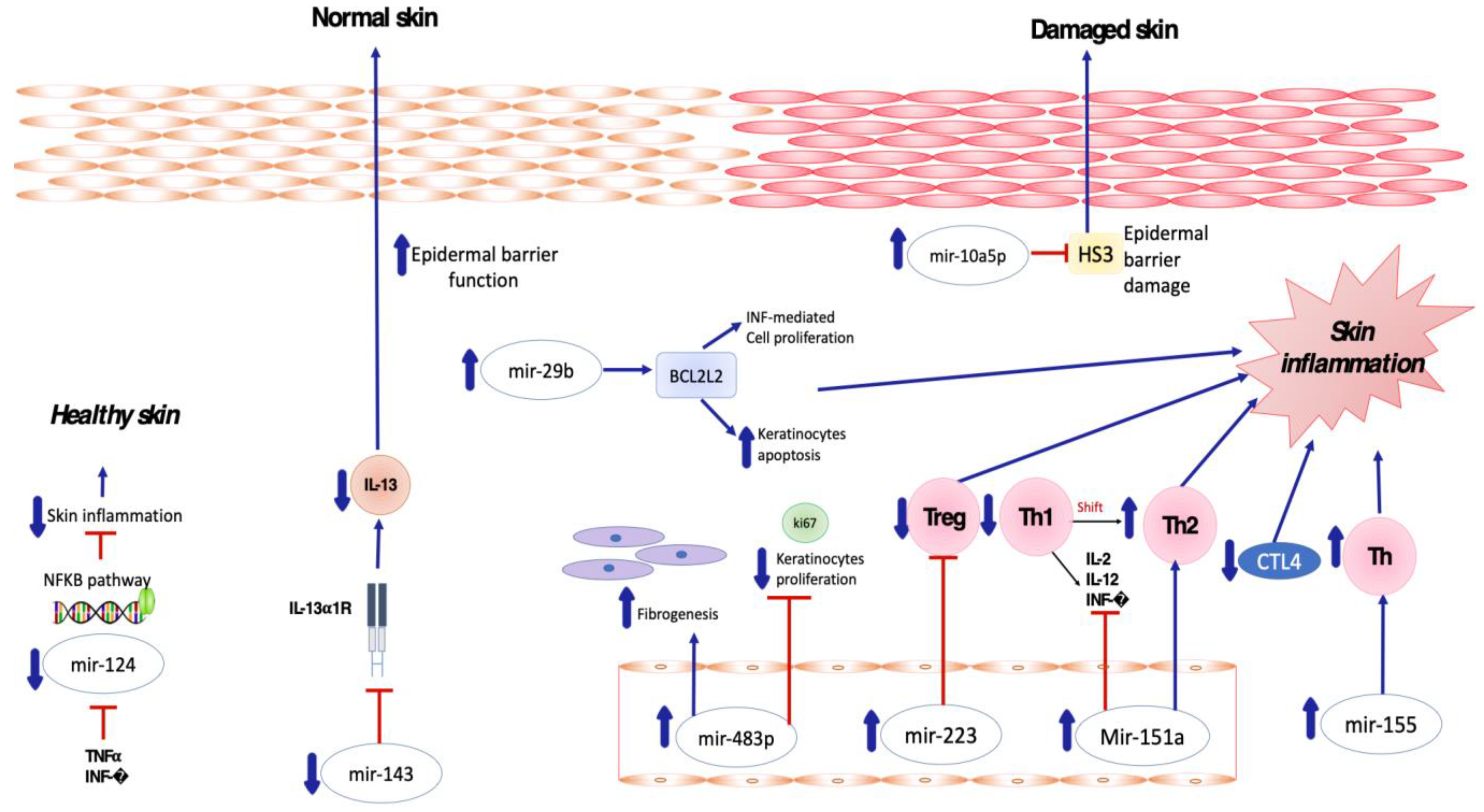
2.1. Pro-Inflammatory and Anti-Inflammatory miRNA in AD

Several miRNAs have been found to be implicated in the crosstalk between inflammatory cells and keratinocytes in patients affected by atopic dermatitis (Figure 1). In particular, Sonkoly et al. [54] reported that mir-155 is one of the most significantly upregulated miRNAs in the lesional skin of patients affected by AD, suggesting its major role in the pathogenesis of the disease. MiR-155 is a multifunctional miRNA that is involved in immune cell maturation and the regulation of innate and adaptive immune systems, playing a critical role in both T- and B-cell responses [55][56][57]. Overexpression of miR-155 decreases CTLA-4 levels and increases the proliferation of T-helper cells, promoting chronic skin inflammation. Rebane et al. [58] demonstrated upregulation of miR-146 in the serum and skin of patients with AD compared to a healthy population, as well as in murine skin specimens obtained from an AD-like mice model in comparison to a control group.
Figure 1. Skewed skin inflammation associated with dysregulated miRNA expression in patients with Atopic Dermatitis.
MiR146a is a known anti-inflammatory and NFκB pathway-dependent miRNA expressed in B cells, T cells, monocytes and dendritic cells [59][60][61]. Its role is to regulate innate and adaptive immunity, regulating antibody production, inflammatory factor secretion and immune cell differentiation [62].
MiR-151a was also found to be overexpressed in the plasma of 500 atopic dermatitis patients by Chen XF et al. [63], whereas its expression in eczematous skin was not determined to be relevant. Upregulation of miR-151a favors the shift toward Th2 cell response, being predominant in AD [64] and suppressing the expression of Th1 cytokines including IL-2, IL-12 and INF-gamma [65].
MiRNA-143 has been shown to decrease IL-13 activity and inflammation by targeting and downregulating IL-13Rα1 in epidermal keratinocytes, thus playing a potential role in decreasing AD-induced skin inflammation. In detail, miR-143 opposes the negative regulation of filaggrin, loricrin and involucrin induced by IL-13 in human keratinocytes, potentially improving epidermal barrier function [66].
MiR-124 has been demonstrated to directly target nuclear factor (NF)-κB in B-cell lymphoma [67]. Yang et al. [68] investigated the role of miR-124 in atopic dermatitis, demonstrating that miR-124 expression was downregulated in chronic AD skin lesions. Tumor necrosis factor (TNF)-α and IFN-γ were also able to inhibit MiR-124 expression. Modulating the NF-κB pathway, miR-124 reduces chronic skin inflammation and inflammatory responses in keratinocytes in AD [68].

2.2. Pro-Inflammatory and Anti-Inflammatory miRNA in ACD

Concerning the involvement of miRNAs in the pathogenesis of ACD, Vennegaard et al. were the first to describe aberrant miRNA expression in this disease, analyzing skin biopsies from subjects who had been sensitized with diphenylcyclopropenone (DPCP). They found that miRNA-21, miR-223, miR-142-3p and miR-142-5p were upregulated in the inflammatory lesions. In addition, they were also upregulated in skin biopsies from mouse models which were sensitized with 2,4-dinitrofluorobenzene, showing that mouse models are valuable tools for further study of the involvement of miRNAs in ACD [69]. Subsequently, in 2015, Gulati et al. [70] confirmed the altered expression of miR-21, miR-223, miR-142-3p and miR-142-5p in skin lesions obtained 3 and 14 days after a challenge with DPCP in sensitized subjects. They also identified 6 miRNAs that were significantly upregulated 120 days after the challenge, demonstrating the long-lasting allergen-mediated immune reaction that occurs in the skin [70]. Anderson et al. demonstrated consistent changes in miRNA expression for miR-21, miR-22, miR-27b, miR-31, miR-126, miR-155, miR-210 and miR-301a during the sensitization phase of an allergic response to TDI in a murine model [71]. In 2021, Werner et al. analyzed miRNA expression data of positive patch test reactions from patients exposed to allergens such as nickel sulphate, epoxy resin and methylochloroisothiazolinone, and to irritants such as sodium lauryl sulfate (SLS) and nonanoic acid. All allergens induced miRNAs expression in human skin, while of the irritants, only SLS did. Eighty-six miRNAs were significantly upregulated or downregulated; miR-142-3p, miR-142-5p, miR-146b and miR-155-5p were differentially expressed across all investigated allergens. Meanwhile, miR-497-5p was significantly expressed only in MCI; miR-22b-3p, miR-99a-5p, miR-193b-3p and miR-199a-3p were significantly expressed in epoxy resin ACD [72].

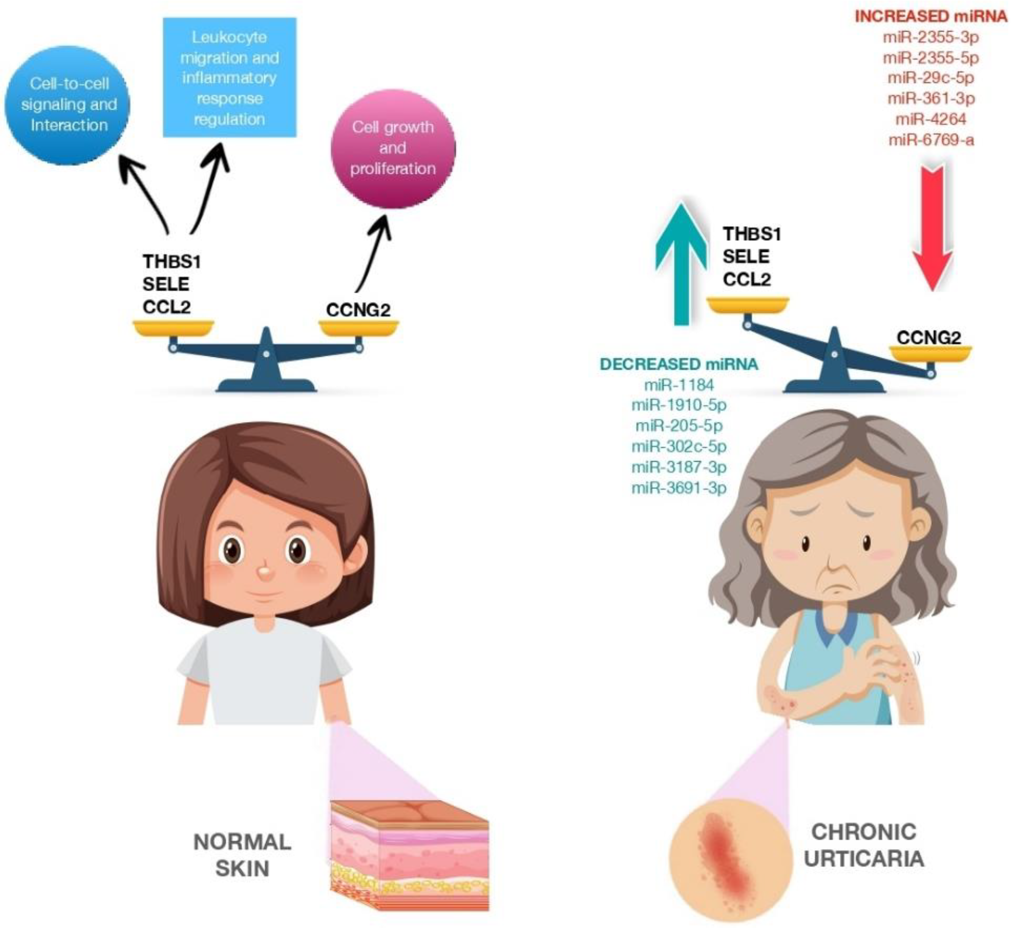
2.3. Pro-Inflammatory and Anti-Inflammatory miRNA in CSU

A few studies have examined the role of several miRNAs in the pathophysiology of CSU. In 2017, Lin et al. studied whether miRNAs are involved in CSU regulation as biomarkers. Their study involved 12 patients who were divided according to according to whether they had active hives or no hives and to the presence or absence of CSU. MiRNAs were isolated from patient plasma, and 16 miRNAs were found to be differentially expressed in patients with active hives. Among these, miR-2355-3p, miR-4264, miR-2355-5p, miR-29c-5p and miR-361-3p were upregulated in exacerbated CSU patients; thus, they could be useful biomarkers for patients with chronic autoimmune urticaria. Then, the researchers compared these targets against urticaria-related genes. Twenty-five genes were found to match, eight of which were significantly downregulated, while the other eight were significantly upregulated.
In addition, 12 genes (Figure 2) did not serve a signaling role, whereas the other 13 were involved in regulatory pathways such as the transforming growth factor beta (TGF-β) signaling pathway (nuclear receptor subfamily 3, group C, member 1, glucocorticoid receptor (NR3C31), kit ligand (KITLG), thrombospondin I (THBS1), chemokine (C-C motif) ligand 2 (CCL2)), glucocorticoid receptor signaling pathway (NR3C1, selectin E (SELE), CCL2), p53 signaling pathway (cyclin G2 (CCNG2), THBS1, CCL2), p21-activated kinase pathway (PAK1 interacting protein 1 (PAK1IP1), KITLG, CCL2), phosphoinositide-3 kinase protein kinase B signaling pathway (KITLG, cholinergic receptors muscarinic (CHRM), THBS1) and neuroactive ligand–receptor interaction (NRC31, histamine receptors H1 (HRH1), CHRM), which could play important roles in CSU [73]. In 2019, Zhang et al. found miR-125a-5p and CCL17 (C-C motif chemokine ligand 17) to be significantly upregulated in the serum of patients with active CSU and decreased in the serum of patients in remission. Furthermore, upregulated expression of miR-125a-5p was observed in refractory CSU, indicating its potential use as a biomarker [33].
Figure 2. CCNG2, THBS1, SELE and CCL2 expression in normal skin and CCNG2, THBS1, SELE and CCL2 expression modulated by miRNA dysregulation in patients with chronic urticaria. The figure is designed by Freepik.

This entry is adapted from the peer-reviewed paper 10.3390/biomedicines11051266

References

  1. Weidinger, S.; Beck, L.A.; Bieber, T.; Kabashima, K.; Irvine, A.D. Atopic Dermatitis. Nat. Rev. Dis. Primers 2018, 4, 1.
  2. Nettis, E.; Patella, V.; Brancaccio, R.; Detoraki, C.; Di Leo, E.; Incorvaia, C.; Macchia, L.; Pellacani, G.; Bonzano, L. Efficacy of Dupilumab in Concomitant Atopic Dermatitis and Chronic Rhinosinusitis With Nasal Polyps: A Preliminary Study. Allergy Asthma Immunol. Res. 2021, 13, 347–349.
  3. Megna, M.; Patruno, C.; Balato, A.; Rongioletti, F.; Stingeni, L.; Balato, N.; Ayala, F.; Brambilla, L.; Congedo, M.; Corazza, M.; et al. An Italian Multicentre Study on Adult Atopic Dermatitis: Persistent versus Adult-Onset Disease. Arch. Derm. Res. 2017, 309, 443–452.
  4. Kraft, M.; Worm, M. Dupilumab in the Treatment of Moderate-to-Severe Atopic Dermatitis. Expert Rev. Clin. Immunol. 2017, 13, 301–310.
  5. Patel, N.; Feldman, S.R. Management of Atopic Dermatitis. Adherence in Atopic Dermatitis. Introduction. Adv. Exp. Med. Biol. 2017, 1027, 139–159.
  6. Elias, P.M.; Steinhoff, M. “Outside-to-inside” (and Now Back to “Outside”) Pathogenic Mechanisms in Atopic Dermatitis. J. Investig. Dermatol. 2008, 128, 1067–1070.
  7. Bertino, L.; Guarneri, F.; Cannavò, S.P.; Casciaro, M.; Pioggia, G.; Gangemi, S. Oxidative Stress and Atopic Dermatitis. Antioxidants 2020, 9, 196.
  8. Nedoszytko, B.; Reszka, E.; Gutowska-Owsiak, D.; Trzeciak, M.; Lange, M.; Jarczak, J.; Niedoszytko, M.; Jablonska, E.; Romantowski, J.; Strapagiel, D.; et al. Genetic and Epigenetic Aspects of Atopic Dermatitis. Int. J. Mol. Sci. 2020, 21, 6484.
  9. Zaniboni, M.C.; Samorano, L.P.; Orfali, R.L.; Aoki, V. Skin Barrier in Atopic Dermatitis: Beyond Filaggrin. Bras Derm. 2016, 91, 472–478.
  10. Katsunuma, T.; Kawahara, H.; Yuki, K.; Akasawa, A.; Saito, H. Impaired Interferon-Gamma Production in a Subset Population of Severe Atopic Dermatitis. Int. Arch. Allergy Immunol. 2004, 134, 240–247.
  11. Malik, K.; Heitmiller, K.D.; Czarnowicki, T. An Update on the Pathophysiology of Atopic Dermatitis. Derm. Clin. 2017, 35, 317–326.
  12. Palmer, C.N.A.; Irvine, A.D.; Terron-Kwiatkowski, A.; Zhao, Y.; Liao, H.; Lee, S.P.; Goudie, D.R.; Sandilands, A.; Campbell, L.E.; Smith, F.J.D.; et al. Common Loss-of-Function Variants of the Epidermal Barrier Protein Filaggrin Are a Major Predisposing Factor for Atopic Dermatitis. Nat. Genet. 2006, 38, 441–446.
  13. Salimi, M.; Barlow, J.L.; Saunders, S.P.; Xue, L.; Gutowska-Owsiak, D.; Wang, X.; Huang, L.C.; Johnson, D.; Scanlon, S.T.; McKenzie, A.N.J.; et al. A Role for IL-25 and IL-33-Driven Type-2 Innate Lymphoid Cells in Atopic Dermatitis. J. Exp. Med. 2013, 210, 2939–2950.
  14. Ito, T.; Wang, Y.H.; Duramad, O.; Hori, T.; Delespesse, G.J.; Watanabe, N.; Qin, F.X.F.; Yao, Z.; Cao, W.; Liu, Y.J. TSLP-Activated Dendritic Cells Induce an Inflammatory T Helper Type 2 Cell Response through OX40 Ligand. J. Exp. Med. 2005, 202, 1213–1223.
  15. Gandhi, N.A.; Bennett, B.L.; Graham, N.M.H.; Pirozzi, G.; Stahl, N.; Yancopoulos, G.D. Targeting Key Proximal Drivers of Type 2 Inflammation in Disease. Nat. Rev. Drug Discov. 2016, 15, 35–50.
  16. Islam, S.A.; Luster, A.D. T Cell Homing to Epithelial Barriers in Allergic Disease. Nat. Med. 2012, 18, 705–715.
  17. Liu, B.; Tai, Y.; Achanta, S.; Kaelberer, M.M.; Caceres, A.I.; Shao, X.; Fang, J.; Jordt, S.E. IL-33/ST2 Signaling Excites Sensory Neurons and Mediates Itch Response in a Mouse Model of Poison Ivy Contact Allergy. Proc. Natl. Acad. Sci. USA 2016, 113, E7572–E7579.
  18. Nassau, S.; Fonacier, L. Allergic Contact Dermatitis. Med. Clin. N. Am. 2020, 104, 61–76.
  19. Zack, B.; Arrandale, V.H.; Holness, D.L. Preventing Occupational Skin Disease: A Review of Training Programs. Dermatitis 2017, 28, 169–182.
  20. Rustemeyer, T.; Van Hoogstraten, I.M.W.; Von Blomberg, B.M.E.; Gibbs, S.; Scheper, R.J. Mechanisms of Irritant and Allergic Contact Dermatitis. Contact Dermat. 2011, 43–90.
  21. Adelman, D.C.; Casale, T.B.; Corren, J.; Ovid Technologies, Inc. Manual of Allergy and Immunology; Lippincott Williams & Wilkins: Philadelphia, PA, USA, 2012; p. 494.
  22. Zuberbier, T.; Abdul Latiff, A.H.; Abuzakouk, M.; Aquilina, S.; Asero, R.; Baker, D.; Ballmer-Weber, B.; Bangert, C.; Ben-Shoshan, M.; Bernstein, J.A.; et al. The International EAACI/GA2LEN/EuroGuiDerm/APAAACI Guideline for the Definition, Classification, Diagnosis, and Management of Urticaria. Allergy Eur. J. Allergy Clin. Immunol. 2022, 77, 734–766.
  23. Nettis, E.; Cegolon, L.; Di Leo, E.; Lodi Rizzini, F.; Detoraki, A.; Canonica, G.W. Omalizumab in Chronic Spontaneous Urticaria: Efficacy, Safety, Predictors of Treatment Outcome, and Time to Response. Ann. Allergy Asthma Immunol. 2018, 121, 474–478.
  24. Vestergaard, C.; Toubi, E.; Maurer, M.; Triggiani, M.; Ballmer-Weber, B.; Marsland, A.; Ferrer, M.; Knulst, A.; Giménez-Arnau, A. Treatment of Chronic Spontaneous Urticaria with an Inadequate Response to H1-Antihistamines: An Expert Opinion. Eur. J. Dermatol. 2017, 27, 10–19.
  25. Bracken, S.J.; Abraham, S.; MacLeod, A.S. Autoimmune Theories of Chronic Spontaneous Urticaria. Front. Immunol. 2019, 10, 627.
  26. Grattan, C.E.H.; Dawn, G.; Gibbs, S.; Francis, D.M. Blood Basophil Numbers in Chronic Ordinary Urticaria and Healthy Controls: Diurnal Variation, Influence of Loratadine and Prednisolone and Relationship to Disease Activity. Clin. Exp. Allergy 2003, 33, 337–341.
  27. Kern, F.; Lichtenstein, L.M. Defective Histamine Release in Chronic Urticaria. J. Clin. Investig. 1976, 57, 1369–1377.
  28. Sun, R.S.; Sui, J.F.; Chen, X.H.; Ran, X.Z.; Yang, Z.F.; Guan, W.D.; Yang, T. Detection of CD4+ CD25+ FOXP3+ Regulatory T Cells in Peripheral Blood of Patients with Chronic Autoimmune Urticaria. Australas J. Dermatol. 2011, 52, e15–e18.
  29. Zhu, Y.; Huang, Y.; Ming, B.; Wu, X.; Chen, Y.; Dong, L. Regulatory T-Cell Levels in Systemic Lupus Erythematosus Patients: A Meta-Analysis. Lupus 2019, 28, 445–454.
  30. Morita, T.; Shima, Y.; Wing, J.B.; Sakaguchi, S.; Ogata, A.; Kumanogoh, A. The Proportion of Regulatory T Cells in Patients with Rheumatoid Arthritis: A Meta-Analysis. PLoS ONE 2016, 11, e0162306.
  31. Vasagar, K.; Vonakis, B.M.; Gober, L.M.; Viksman, A.; Gibbons, S.P.; Saini, S.S. Evidence of in Vivo Basophil Activation in Chronic Idiopathic Urticaria. Clin. Exp. Allergy 2006, 36, 770–776.
  32. Ulambayar, B.; Chen, Y.H.; Ban, G.Y.; Lee, J.H.; Jung, C.G.; Yang, E.M.; Park, H.S.; Ye, Y.M. Detection of Circulating IgG Autoantibody to FcεRIα in Sera from Chronic Spontaneous Urticaria Patients. J. Microbiol. Immunol. Infect. 2020, 53, 141–147.
  33. Zhang, L.; Qi, R.; Yang, Y.; Gao, X.; Chen, H.; Xiao, T. Serum MiR-125a-5p and CCL17 Upregulated in Chronic Spontaneous Urticaria and Correlated with Treatment Response. Acta Derm. Venereol. 2019, 99, 571–578.
  34. Ambros, V.; Lee, R.C.; Lavanway, A.; Williams, P.T.; Jewell, D. MicroRNAs and Other Tiny Endogenous RNAs in C. Elegans. Curr. Biol. 2003, 13, 807–818.
  35. Setoyama, T.; Ling, H.; Natsugoe, S.; Calin, G.A. Non-Coding RNAs for Medical Practice in Oncology. Keio J. Med. 2011, 60, 106–113.
  36. Friedman, R.C.; Farh, K.K.H.; Burge, C.B.; Bartel, D.P. Most Mammalian MRNAs Are Conserved Targets of MicroRNAs. Genome Res. 2009, 19, 92–105.
  37. Borchert, G.M.; Lanier, W.; Davidson, B.L. RNA Polymerase III Transcribes Human MicroRNAs. Nat. Struct Mol. Biol. 2006, 13, 1097–1101.
  38. Pasquinelli, A.E.; Reinhart, B.J.; Slack, F.; Martindale, M.Q.; Kuroda, M.I.; Maller, B.; Hayward, D.C.; Ball, E.E.; Degnan, B.; Müller, P.; et al. Conservation of the Sequence and Temporal Expression of Let-7 Heterochronic Regulatory RNA. Nature 2000, 408, 86–89.
  39. Hashimoto, Y.; Akiyama, Y.; Yuasa, Y. Multiple-to-multiple relationships between microRNAs and target genes in gastric cancer. PLoS ONE 2013, 8, e62589.
  40. Kozomara, A.; Griffiths-Jones, S. MiRBase: Annotating High Confidence MicroRNAs Using Deep Sequencing Data. Nucleic. Acids Res. 2014, 42, D68–D73.
  41. Sayed, D.; Abdellatif, M. MicroRNAs in Development and Disease. Physiol. Rev. 2011, 91, 827–887.
  42. Hutvágner, G.; Zamore, P.D. A MicroRNA in a Multiple-Turnover RNAi Enzyme Complex. Science 2002, 297, 2056–2060.
  43. Lewis, B.P.; Burge, C.B.; Bartel, D.P. Conserved Seed Pairing, Often Flanked by Adenosines, Indicates That Thousands of Human Genes Are MicroRNA Targets. Cell 2005, 120, 15–20.
  44. Hu, H.Y.; Yan, Z.; Xu, Y.; Hu, H.; Menzel, C.; Zhou, Y.H.; Chen, W.; Khaitovich, P. Sequence Features Associated with MicroRNA Strand Selection in Humans and Flies. BMC Genom. 2009, 10, 413.
  45. Fabian, M.R.; Sonenberg, N.; Filipowicz, W. Regulation of MRNA Translation and Stability by MicroRNAs. Annu. Rev. Biochem. 2010, 79, 351–379.
  46. Zhou, W.Y.; Cai, Z.R.; Liu, J.; Wang, D.S.; Ju, H.Q.; Xu, R.H. Circular RNA: Metabolism, functions and interactions with proteins. Mol. Cancer 2020, 14, 172.
  47. Tong, K.L.; Tan, K.E.; Lim, Y.Y.; Tien, X.Y.; Wong, P.F. CircRNA-miRNA interactions in atherogenesis. Mol. Cell. Biochem. 2022, 477, 2703–2733.
  48. Huntzinger, E.; Izaurralde, E. Gene Silencing by MicroRNAs: Contributions of Translational Repression and MRNA Decay. Nat. Rev. Genet. 2011, 12, 99–110.
  49. Ambros, V. The Functions of Animal MicroRNAs. Nature 2004, 431, 350–355.
  50. Fiorucci, G.; Chiantore, M.V.; Mangino, G.; Percario, Z.A.; Affabris, E.; Romeo, G. Cancer Regulator MicroRNA: Potential Relevance in Diagnosis, Prognosis and Treatment of Cancer. Curr. Med. Chem. 2012, 19, 461–474.
  51. Shelburne, C.P.; Ryan, J.J. The Role of Th2 Cytokines in Mast Cell Homeostasis. Immunol. Rev. 2001, 179, 82–93.
  52. Banerjee, J.; Sen, C.K. MicroRNAs in skin and wound healing. Methods Mol. Biol. 2013, 936, 343–356.
  53. Frankel, H.C.; Qureshi, A.A. Comparative Effectiveness of Topical Calcineurin Inhibitors in Adult Patients with Atopic Dermatitis. Am. J. Clin. Dermatol. 2012, 13, 113–123.
  54. Sonkoly, E.; Janson, P.; Majuri, M.L.; Savinko, T.; Fyhrquist, N.; Eidsmo, L.; Xu, N.; Meisgen, F.; Wei, T.; Bradley, M.; et al. MiR-155 Is Overexpressed in Patients with Atopic Dermatitis and Modulates T-Cell Proliferative Responses by Targeting Cytotoxic T Lymphocyte-Associated Antigen 4. J. Allergy Clin. Immunol. 2010, 126, 581–589.e20.
  55. Sonkoly, E.; Pivarcsi, A. Advances in MicroRNAs: Implications for Immunity and Inflammatory Diseases. J. Cell Mol. Med. 2009, 13, 24–38.
  56. Rodriguez, A.; Vigorito, E.; Clare, S.; Warren, M.V.; Couttet, P.; Soond, D.R.; Van Dongen, S.; Grocock, R.J.; Das, P.P.; Miska, E.A.; et al. Requirement of Bic/MicroRNA-155 for Normal Immune Function. Science 2007, 316, 608–611.
  57. Thai, T.H.; Calado, D.P.; Casola, S.; Ansel, K.M.; Xiao, C.; Xue, Y.; Murphy, A.; Frendewey, D.; Valenzuela, D.; Kutok, J.L.; et al. Regulation of the Germinal Center Response by MicroRNA-155. Science 2007, 316, 604–608.
  58. Rebane, A.; Runnel, T.; Aab, A.; Maslovskaja, J.; Rückert, B.; Zimmermann, M.; Plaas, M.; Kärner, J.; Treis, A.; Pihlap, M.; et al. MicroRNA-146a Alleviates Chronic Skin Inflammation in Atopic Dermatitis through Suppression of Innate Immune Responses in Keratinocytes. J. Allergy Clin. Immunol. 2014, 134, 836–847.e11.
  59. West, C.; McDermott, M.F. Effects of MicroRNA-146a on the Proliferation and Apoptosis of Human Osteochondrocytes by Targeting TRAF6 through the NF- ΚB Signalling Pathway. Biosci. Rep. 2017, 37, 180.
  60. Park, H.; Huang, X.; Lu, C.; Cairo, M.S.; Zhou, X. MicroRNA-146a and MicroRNA-146b Regulate Human Dendritic Cell Apoptosis and Cytokine Production by Targeting TRAF6 and IRAK1 Proteins. J. Biol. Chem. 2015, 290, 2831–2841.
  61. Lindner, J.M.; Kayo, H.; Hedlund, S.; Fukuda, Y.; Fukao, T.; Nielsen, P.J. Cutting Edge: The Transcription Factor Bob1 Counteracts B Cell Activation and Regulates MiR-146a in B Cells. J. Immunol. 2014, 192, 4483–4486.
  62. Williams, A.E.; Perry, M.M.; Moschos, S.A.; Larner-Svensson, H.M.; Lindsay, M.A. Role of MiRNA-146a in the Regulation of the Innate Immune Response and Cancer. Biochem. Soc. Trans. 2008, 36, 1211–1215.
  63. Chen, X.F.; Zhang, L.J.; Zhang, J.; Dou, X.; Shao, Y.; Jia, X.J.; Zhang, W.; Yu, B. MiR-151a Is Involved in the Pathogenesis of Atopic Dermatitis by Regulating Interleukin-12 Receptor Β2. Exp. Dermatol. 2018, 27, 427–432.
  64. Kim, C.H.; Park, C.D.; Lee, A.Y. Administration of Poly(I:C) Improved Dermatophagoides Farinae-Induced Atopic Dermatitis-like Skin Lesions in NC/Nga Mice by the Regulation of Th1/Th2 Balance. Vaccine 2012, 30, 2405–2410.
  65. Guo, H.W.; Yun, C.X.; Hou, G.H.; Du, J.; Huang, X.; Lu, Y.; Keller, E.T.; Zhang, J.; Deng, J.G. Mangiferin Attenuates TH1/TH2 Cytokine Imbalance in an Ovalbumin-Induced Asthmatic Mouse Model. PLoS ONE 2014, 9, e100394.
  66. Zeng, Y.P.; Nguyen, G.H.; Jin, H.Z. MicroRNA-143 Inhibits IL-13-Induced Dysregulation of the Epidermal Barrier-Related Proteins in Skin Keratinocytes via Targeting to IL-13Rα1. Mol. Cell Biochem. 2016, 416, 63–70.
  67. Jeong, D.; Kim, J.; Nam, J.; Sun, H.; Lee, Y.H.; Lee, T.J.; Aguiar, R.C.T.; Kim, S.W. MicroRNA-124 Links P53 to the NF-ΚB Pathway in B-Cell Lymphomas. Leukemia 2015, 29, 1868–1874.
  68. Yang, Z.; Zeng, B.; Wang, C.; Wang, H.; Huang, P.; Pan, Y. MicroRNA-124 Alleviates Chronic Skin Inflammation in Atopic Eczema via Suppressing Innate Immune Responses in Keratinocytes. Cell Immunol. 2017, 319, 53–60.
  69. Vennegaard, M.T.; Bonefeld, C.M.; Hagedorn, P.H.; Bangsgaard, N.; Løvendorf, M.B.; Odum, N.; Woetmann, A.; Geisler, C.; Skov, L. Allergic Contact Dermatitis Induces Upregulation of Identical MicroRNAs in Humans and Mice. Contact Dermat. 2012, 67, 298–305.
  70. Gulati, N.; Løvendorf, M.B.; Zibert, J.R.; Akat, K.M.; Renwick, N.; Tuschl, T.; Krueger, J.G. Unique MicroRNAs Appear at Different Times during the Course of a Delayed-Type Hypersensitivity Reaction in Human Skin. Exp. Dermatol. 2015, 24, 953–957.
  71. Anderson, S.E.; Beezhold, K.; Lukomska, E.; Richardson, J.; Long, C.; Anderson, K.; Franko, J.; Meade, B.J.; Beezhold, D.H. Expression Kinetics of MiRNA Involved in Dermal Toluene 2,4-Diisocyanate Sensitization. J. Immunotoxicol. 2014, 11, 250–259.
  72. Werner, P.; Wisgrill, L.; Riskumäki, M.; Jalonen, E.; Vendelin, J.; Suomela, S.; Lauerma, A.; Alenius, H.; Fyhrquist, N. Identification of Novel MiRNA-MRNA Regulatory Networks in Contact Dermatitis by Integrated Microarray Analysis. Allergy 2021, 76, 1257–1261.
  73. Lin, C.K.E.; Kaptein, J.S.; Sheikh, J. Differential Expression of MicroRNAs and Their Possible Roles in Patients with Chronic Idiopathic Urticaria and Active Hives. Allergy Rhinol. 2017, 8, e67–e80.
More
This entry is offline, you can click here to edit this entry!
Video Production Service