Pathogenesis and Diagnosis of Immature Teratoma: History
Please note this is an old version of this entry, which may differ significantly from the current revision.

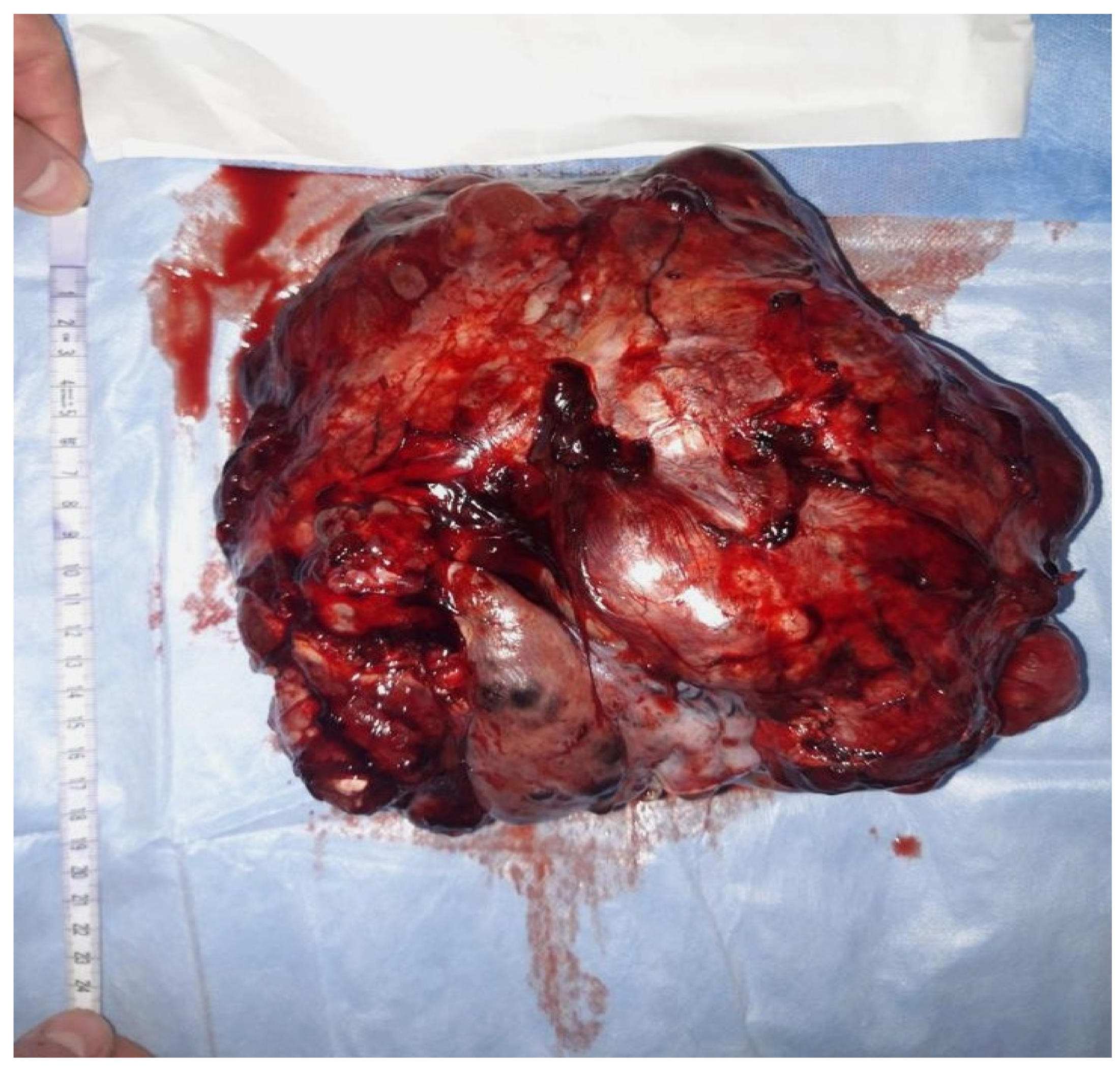
An immature teratoma is a germinal malignant tumor that is composed of three germ cell layers (the ectoderm, endoderm, and mesoderm), and it is histologically characterized by immature tissue, most frequently, neuroepithelial tissue. The most frequently affected individuals are adolescents and young adults in their reproductive age.

  • immature teratoma
  • germ cell
  • oocyte

1. Introduction 

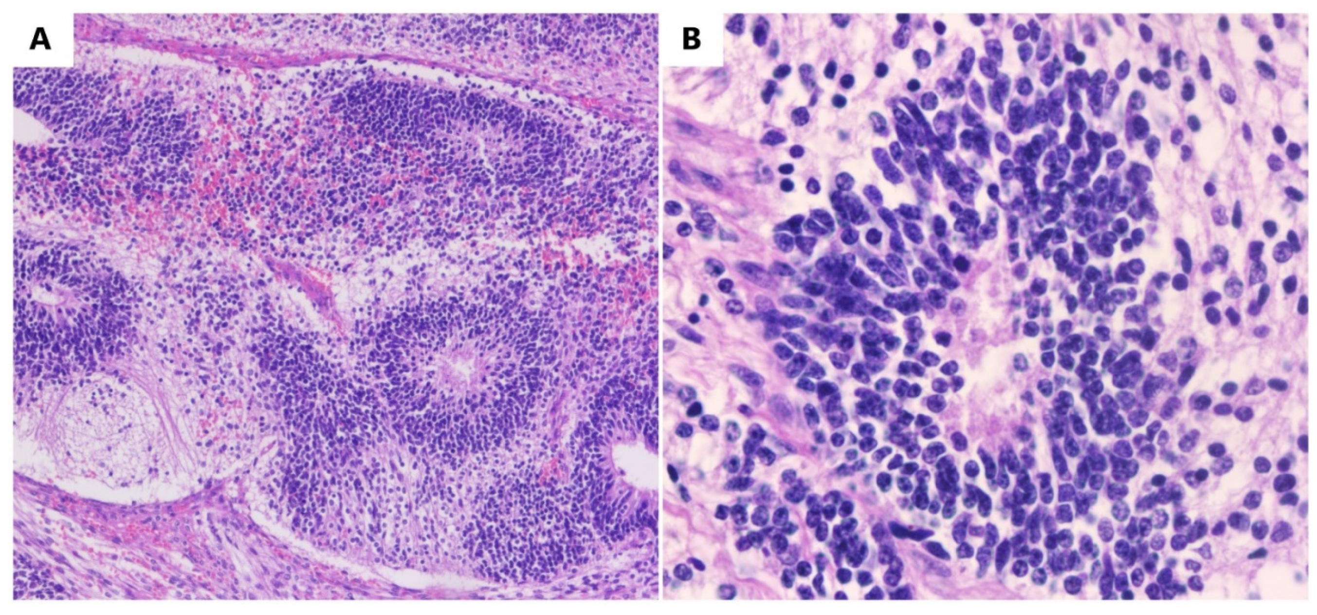
An immature teratoma is the second most frequent, after dysgerminoma, among malignant germinal tumors [1]. An immature teratoma is the only neoplasm with germ cells that are histologically graded depending on the immature neural elements, and this is a prognostic factor for overall survival [2]. This grading system is called the Norris grading system [3]. Immature teratomas represent <1% of ovarian cancers, occurring more frequently in young women [4]. Statistically, its incidence is 2.3 per 100,000 patients [5]. It represents 35.6% of all germ tumors in young women.

2. Pathogenesis

The etiopathogenesis of these tumors is not clear. There are several hypotheses, such as their development from trapped oocytes following defective folliculogenesis [6]. They can also develop from pluripotent stem cells or residual fetal cells. They undergo a process of metaplasia under the influence of inflammatory factors that act as triggers [7]. However, these are all merely assumptions [7]. Their molecular and genetic etiopathogenesis is not known and remains unstudied [6]. The origin of immature teratomas has not yet been investigated. Mature teratomas are parthenogenetic tumors containing only maternal genomes. Mature and immature teratomas showed similar methylation levels, but there are some aberrant methylation levels in immature teratomas that differ from that in mature teratomas. DNA methylation is a mechanism of genome imprinting, and this situation suggests different pathogenic mechanisms. Such lesions correspond to mixed germ cell tumors [8], and immature teratomas are a subtype of malignant germ cell tumors of the ovary [9]. Diverse errors are the key to the formation of immature teratomas, and epigenetic differences are responses key to the variation in differentiation patterns in teratomas and for the transformation from benign to malignant tumors [9].
Multiregional exome sequencing of ovarian immature teratomas reveals a paucity of somatic mutations and extensive allelic imbalances, and a somatic mutation evaluation has been performed by The Cancer Genome Atlas Research Network. The existence of an extensive loss of heterozygosity and the absence of known oncogenic variants has been demonstrated. Heskett et al. in their study used genome-level sequencing to explain the pathogenic mechanism of immature teratomas for the first time and highlighted that meiotic nondisjunction events producing the 2Nnear-diploid genome and allelic imbalance are responsible for the arise of immature teratomas [9].
Homozygosity is well-known in mature teratomas, but the zygosity in immature teratomas is not well studied [10]. While a mature teratoma develops from the oocyte after the completion of meiosis I, with the failure of meiosis II, containing homozygous chromosomes, an immature teratoma has a different pathogenic pathway [10]. However, in immature teratomas, compared with mixed germ cell tumors, fewer genetic alterations were found.
The difference in the rate of homozygosity between pure immature teratomas and mixed germ cell tumors suggests different pathogenic pathways and biological behaviors. Pure immature teratomas harbor fewer detectable genetic alterations and lack the somatic abnormalities seen in mixed germ cell tumors. Further research is needed to provide additional insights into the pathogenesis of immature teratomas [10].

3. Methods of Diagnosis

As for the methods of diagnosing these ovarian tumors, according to data from the literature, several elements can help, such as biomarkers, clinical manifestations of the disease, or imaging data, but a definitive diagnosis is histological, with surgical staging.
(A)
 Signs and symptoms
The symptoms are not specific. Clinically, a pelvic tumor mass with pain is more frequently observed. Symptoms can become acute in the case of complications and include rupture, torsion, hemorrhage, and superinfection with peritonitis [11][12][13][14][15]. However, the specificity of these manifestations is low, and they also accompany other ovarian tumors [14][15];
    (B) Biomarkers
Both in children and adults, modified markers appear, and here, we discuss CA-125, AFP, and beta-HCG, though not to the same extent as those in other germ cell tumors. Alpha-fetoprotein levels are elevated (AFP) but not above 1000 ng/mL [2][12]. However, these markers can also have normal values [13]. In children, the use of biomarkers has been abandoned, considering that values above 100 ng/mL are correlated with the presence of yolk sac elements [5]. This fact emphasizes the need for surgical intervention to make a correct diagnosis;
     (C) Imaging
Ultrasonography is usually used, CT scans, and MRIs are used in cases where the immature teratoma imaging result is less specific, but these methods are useful in making a therapeutic decision. A mixed appearance, both solid and cystic, with calcifications may be presented [12][16]. More recently, 18-FDG PET has been used, which, compared to a CT scan, is superior for visualizing lymph nodes, and it is used for locating samples (the locations of areas to be biopsied). This method helps to choose the correct therapeutic strategy and is better for staging, monitoring, and detecting recurrences or metastases, but it is expensive. It is preferable where CT and MRI have failed [4][16];
     (D) Anatomy and Histology
The ovaries are paired organs, representing the female gonads, located in the peritoneal cavity on the side wall in a depression called the ovarian fossa on one side and the uterus on the other. The peritoneum on the surface is missing, being replaced by a single-layered germinal epithelium under which a tunic is located (i.e., the albuginea). This tunic comes into contact with the cortical, a glandular portion that comes into contact with the medullary, which is constituted by loose connective tissue. The medulla is crisscrossed by blood vessels, lymphatics, and nerve fibers. In the cortex, the ovarian follicles develop in various developmental stages, and they are included in the fibrous conjunctival stroma. Macroscopically, it appears as a soft yellow tumor [16];
      (E) Histopathology
Histopathologically, immature teratomas form as islands of poorly differentiated (primitive), immature, and blast-like cells. In most immature teratomas, the neuroectodermal elements are immature and dominant, and these are the easiest elements to recognize and quantify. Immature neuroectodermal tissues include islets and nests of neuroblasts, hypercellular and mitotically active immature glia, and primitive, melanic pigmented retinal tissue. Occasionally, there may be associated benign vascular proliferations associated with neural elements, it can be confused with a vascular tumor [17][18][19].
Macroscopically, an immature teratoma is predominantly or entirely solid and, typically, unilateral, and its dimensions are variable, with a typical diameter of approximately 15 cm, which is nearly double the average diameter of a mature teratoma (Figure 1).
Figure 1. Macroscopic aspects of a surgical specimen from a 23-year-old nulliparous patient presenting with an abdominal tumor accompanied by gastrointestinal disorders. A surgical intervention was performed in order to remove the adherent tumor of the right ovary to the uterus and bladder on 24 December 2021.
On a section, the solid areas appear as white, copper, gray, or brown, and the consistency of the tissue is variable (soft or firm) where cartilage or bone is present. The contralateral ovary to the one with an immature teratoma can present a dermoid cyst in 7.1% of cases [20].
Microscopically, immature mesodermal tissue is hypercellular, consisting of spindle-shaped, small cells with hyperchromatic nuclei (Figure 2), which are mitotically active, and they may contain the foci of immature cartilage tissue, immature adipose tissue, osteoids, or even rhabdomyoblasts. Endodermal tissues are usually less represented and include primitive glands lined by columnar cells with vacuoles, resulting in an enteroblastic appearance. Immature renal (metanephrogenic) tissue is another rare type of immature tissue found in immature teratomas. The differential diagnosis of immature teratomas is made with mature teratomas, mixed germinal tumors, and mixed mesodermal tumors. Differentiating between a mature and an immature teratoma can be difficult, as a teratoma that has only a small amount of immature tissue is difficult to frame. It is recommended that these tumors be referred to as mature teratomas with microscopic foci of immature tissue [17][20][21][22].
Figure 2. Immature teratomas. (A) Immature neuroectodermal tissue with rosettes (HE, ob. 10×). (B) Detail of the neuroectodermal tissue (HE, ob. 40×).
In some immature teratomas, the microscopic foci of yolk sac tumors are observed. As long as these rare foci are less than 3 mm in diameter, they do not appear to have a negative impact on prognosis, and they do not justify the diagnosis of a mixed germ cell tumor. Extraovarian tumor implants located in the omentum, peritoneum, and lymph nodes are more frequently detected in patients with immature teratomas and only occasionally in those with mature teratomas. In the literature, approximately 100 such cases were detected. Immature tissue may be present in an implant, being mainly made up of mature neural tissue (Figure 2), which causes the implant to be designated as a gliomatosis. Although astroglia is the main component, the deposits are made up of glial cells and, invariably, neurons, neurofilaments, and other benign mesenchymal and epithelial elements are also present [21][22][23].
As an example, researchers present some macroscopic and microscopic images of a new immature teratoma case (Figure 1 and Figure 2). It is a 23-year-old nulliparous patient presenting with an abdominal tumor accompanied by gastrointestinal disorders. A CT scan confirmed the tumor, and it was followed by surgical intervention in order to remove the adherent tumor of the right ovary to the uterus and bladder on 24 December 2021. Adjuvant chemotherapy was indicated in this situation.

This entry is adapted from the peer-reviewed paper 10.3390/diagnostics13091516

References

  1. Chai, Y.; Woo, C.G.; Kim, J.Y.; Kim, C.J.; Khang, S.K.; Kim, J.; Park, I.A.; Kim, E.N.; Kim, K.R. Diagnostic Significance of Cellular Neuroglial Tissue in Ovarian Immature Teratoma. J. Pathol. Transl. Med. 2017, 51, 49–55.
  2. Alwazzan, A.B.; Popowich, S.; Dean, E.; Robinson, C.; Lotocki, R.; Altman, A.D. Pure Immature Teratoma of the Ovary in Adults: Thirty-Year Experience of a Single Tertiary Care Center. Int. J. Gynecol. Cancer 2015, 25, 1616–1622.
  3. Li, X.; Zhu, D.; Lv, L.I.; Yu, J. An uncommon recurrence of an immature teratoma: A case report. Oncol. Lett. 2016, 11, 2453–2456.
  4. Wang, M.; Jiang, S.; Zhang, Y.; Jiang, C.; Xia, F.; Lyu, W.; Ma, X. The application of 18F-FDG PET/CT in ovarian immature teratomas when pathological examination results contradict clinical observations: A case report. Medicine 2017, 96, e9171.
  5. Shinkai, T.; Masumoto, K.; Chiba, F.; Shirane, K.; Tanaka, Y.; Aiyoshi, T.; Sasaki, T.; Ono, K.; Gotoh, C.; Urita, Y.; et al. Pediatric ovarian immature teratoma: Histological grading and clinical characteristics. J. Pediatr. Surg. 2020, 55, 707–710.
  6. Balakrishnan, A.; Chaillet, J.R. Role of the inositol polyphosphate-4-phosphatase type II Inpp4b in the generation of ovarian teratomas. Dev. Biol. 2013, 373, 118–129.
  7. Stolnicu, S.; Szekely, E.; Molnar, C.; Molnar, C.V.; Barsan, I.; D’Alfonso, V.; Moldovan, C.; Zheng, G.; Ronnett, B.M.; Soslow, R.A. Mature and Immature Solid Teratomas Involving Uterine Corpus, Cervix, and Ovary. Int. J. Gynecol. Pathol. 2017, 36, 222–227.
  8. Kato, N.; Kamataki, A.; Kurotaki, H. Mature and immature ovarian teratomas share methylation profiles of imprinted genes: A MS-MLPA analysis. Virchows Arch. 2023, 482, 561–566.
  9. Heskett, M.B.; Sanborn, J.Z.; Boniface, C.; Goode, B.; Chapman, J.; Garg, K.; Rabban, J.T.; Zaloudek, C.; Benz, S.C.; Spellman, P.T.; et al. Multiregion exome sequencing of ovarian immature teratomas reveals 2N near-diploid genomes, paucity of somatic mutations, and extensive allelic imbalances shared across mature, immature, and disseminated components. Mod. Pathol. 2020, 33, 1193–1206.
  10. Talukdar, S.; Kumar, S.; Bhatla, N.; Mathur, S.; Thulkar, S.; Kumar, L. Neo-adjuvant chemotherapy in the treatment of advanced malignant germ cell tumors of ovary. Gynecol. Oncol. 2014, 132, 28–32.
  11. Khanna, S.; Srivastava, V.; Saroj, S.; Mishra, S.P.; Gupta, S.K. An unusual presentation of ovarian teratoma: A case report. Case Rep. Emerg. Med. 2012, 2012, 845198.
  12. Saleh, M.; Bhosale, P.; Menias, C.O.; Ramalingam, P.; Jensen, C.; Iyer, R.; Ganeshan, D. Ovarian teratomas: Clinical features, imaging findings and management. Abdom. Radiol. 2021, 46, 2293–2307.
  13. Al-Hazaimeh, M.; Jaradat, M.; El-Sadoni, M.; Smadi, T.; Shannaq, R.; Bani Hani, O.; Alhesa, A.; Abu Shahin, N.; Saleh, T. An Uncommon Recurrent Metastasis of Ovarian Immature Teratoma to the Small Bowel. Case Rep. Oncol. 2021, 14, 1834–1840.
  14. Hasdemir, P.S.; Guvenal, T.; Menekse, S.; Solmaz, U.; Kandiloglu, A.R.; Koyuncu, F.; Ayhan, A. Ovarian Immature Teratoma Detected During Pregnancy. Med. Sci. Discov. 2016, 3, 1–6.
  15. Reddihalli, P.V.; Subbian, A.; Umadevi, K.; Rathod, P.S.; Krishnappa, S.; Nanaiah, S.P.; Bafna, U.D. Immature teratoma of ovary--outcome following primary and secondary surgery: Study of a single institution cohort. Eur. J. Obstet. Gynecol. Reprod. Biol. 2015, 192, 17–21.
  16. Zhao, Y.; Xu, T.; Bu, X.; Yuan, D.; Wu, Y.; Qian, H. Immature teratoma arising from uterine corpus in an 11-year-old girl: Case report and review of the literature. J. Obstet. Gynaecol. Res. 2021, 47, 452–455.
  17. Gkrozou, F.; Tsonis, O.; Vatopoulou, A.; Galaziou, G.; Paschopoulos, M. Ovarian Teratomas in Children and Adolescents: Our Own Experience and Review of Literature. Children 2022, 9, 1571.
  18. Kaijser, J.; Bourne, T.; Valentin, L.; Sayasneh, A.; Van Holsbeke, C.; Vergote, I.; Testa, A.C.; Franchi, D.; Van Calster, B.; Timmerman, D. Improving strategies for diagnosing ovarian cancer: A summary of the International Ovarian Tumor Analysis (IOTA) studies. Ultrasound Obstet. Gynecol. 2013, 41, 9–20.
  19. Pashankar, F.; Hale, J.P.; Dang, H.; Krailo, M.; Brady, W.E.; Rodriguez-Galindo, C.; Nicholson, J.C.; Murray, M.J.; Bilmire, D.F.; Stoneham, S.; et al. Is adjuvant chemotherapy indicated in ovarian immature teratomas? A combined data analysis from the Malignant Germ Cell Tumor International Collaborative. Cancer 2016, 122, 230–237.
  20. Ghosh, A.; Ghartimagar, D.; Thapa, S.; Sathian, B.; Narasimhan, R.; Talwar, O.P. Ovarian Tumors: Pattern of Histomorphological Types-A 10 Years Study in a Tertiary Referral Center and Review of Literature. Kathmandu Univ. Med. J. 2016, 14, 153–158.
  21. Nasioudis, D.; Chapman-Davis, E.; Frey, M.K.; Caputo, T.A.; Holcomb, K. Management and prognosis of ovarian yolk sac tumors; an analysis of the National Cancer Data Base. Gynecol. Oncol. 2017, 147, 296–301.
  22. Kiely, D.; Lewis, C.; Gray, J.; Hall, N. Prevalence of metachronous contralateral mature ovarian teratoma: A systematic review. Pediatr. Blood Cancer 2021, 68, e29237.
  23. Sauer, M.A.; Coy, S.; Quade, B.J.; Nucci, M.R. Well-developed Cerebellum in an Ovarian Mature Teratoma From a Pregnant Female. Int. J. Gynecol. Pathol. 2022, 41, 615–621.
More
This entry is offline, you can click here to edit this entry!