Anterior Knee Displacement during Different Barbell Squat Techniques: History
Please note this is an old version of this entry, which may differ significantly from the current revision.

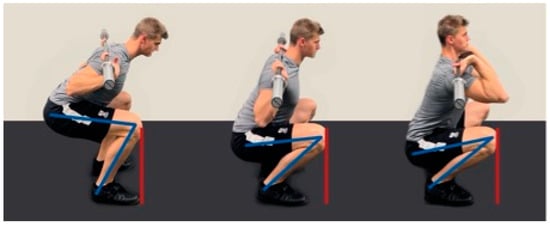
Based on seminal research from the 1970s and 1980s, the myth that the knees should only move as far anterior during the barbell squat until they vertically align with the tips of the feet in the sagittal plane still exists. However, the role of both the hip joint and the lumbar spine, which are exposed to high peak torques during this deliberate restriction in range of motion, has remained largely unnoticed in the traditional literature. More recent anthropometric and biomechanical studies have found disparate results regarding anterior knee displacement during barbell squatting. For a large number of athletes, it may be favorable or even necessary to allow a certain degree of anterior knee displacement in order to achieve optimal training outcomes and minimize the biomechanical stress imparted on the lumbar spine and hip. Overall, restricting this natural movement is likely not an effective strategy for healthy trained individuals.

  • anterior knee translation
  • back squat
  • restricted squat
  • unrestricted squat
  • knee rehabilitation

1. Introduction

Since the 1980s, there has been a widespread perception that the barbell squat should be performed with an upright posture and that the knees should not be moved beyond the tips of the toes. According to this traditional dogma, anterior knee movement should be limited in the sagittal plane once a vertical line with the tips of the feet is achieved [1][2]. Widely accepted instructions for proper knee positioning during barbell squats [2][3] are based on previous studies [4][5][6] that showed that anterior knee displacement (AKD) past the toes is associated with greater shearing forces in the knees, specifically the tibiofemoral joints [4], and that moving the knees anteriorly to a lesser extent during squatting generates lower knee extensor torque [5]. Based on these findings, it has become standard practice to maintain the shin as vertical as possible and that “maximal forward movement of the knees should place them no more than slightly in front of the toes” when squatting in order to lessen the shear stress placed on the knee [3]. From a practical standpoint, these guidelines advise against allowing the knees to displace anteriorly past the toes [1][2][3]. Although not reported in the literature, the recommendation to not or only slightly push the knees over the tips of the toes seems to be a very vague statement, as disproportionately large feet in relation to the lower extremities of an individual will likely not lead to reduced shearing forces. In addition, the recommendation to limit AKD results in altered knee and hip coordination [7], with a stronger upper body inclination [2][8][9], enhanced trunk flexion in the thoracic and lumbar spine [10], and reduced squatting depth [9]. Moreover, using a moderate foot stance, which represents an outward-directed foot angle of approximately 20° with the toes pointed laterally in combination with a shoulder-width stance [11], avoiding AKD cannot be achieved by most athletes performing different barbell squat techniques [7][9][11][12]. Given that deep barbell squat variations, such as deep high-bar back squats (DHBBSs) and deep front squats (DFSs), provide several fundamental benefits, including greater muscle activation, improved functional capacity, and higher athletic performance, as well as performance-enhancing transfer effects of dynamic maximal strength to dynamic speed-strength of hip and knee extensors [13][14][15], they are likely to be preferred over variations where the range of motion is deliberately limited under most circumstances. The center of gravity must remain vertically above the supporting surface during all barbell squat techniques, otherwise balance cannot be maintained [10][16]. To ensure this, anthropometry and biomechanics require the majority of exercisers to move the knee joints anteriorly over the toes during deep barbell squats. This is considered a normal and a required part of the squat movement, which should be encouraged in healthy individuals [17]. Even more so, the American College of Sports Medicine (ACSM) advised that healthy adults should perform every exercise through a full range of motion [18]. Exercising over the entire range of motion enables strength adaptations to take place at every angle the joint traverses, which may lower the risk of injury in those ranges [19]. Conversely, it has been demonstrated that spinal flexion and extension have a major impact on joint kinetics when performing squats [20]. To lower the risk of lumbar spine injuries, it is crucial to maintain a neutral spine position when lifting objects [21]. Practice guidelines concur that, in order to lower the risk of injury, both the spine and pelvis should remain in a neutral position with no relative movement during the squatting motion [20][22][23]. This is particularly true at the bottom position of the squatting movement, when the maximal angular displacement of the trunk and pelvis is greatest [21]. Deep squatting therefore seems to be a safe exercise when a neutral spine is maintained, although AKD increases with the depth of the squat [7].

2. Restricted vs. Unrestricted Barbell Squats

From a biomechanical perspective, AKD past the toes during squatting is associated with greater shearing forces on the knee [4]. It was therefore suggested that the resulting knee excursion could contribute to knee injury and that the shin should be kept as vertical as possible in order to reduce the shear stress on the knee [3][4]. However, the impact on the hip joint and the lumbar spine was not considered by these authors.
Research by Fry et al. [2] confirmed that knee torque increased by about 28% on average when exercisers performed unrestricted parallel high-bar back squats (PHBBSs; knee torque unrestricted squat 150.1 ± 50.8 Nm vs. knee torque restricted squat 117.3 ± 34.2 Nm), which are defined by a knee angle of 60–70° [16] and the knees were allowed to move beyond the toes. It was previously reported that patellofemoral joint stress (PFJS) during squatting increases during the lowering phase, continues to slightly increase during the rising phase, and finally decreases as the knee becomes more extended [24]. Additionally, peak PFJS and patellofemoral joint reaction forces rise as AKD, knee flexion angle, and external resistance increase [24][25]. The force of the quadriceps muscle might lead to anterior tibial displacement, particularly close to complete knee extension [26]. This displacement is counteracted by the quadriceps/hamstring co-contraction during squatting [27][28][29][30][31]. It is assumed that this co-contraction helps to neutralize the tibiofemoral shear forces imparted by the quadriceps [31], thus providing a stabilizing force at the knee during squatting [28].
Conversely, it was also shown that the external torque on the hip joints increased by approximately 973% (hip torque unrestricted squat 28.2 ± 65.0 Nm vs. hip torque restricted 302.7 ± 71.2 Nm) when the AKD was deliberately limited to toe-off height [2]. Biomechanically, this is an enormously high torque on the hip joint and likely much less favorable than the slightly increased torque on the knee joints. Assuming a simple kinematic chain model with a similar ground reaction force at the deepest position, the approximately 10-fold larger observed momentum in the hip during restricted high-bar back squats further suggest higher torque on the lower back [2][9]. The distance related to the moment of the muscle force around the screw axis of the joint is known as the moment arm, which has been demonstrated to increase in the hip joints when restricting AKD [2]. Conversely, a greater dorsiflexion of the ankle joints, which results in larger moment arms for the ground reaction force, explains why the maximum moment in the knees during unrestricted squats is greater than that during limited squats with larger knee flexion angles [9]. The idea of the moment arm appears to be crucial in practice, as the LBBS is particularly recommended when the primary goal is to lift weights as heavy as possible [32]. By placing the barbell lower on the back, the LBBS decreases the moment arm in key anatomical compartments and facilitates enhanced biomechanical working conditions for the hip extensor muscles, ultimately allowing for heavier weights to be used during squatting [33]. While in LBBSs, the moment arms in the hips are greater than when performing high-bar squats; the latter exercise affords moment arms in the knees that are relatively higher than in the hips [34].
What is more, the difference in maximum hip momentum during restricted and unrestricted high-bar back squats increases with increased barbell load, being 6.9% higher for restricted high-bar back squats with bodyweight only, 11.3% higher with one and one-quarter of the bodyweight, and 14.6% higher with one and one-half of the bodyweight [9]. It was therefore concluded that although deliberate restriction of the AKD lowers the torque on the knee, disproportionately high forces occur in the hips [2], which are likely transferred towards the lower back [2][9]. Adequate joint loading during barbell squatting consequently requires movement of the knees beyond the toes [2][16].
Restricting the natural AKD leads to altered knee–hip coordination [6], which is associated with increased trunk flexion in the thoracic and lumbar spine [10]. This form of evasive movement can lead to increased tensile stress on the intervertebral ligaments [35][36] and has been consequently discouraged by various authors [37][38]. It was demonstrated that the torque in the knee joints is significantly higher in the unrestricted high-bar back squat (maximal knee flexion angle of 85 ± 11°) than in the restricted high-bar back squat (maximal knee flexion angle of 106 ± 10°) [9]. However, the resulting torque in the hip joints behaved in an inverse manner. The higher torque in the hips with restricted high-bar back squats indicated a higher load on the lower back, which is why the authors argued that unrestricted high-bar back squats should be the preferred technique. Accordingly, subsequent studies found that unrestricted barbell squat variations are more suitable for stimulating the lower extremities [39], minimizing strain on the lower lumbar spine and lower back in comparison with restricted techniques [8][9], and are therefore the recommended technique to improve athletic performance based on current evidence [40].

3. Barbell Squat Technique Variations and AKD

To avoid AKD, without performing a technically inaccurate barbell squat in which the peak loads shift towards the lumbar spine, reducing the depth of the squat is an applicable strategy. While in clinical settings full knee extension is typically defined as 0°, the studies discussed here utilize a 180° knee angle to define full knee extension (i.e., 180° being equivalent to a straight stance) [41]. Accordingly, in “quarter” high-bar back squats (QHBBSs) and “half” high-bar back squats (HHBBSs, defined by a knee angle of approximately 110–140° and 80–100°, respectively), the knees of most athletes are not or only slightly pushed anterior over the toes [13][41][42].
Subjects performing a QHBBS were able to move an average load 4.02 (±1.59) times greater than with a DHBBS (defined by a knee angle of approximately 40–45°) [13][14][34][38]. However, such weights are not used in routine training practices for advanced athletes given that such high loads cannot be stabilized by the trunk or spine. Furthermore, with heavier weights, the compression forces on the vertebral bodies and the intradiscal pressure on the intervertebral discs also increase [43][44]. Heavier weight loads also lead to a significant augmentation of tibiofemoral [26] and patellofemoral compression forces [25]. As reviewed by Hartmann and Wirth [38], these relationships are often not considered when examining spinal [20] and knee joint loads at different knee flexion depths [20][45]. QHBBSs and HHBBSs with relatively supramaximal loads as compared with DHBBSs increase the risk for long-term degenerative alterations in knee joints and the spine [41]. Contrary to frequently expressed concerns, it has been shown that DHBBSs present an effective exercise for protection against back and hip injuries and do not contribute to an increased risk of knee injury [41], even though the AKD has been reported to range from 63.8 to 64.7 mm in men when squatting to a knee angle of 59.1 ± 2.0° and 93.2 to 96.6 mm in women when doing so to a knee angle of 72.4 ± 2.9° [7]. Taken together, these studies indicate that eliminating or minimizing AKD by reducing the squat depth with QHBBSs and HHBBSs may not be the most optimal choice for healthy individuals aiming to strengthen the muscles of the lower extremity. It should be noted, however, that squatting with a range of motion between 180° (upright stance) and 130° of knee flexion has been shown to minimize patellofemoral compressive forces [46] and may therefore be an appropriate option for knee rehabilitation patients, using no or very little additional load. A flexion depth of 130° corresponds to a “quarter” squat (QS) where the knees do not move past the toes. In this functional range, patellofemoral compressive forces and tibiofemoral compressive and shear forces in the knee joints are minimal [45], and deliberate limitation of AKD can therefore be an adequate strategy for rehabilitation [24].

4. Barbell Placement and AKD

AKD is directly related to the positioning of the barbell on the neck, as differences in the placement of the bar lead to a change in the overall center of gravity of the body [33]. Consequently, in order to maintain balance in an upright position, the positioning and joint angles associated with the lower extremities and the trunk also change. The more anteriorly the barbell is placed, the more upright the upper body can be positioned during a squatting movement. A more upright upper body or a greater trunk segment angle (TSA) afforded by a DFS (TSA: 63.6 ± 4,2; Figure 1, right) and a DHBBS (TSA: 46.3 ± 4,8; Figure 1, center) is associated with greater AKD than a low-bar back squat (LBBS—Figure 1, left), which has a more restricted relative to horizontal TSA of 40.7 ± 5.8 [16]. Moreover, a DHBBS allows for a greater degree of squatting depth than a LBBS because it permits more knee flexion, and this also impacts AKD (Figure 1) [38][47].
Figure 1. Squat variations and AKD: Low-bar back squat (LBBS, left), deep high-bar back squat (DHBBS, center), and deep front squat (DFS, right). AKD (red lines) and knee angle (formed by the lines in blue) vary between different barbell squat techniques.

5. Ankle Mobility, Weightlifting Shoes, and AKD

A sufficient degree of ankle dorsiflexion (ADF) forms the basis for a technically correct squat, regardless of the specific variations in technique [48]. Limited ankle mobility has been demonstrated to have a negative effect on squat exercise biomechanics [49][50] and, specific to young athletes, might be a significant contributor to the development of Osgood–Schlatter disease [51]. The ankle joints therefore need to have adequate closed kinematic chain range of motion to meet the technical requirements for lowering and elevating the center of mass vertically [37][52]. When performing a barbell squat, a sufficient amount of ADF is most important at the bottom of the descent phase [53]. As the hips, knees, and ankles flex, limitations in ankle flexibility and the consequently limited amount of AKD may arise as compensatory movements as complete joint range of motion is reached [7]. Numerous studies have revealed that the greatest restriction for compensating squatting mechanics is the ankle joints [48][54][55][56]. While attempting to lower the center of mass during the descent phase of the squatting movement, an individual may develop compensatory movements due to constraints in their ADF range of motion (ADF-ROM) [56]. It has been demonstrated that greater active ADF-ROM is associated with greater knee flexion and ADF displacement during squatting and that simulating reduced ADF with a wedge (12° forefoot angle) decreases peak knee flexion [48][54]. Moreover, it was shown that average ADF was 23.4–25.9° (measured as the angle between the line connecting the fibula head and the calcaneus and the line connecting the base and head of the fifth metatarsal bone) when deep bodyweight squats (56°) were performed and suggested that ADF is an important factor that determines the ROM of deep squats (DS) [57].

This entry is adapted from the peer-reviewed paper 10.3390/jcm12082955

References

  1. Dunn, B.; Klein, K.; Kroll, B.; McLaughun, T.; O’Shea, P.; Wathen, D. Coaches roundtable: The squat and its application to athletic performance. Strength Cond. J. 1984, 6, 10–22.
  2. Fry, A.C.; Smith, J.C.; Schilling, B.K. Effect of knee position on hip and knee torques during the barbell squat. J. Strength Cond. Res. 2003, 17, 629–633.
  3. Chandler, T.J.; Stone, M.H. The squat exercise in athletic conditioning: A position statement and review of the literature. Chiropr. Sports Med. 1992, 6, 105.
  4. Ariel, B. Biomechanical analysis of the knee joint during deep knee bends with heavy load. In Biomechanics IV; Springer: Berlin/Heidelberg, Germany, 1974; pp. 44–52.
  5. McLaughlin, T.M.; Lardner, T.J.; Dillman, C.J. Kinetics of the parallel squat. Res. Q. 1978, 49, 175–189.
  6. McLaughlin, T.M.; Dillman, C.J.; Lardner, T.J. A kinematic model of performance in the parallel squat by champion powerlifers. Med. Sci. 1977, 9, 128–133.
  7. McKean, M.R.; Dunn, P.K.; Burkett, B.J. Quantifying the movement and the influence of load in the back squat exercise. J. Strength Cond. Res. 2010, 24, 1671–1679.
  8. Hebling Campos, M.; Furtado Alaman, L.I.; Seffrin-Neto, A.A.; Vieira, C.A.; Costa de Paula, M.; Barbosa de Lira, C.A. The geometric curvature of the lumbar spine during restricted and unrestricted squats. J. Sports Med. Phys. Fit. 2017, 57, 773–781.
  9. Lorenzetti, S.; Gülay, T.; Stoop, M.; List, R.; Gerber, H.; Schellenberg, F.; Stüssi, E. Comparison of the angles and corresponding moments in the knee and hip during restricted and unrestricted squats. J. Strength Cond. Res. 2012, 26, 2829–2836.
  10. List, R.; Gülay, T.; Stoop, M.; Lorenzetti, S. Kinematics of the trunk and the lower extremities during restricted and unrestricted squats. J. Strength Cond. Res. 2013, 27, 1529–1538.
  11. Lorenzetti, S.; Ostermann, M.; Zeidler, F.; Zimmer, P.; Jentsch, L.; List, R.; Taylor, W.R.; Schellenberg, F. How to squat? Effects of various stance widths, foot placement angles and level of experience on knee, hip and trunk motion and loading. BMC Sports Sci. Med. Rehabil. 2018, 10, 14.
  12. Swinton, P.A.; Lloyd, R.; Keogh, J.W.; Agouris, I.; Stewart, A.D. A biomechanical comparison of the traditional squat, powerlifting squat, and box squat. J. Strength Cond. Res. 2012, 26, 1805–1816.
  13. Caterisano, A.; Moss, R.F.; Pellinger, T.K.; Woodruff, K.; Lewis, V.C.; Booth, W.; Khadra, T. The effect of back squat depth on the EMG activity of 4 superficial hip and thigh muscles. J. Strength Cond. Res. 2002, 16, 428–432.
  14. Hartmann, H.; Wirth, K.; Klusemann, M.; Dalic, J.; Matuschek, C.; Schmidtbleicher, D. Influence of squatting depth on jumping performance. J. Strength Cond. Res. 2012, 26, 3243–3261.
  15. Weiss, L.W.; FRX, A.C.; Wood, L.E.; Relyea, G.E.; Melton, C. Comparative effects of deep versus shallow squat and leg-press training on vertical jumping ability and related factors. J. Strength Cond. Res. 2000, 14, 241–247.
  16. Fry, A.; Aro, T.; Bauer, J.; Kraemer, W. A comparison of methods for determining kinematic properties of three barbell squat exercises. J. Hum. Mov. Stud. 1993, 24, 83.
  17. McKean, M.R.; Burkett, B.J. Knee behaviour in squatting. J. Aust. Strength Cond. 2012, 20, 23–36.
  18. Balady, G.J. ACSM’s Guidelines for Exercise Testing and Prescription; American College of Sports Medicine: Indianapolis, IN, USA, 2000.
  19. Cotter, J.A.; Chaudhari, A.M.; Jamison, S.T.; Devor, S.T. Knee joint kinetics in relation to commonly prescribed squat loads and depths. J. Strength Cond. Res. 2013, 27, 1765–1774.
  20. Schoenfeld, B.J. Squatting kinematics and kinetics and their application to exercise performance. J. Strength Cond. Res. 2010, 24, 3497–3506.
  21. Charlton, J.M.; Hammond, C.A.; Cochrane, C.K.; Hatfield, G.L.; Hunt, M.A. The Effects of a Heel Wedge on Hip, Pelvis and Trunk Biomechanics During Squatting in Resistance Trained Individuals. J. Strength Cond. Res. 2017, 31, 1678–1687.
  22. Maduri, A.; Pearson, B.L.; Wilson, S.E. Lumbar-pelvic range and coordination during lifting tasks. J. Electromyogr. Kinesiol. 2008, 18, 807–814.
  23. McGill, S. Low Back Disorders: Evidence-Based Prevention and Rehabilitation; Human Kinetics: Champaign, IL, USA, 2015.
  24. Kernozek, T.W.; Gheidi, N.; Zellmer, M.; Hove, J.; Heinert, B.L.; Torry, M.R. Effects of Anterior Knee Displacement During Squatting on Patellofemoral Joint Stress. J. Sports Rehabil. 2018, 27, 237–243.
  25. Wallace, D.A.; Salem, G.J.; Salinas, R.; Powers, C.M. Patellofemoral joint kinetics while squatting with and without an external load. J. Orthop. Sports Phys. Ther. 2002, 32, 141–148.
  26. Sahli, S.; Rebai, H.; Elleuch, M.H.; Tabka, Z.; Poumarat, G. Tibiofemoral joint kinetics during squatting with increasing external load. J. Sports Rehabil. 2008, 17, 300–315.
  27. Palmitier, R.A.; An, K.N.; Scott, S.G.; Chao, E.Y. Kinetic chain exercise in knee rehabilitation. Sports Med. 1991, 11, 402–413.
  28. Sear, J.A., Jr.; Erickson, J.C.; Worrell, T.W. EMG analysis of lower extremity muscle recruitment patterns during an unloaded squat. Med. Sci. Sports Exerc. 1997, 29, 532–539.
  29. Ninos, J.C.; Irrgang, J.J.; Burdett, R.; Weiss, J.R. Electromyographic analysis of the squat performed in self-selected lower extremity neutral rotation and 30 degrees of lower extremity turn-out from the self-selected neutral position. J. Orthop. Sports Phys. Ther. 1997, 25, 307–315.
  30. Wilk, K.E.; Escamilla, R.F.; Fleisig, G.S.; Barrentine, S.W.; Andrews, J.R.; Boyd, M.L. A comparison of tibiofemoral joint forces and electromyographic activity during open and closed kinetic chain exercises. Am. J. Sports Med. 1996, 24, 518–527.
  31. Stuart, M.J.; Meglan, D.A.; Lutz, G.E.; Growney, E.S.; An, K.N. Comparison of intersegmental tibiofemoral joint forces and muscle activity during various closed kinetic chain exercises. Am. J. Sports Med. 1996, 24, 792–799.
  32. Glassbrook, D.J.; Brown, S.R.; Helms, E.R.; Duncan, S.; Storey, A.G. The High-Bar and Low-Bar Back-Squats: A Biomechanical Analysis. J. Strength Cond. Res. 2019, 33 (Suppl. S1), S1–S18.
  33. Glassbrook, D.J.; Helms, E.R.; Brown, S.R.; Storey, A.G. A Review of the Biomechanical Differences between the High-Bar and Low-Bar Back-Squat. J. Strength Cond. Res. 2017, 31, 2618–2634.
  34. Wretenberg, P.; Feng, Y.; Arborelius, U.P. High- and low-bar squatting techniques during weight-training. Med. Sci. Sports Exerc. 1996, 28, 218–224.
  35. McGill, S.M. The biomechanics of low back injury: Implications on current practice in industry and the clinic. J. Biomech. 1997, 30, 465–475.
  36. Potvin, J.R.; McGill, S.M.; Norman, R.W. Trunk muscle and lumbar ligament contributions to dynamic lifts with varying degrees of trunk flexion. Spine 1991, 16, 1099–1107.
  37. Myer, G.D.; Kushner, A.M.; Brent, J.L.; Schoenfeld, B.J.; Hugentobler, J.; Lloyd, R.S.; Vermeil, A.; Chu, D.A.; Harbin, J.; McGill, S.M. The back squat: A proposed assessment of functional deficits and technical factors that limit performance. Strength Cond. J. 2014, 36, 4–27.
  38. Hartmann, H.; Wirth, K. Literature-based load analysis of different squat variations considering possible overuse injuries and adaptation effects. Schweiz. Z. fur Sportmed. und Sport. 2014, 62, 6–23.
  39. Chiu, L.Z.; vonGaza, G.L.; Jean, L.M. Net joint moments and muscle activation in barbell squats without and with restricted anterior leg rotation. J. Sports Sci. 2017, 35, 35–43.
  40. Comfort, P.; McMahon, J.J.; Suchomel, T.J. Optimizing squat technique—Revisited. Strength Cond. J. 2018, 40, 68–74.
  41. Hartmann, H.; Wirth, K.; Klusemann, M. Analysis of the load on the knee joint and vertebral column with changes in squatting depth and weight load. Sports Med. 2013, 43, 993–1008.
  42. Wilson, G.J. Strength and power in sport. In Applied Anatomy and Biomechanics in Sport; Human Kinetics: Champaign, IL, USA, 1994; pp. 110–208.
  43. Cappozzo, A.; Felici, F.; Figura, F.; Gazzani, F. Lumbar spine loading during half-squat exercises. Med. Sci. Sports Exerc. 1985, 17, 613–620.
  44. Kuo, C.S.; Hu, H.T.; Lin, R.M.; Huang, K.Y.; Lin, P.C.; Zhong, Z.C.; Hseih, M.L. Biomechanical analysis of the lumbar spine on facet joint force and intradiscal pressure—A finite element study. BMC Musculoskelet. Disord. 2010, 11, 151.
  45. Escamilla, R.F. Knee biomechanics of the dynamic squat exercise. Med. Sci. Sports Exerc. 2001, 33, 127–141.
  46. Escamilla, R.F.; Fleisig, G.S.; Zheng, N.; Lander, J.E.; Barrentine, S.W.; Andrews, J.R.; Bergemann, B.W.; Moorman, C.T., 3rd. Effects of technique variations on knee biomechanics during the squat and leg press. Med. Sci. Sports Exerc. 2001, 33, 1552–1566.
  47. Park, J.H.; Lee, S.J.; Shin, H.J.; Cho, H.Y. Influence of Loads and Loading Position on the Muscle Activity of the Trunk and Lower Extremity during Squat Exercise. Int. J. Environ. Res. Public. Health 2022, 19, 13480.
  48. Macrum, E.; Bell, D.R.; Boling, M.; Lewek, M.; Padua, D. Effect of limiting ankle-dorsiflexion range of motion on lower extremity kinematics and muscle-activation patterns during a squat. J. Sports Rehabil. 2012, 21, 144–150.
  49. Brooks, T.; Cressey, E. Mobility training for the young athlete. Strength Cond. J. 2013, 35, 27–33.
  50. Crowe, M.A.; Bampouras, T.M.; Walker-Small, K.; Howe, L.P. Restricted Unilateral Ankle Dorsiflexion Movement Increases Interlimb Vertical Force Asymmetries in Bilateral Bodyweight Squatting. J. Strength Cond. Res. 2020, 34, 332–336.
  51. Sarcević, Z. Limited ankle dorsiflexion: A predisposing factor to Morbus Osgood Schlatter? Knee Surg. Sports Traumatol. Arthrosc. 2008, 16, 726–728.
  52. Kasuyama, T.; Sakamoto, M.; Nakazawa, R. Ankle joint dorsiflexion measurement using the deep squatting posture. J. Phys. Ther. Sci. 2009, 21, 195–199.
  53. Mauntel, T.C.; Begalle, R.L.; Cram, T.R.; Frank, B.S.; Hirth, C.J.; Blackburn, T.; Padua, D.A. The effects of lower extremity muscle activation and passive range of motion on single leg squat performance. J. Strength Cond. Res. 2013, 27, 1813–1823.
  54. Dill, K.E.; Begalle, R.L.; Frank, B.S.; Zinder, S.M.; Padua, D.A. Altered knee and ankle kinematics during squatting in those with limited weight-bearing-lunge ankle-dorsiflexion range of motion. J. Athl. Train. 2014, 49, 723–732.
  55. Malloy, P.; Morgan, A.; Meinerz, C.; Geiser, C.; Kipp, K. The association of dorsiflexion flexibility on knee kinematics and kinetics during a drop vertical jump in healthy female athletes. Knee Surg. Sports Traumatol. Arthrosc. 2015, 23, 3550–3555.
  56. Padua, D.A.; Bell, D.R.; Clark, M.A. Neuromuscular characteristics of individuals displaying excessive medial knee displacement. J. Athl. Train. 2012, 47, 525–536.
  57. Endo, Y.; Miura, M.; Sakamoto, M. The relationship between the deep squat movement and the hip, knee and ankle range of motion and muscle strength. J. Phys. Ther. Sci. 2020, 32, 391–394.
More
This entry is offline, you can click here to edit this entry!
Video Production Service