1. Nitric Oxide (NO∙)
The role of NO
∙ as a signalling molecule has been extensively studied in the past [
126]. The first direct target of NO
∙ to be identified was the hemoprotein soluble guanylate cyclase (sGC) [
127]. NO
∙ binds to the heme iron of sGC forming a heme-nitrosyl complex and enzyme activation leading to the formation of cGMP, which in the endothelium, is a highly potent vasodilator [
128].
As well as binding to metal ions, NO
∙ can also covalently bind to cysteines and tyrosines to form nitrosocysteine- and nitrotyrosine-modified proteins, which may cause a change in enzyme activity and function. By using the novel and sensitive methodology of mass spectrometry, multiple new proteins were identified to be modified by NO
∙ causing dysfunctional signalling and contributing to diseases including cancer and inflammation [
129,
130,
131,
132].
Interestingly, when analysing pituitary adenoma tissue in a nitrosoproteome approach, the S1P lyase (SPL,
Sgpl1) was identified as tyrosine nitrated on two residues, Tyr
356 and Tyr
366 [
131]. Since these two sites are within the catalytic domain of Sgpl1, NO
∙ modification might affect its catalytic activity, although this has not been proven yet.
Another direct target of NO
∙ and a key signalling factor is the small G protein p21
ras, which is cysteine nitrosated in the presence of a NO
∙ donor resulting in more active p21
ras and downstream signalling, such as NFκB activation [
133] and mitogen- and stress-activated protein kinase SAPK activation [
134,
135,
136,
137].
By affecting these fundamental signalling cascades, which in turn regulate many transcription factors, NO
∙ can interfere with gene transcription of many genes, including sphingolipid-regulating enzymes, and thereby alter cell responses. Indeed, NO
∙ was shown to affect sphingolipid signalling in different cell types. Thus, treatment of renal mesangial cells [
138] or endothelial cells [
139] with NO
∙ donors caused a concentration-dependent increase in cellular ceramide formation. This effect mechanistically involved upregulated activities of both aSMase and nSMase, and may occur in a similar manner as reported for ROS-activated SMase activity [
94], i.e., directly by cysteine modification, or indirectly by GSH depletion.
In contrast to these studies, Falcone et al. [
140] rather suggested an inhibitory effect of NO
∙ on aSMase. They showed that apoptosis, induced either in dendritic cells by LPS [
140], or in monocytic U937 cells with TNFα [
141,
142], involves aSMase activation and ceramide formation. This apoptotic effect of LPS and TNFα was blocked by NO
∙, cGMP, and cGMP-dependent protein kinase (PKG, cGK) activation [
140,
141,
142]. Such an anti-apoptotic effect of NO
∙ through aSMase inhibition in tumour-associated macrophages seems also to be relevant in cancer therapy where it leads to chemoresistance [
143]. However, the detailed mechanism of aSMase inhibition by PKG is still not clear, but at some level must involve a phosphorylation step by PKG. While many substrates of PKG have been described [
144], aSMase has so far not been confirmed as a direct substrate of PKG. Therefore, phosphorylation of an upstream factor responsible for aSMase inhibition seems likely.
Another level of regulation of aSMase by NO
∙ may exist by protein-protein interactions through nitrosated residues. In this regard, it was shown that nitrosylation of procaspase-3 promoted the direct interaction of procaspase-3 with aSMase, and this interaction had an inhibitory effect on procaspase-3 function thus inhibiting downstream caspase-8 activation and thereby reducing cell death [
145].
Clearly, there are more factors than the SMases that determine whether cellular ceramides increase and especially the balance between SMases and CDase activities is crucial as well. Therefore, in situations when both activities of SMase and CDase increase, ceramide may not accumulate. This situation was shown for mesangial cells. When exposed to the pro-inflammatory cytokines IL-1β and TNFα, SMase and CDase activities increased in parallel thus resulting in a net unaltered ceramide [
138,
146]. In the case of NO
∙, ceramide accumulates because SMase activities are increased while CDase activities are reduced [
138,
147]. This was mechanistically further approached and it seems that NO
∙ causes proteasomal degradation of neutral CDase [
148].
2. Carbon Monoxide (CO)
CO is structurally very similar to NO
∙, but it is not a radical and therefore is more stable than NO
∙ and diffuses freely. Endogenously, it is mainly generated as a side product of heme degradation to bilirubin by the catalytic action of heme oxygenases [
149,
150]. Consequently, most of the CO derives from hemoglobin and there is a constant generation of this small molecule as a result of red blood cell turnover. In addition, a small part also derives from other heme-containing proteins (hemoproteins) such as myoglobin, cytochrome c oxidase, cytochrome P450, and even nitric oxide synthases [
150,
151]. CO produced by nitric oxide synthases is thought to regulate neurotransmission and blood flow in the central nervous system.
In biological systems, CO reacts with reduced transition metals such as iron in hemoproteins [
151]. High concentrations of CO are toxic and this is due to its binding to hemoglobin which occurs with many-fold higher affinity than oxygen. Consequently, CO displaces oxygen from the heme-binding resulting in impaired respiration and tissue hypoxia [
151]. However, it is now appreciated that low concentrations of CO, as it steadily arises from red blood cell turnover, are cytoprotective. This has led to the development of CO-releasing molecules (CORM) for various therapeutic purposes [
152] such as regulation of vascular tone, platelet aggregation, and inflammation. Mechanisms that mediate this protective effect of CO include the direct binding of CO to the various hemoproteins including sGC, cytochrome P450 proteins, cytochrome c oxidase, NADPH oxidase, and nitric oxide synthase [
153]. Indeed, it was shown that CO and cigarette smoke, similar to NO
∙, can activate the sGC yielding increased cGMP levels and endothelial relaxation [
154,
155,
156].
The influence of CO on sphingolipids has only been poorly studied over the years. In one early case report of CO poisoning causing a myelinopathy, manifested by demyelination of neuronal cells, lipid analysis in the brain revealed decreased levels of total phospholipids, cerebrosides, sphingomyelins, and free cholesterol with enhanced cholesterol esters in the white matter. No changes occurred in the grey matter of the cerebral cortex [
157]. Similarly, in a rat model of experimental acute CO poisoning, decreased brain gangliosides were detected together with changes in myelin [
158].
Multiple stimuli can induce HO-1 expression and thereby also generate CO [
153]. Among those, S1P was identified as an inducer of HO-1 in primary human macrophages [
159]. This effect was mediated by the S1P
1 receptor and resulted in a polarisation of macrophages to an M2 phenotype and an anti-inflammatory and anti-apoptotic reaction [
159]. However, in other cells, such as in human leukemia cells, S1P and its acylated form, ceramide 1-phosphate (C1P), both downregulated HO-1 which was proposed to be an important mechanism in the pro-metastatic effect of these lipids on leukemia cells [
160]. Moreover, short-chain C2-ceramide induced HO-1 in rat primary astrocytes [
161]. This occurred through the activation of the AMPK and MAPK signalling cascades.
These few data vaguely propose that there may exist a bidirectional regulation of the sphingolipid rheostat and HO-1/CO, although a therapeutic use of this regulatory setting is not yet clear.
3. Hydrogen Disulfide
To date, only few data are available to show a direct link between H
2S and sphingolipids. Interestingly, among the three H
2S-producing enzymes, only CBS is converting L-cysteine to L-serine and H
2S. L-serine is also the precursor in the de-novo biosynthesis of sphingolipids, and is directly used by the SPT for condensation with palmitoyl-CoA. Thus, changing the CBS expression or activity may have an impact on sphingolipid synthesis. In a recent study on isolated mouse aorta rings, it was shown that L-cysteine and L-serine have a vasorelaxant effect [
162]. The vascular effect of both L-cysteine and L-serine was reduced by a NOS inhibitor, but also by the SPT inhibitor myriocin and the S1P
1 receptor antagonist W146, suggesting the involvement of both NO
∙ and S1P in the relaxant effect. It was speculated that this mechanism could be involved in the marked dysregulation of vascular tone in hyperhomocysteinemic patients (CBS deficiency) and may represent a feasible therapeutic target [
162].
Since there is a well-studied bidirectional cross-talk between NO
∙ and H
2S [
162,
163,
164], and in view of the cross-regulation of NO
∙ on sphingolipid signalling (see
Section 2.4.1), it seems very obvious that there is also a cross-talk between H
2S-generating enzymes and sphingolipids.
In multiple myeloma cells, H
2S donors synergistically enhanced apoptosis of cells induced by the green tea polyphenol (-)-epigallocatechin-3-O-gallate (EGCG), and thereby, potentiated the anti-cancer effect of EGCG in a mouse xenograft model [
165]. This study further showed that in the presence of H
2S donors, EGCG enhanced acid sphingomyelinase activity which was not seen by either substance alone. It was speculated that this effect on aSMase resulting in enhanced ceramide formation, is responsible for the increased apoptosis of cells [
165].
Another interesting study showed that in human fibroblasts undergoing AKT-induced senescence, the expression of the enzyme cystathionine-β-synthase (CBS) was enhanced leading to increased H
2S and GSH production, and consequently protected senescent cells from oxidative stress-induced cell death [
166]. Especially in view of the fact that GSH is an endogenous inhibitor of nSMases, it could even be speculated that the protective effect of CBS on cell death, besides a direct protective effect of H
2S, is additionally mediated by reduced ceramide formation through blocked nSMase activity.
Finally, a recent study suggested an indirect link between H
2S and sphingolipids in a rat model of cerebral ischemia. In that study, the authors orally applied berberine to ischemic rats which improved the neuroinflammation and disease scores in tMCAO-induced cerebral ischemia [
167]. It was shown that oral berberine acted on the gut microbiota and stimulated H
2S production in the intestine, which subsequently activated the vagus nerve to subsequently alter the cerebral microenvironment resulting in less microglia activation and reduced neuroinflammation. Metabolomics analysis of various brain regions revealed changes in sphingolipid metabolism which may mediate the neuroprotection following vagus nerve activation. Notably, sphingosine was strongly increased in the ischemic rat cortex and downregulated by berberine treatment [
167]. These data suggest that microbiota H
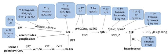
2S production affects cerebral sphingolipid metabolism through vagus nerve activation. Altogether, the critical issues regarding the complex interaction of redox- and lipid-signalling described in this review article are summarized in
Figure 3 and
Table 1.
Condition/
Enzyme |
Hypoxia |
Hyperoxia |
ROS |
NO∙ |
CO |
H2S |
| Ceramides |
↑OL [52], CM [53], HC [54],
PA [55]
↓VSMC [56] |
|
↑cancer cells [82,83,89],
EC [90,91], MC [91], CM [53] |
↑MC [91,138], EC [91,139]
↓DC [140], U937 [141,142] |
|
↑cancer cells [165] |
| Sphingosine |
|
|
|
|
|
↑cortex [167] |
| S1P |
↑VSMC [56], EC [59], cancer cells [58,61] |
↑mouse lung [77],
human lung [78],
EC [77] |
↓CM [121] |
|
|
|
| SM, Gangliosides |
|
|
|
|
↓brain [157,158] |
|
| Cholesterolesters |
|
|
|
|
↑brain [157] |
|
| Cerebrosides |
↓OL [52] |
|
|
|
↓brain [157] |
|
| nSMase |
↑CM [53], PA [55] |
|
↑EC [90], CM [53] |
↑MC [138] |
|
|
| aSMase |
↑HC [54] |
|
↑EC [90] |
↑MC [138]
↓DC [140], U937 [141,142] |
|
↑cancer cells [165] |
| nCDase, aCDase |
|
|
|
↓MC [138,147] |
|
|
| ACER2 |
↑adipocytes [57] |
|
|
|
|
|
| Sphk1 |
↑EC [59], PSMC [60], cancer cells [58,62] |
↑mouse lung [77,79] |
↓CM [121] |
|
|
|
| Sphk2 |
↑cancer cells [61], PSMC [60]
↓EC [59] |
|
|
|
|
|
| SPL |
|
|
|
Tyr nitration [131] |
|
|
| SPT2 |
↑neuroblastoma cells [69] |
|
|
|
|
|
This entry is adapted from the peer-reviewed paper 10.3390/metabo13030426