Pathogenesis of Endometriosis: The Current Spectrum of Theories: History
Please note this is an old version of this entry, which may differ significantly from the current revision.
Contributor: , ,

Endometriosis is a chronic disease, defined by abnormal presence of non-neoplastic endometrial glands and endometrial stroma outside the uterine mucosa. Most frequently it affects pelvic organs, followed by abdominal locations: ovaries, fallopian tubes, urinary bladder, intestines or peritoneum [1,2]. Occasionally, endometriotic foci are localized in distant organs far outside the pelvis: diaphragm, pleura or lungs, abdominal wall, central or peripheral nervous system [2]. According to the World Health Organization data, approximately 10% of reproductive-aged women (190 million) are diagnosed with this condition worldwide [1]. Endometriosis is mainly found in girls and ladies of reproductive age. The number of diagnosed cases peaks between 25 and 45 years of age [3], but it may take up to 8–10 years to reach the definitive diagnosis, as the clinical manifestations are diverse, non-specific and not always recognized as a pathology even by the patient herself. Endometriosis has a considerable impact on worldwide economics as well – it costs the world over 80 billion USD per year [5].

Endometriosis has a wide range of manifestations – from accidentally found asymptomatic lesions to a severe condition [3]. The main symptoms caused by endometriosis are chronic pelvic pain, severely painful menstrual periods, dyspareunia, dysuria and/or painful defecation, abdominal bloating and constipation. Infertility represents another endometriosis-related major problem: 40–50% of infertile women are diagnosed with endometriosis [2,5]. There are different mechanisms how endometriosis can decrease fertility: distorted anatomy of the pelvic cavity, development of the adhesions, fibrosis of the fallopian tubes, local inflammation of the pelvic organs and tissues, systemic and local (i.e., endometrial) immune dysregulation, changes in hormonal environment inside the uterus and/or impaired implantation of the embryo [3]. In addition, the disease has a significant negative impact on the quality of life and social well-being of patients – due to pain and other symptoms, e.g., fatigue, severe bleeding or mood swings, women have to skip their studies or work and might tend to avoid sex. Endometriosis may increase the risk of secondary mental health issues, such as anxiety and depression.

Three subtypes of endometriosis are recognised: superficial peritoneal endometriosis, ovarian endometriotic cysts and deep infiltrating endometriosis. Superficial peritoneal endometriosis is found on the surface of pelvic organs and the peritoneum. Ovarian endometriotic cysts are fluid-filled cavities, known also as endometriomas or “chocolate cysts”. Deep infiltrating endometriosis can invade pelvic or extrapelvic viscera to the depth of 5 mm or more, distorting the local anatomy [6].

Since the disease was described, several pathogenetic pathways have been considered, including retrograde menstruation, so-called benign metastasis, immune dysregulation, coelomic metaplasia, hormonal disbalance, involvement of stem cells and alterations in epigenetic regulation. These theories are highlighted in the given entry. 

  • endometriosis
  • pathogenesis
  • immune regulation
  • oestrogen
  • progesterone
  • stem cells
  • metaplasia
  • epigenetics
  • carcinogenesis
  • retrograde menstruation

1. Retrograde Menstruation

The theory of retrograde menstruation is known as Sampson’s theory. It remains relevant since it was proposed in 1925. The main idea of it is that menstrual blood containing endometrial cells regurgitate via patent fallopian tubes into the peritoneal cavity, where the implantation of these cells might occur [7,8]. After implantation, development and growth of the lesion is supported by angiogenesis [9]. It is possible because of activated peritoneal macrophages, which produce angiogenic factors, e.g., vascular endothelial growth factor (VEGF) [10]. 
Retrograde menstruation might explain the pathogenesis of ovarian and superficial peritoneal endometriosis. The weakness of this theory is that it cannot account for deep infiltrating endometriosis or lesions outside the peritoneal cavity [6,7,8,9]. Further, several studies show that reflux of menstrual blood is physiological for women with patent fallopian tubes, and most of them (76–90%) experience retrograde menstruation without further endometriosis [9,11]. The cases, when endometriosis develops in women, who have retrograde menstruation, could be explained by epidemiological studies which expose the risk factors of endometriosis – short menstrual cycle, longer menstrual flow and uterine outflow obstruction. These factors increase the quantity of retrogradely flushed cells [11,12]. Most likely, there is a correlation between retrograde menstruation and the development of endometriosis, but other pathogenetic factors, e.g., immune or hormonal dysregulation (see further, please), might be necessary to ensure the implantation and successful growth of retrogradely travelling endometrial cells [11].

2. Benign Metastasis

In 1927, John A. Sampson suggested an additional pathogenetic mechanism – theory of metastatic endometriosis. This concept assumes that a small amount of the endometrial tissue can be disseminated through the uterine-draining lymph vessels during menstruation. The idea is based on Sampson's observation: there was an endometrial polyp projecting into the lumen of a lymph vessel [13]. The benefit of this theory is that lymphogenic dissemination can explain the occurrence of endometriosis in lymphatic nodes and distant locations such as lungs, because lymphatic capillaries are found in almost all organs [14]. Nowadays, there are some reports of lymph node endometriosis, confirmed by histopathological examination that shows the presence of endometrial glandular and stromal cells in lymph nodes, and immunohistochemistry that is positive for oestrogen and progesterone receptors, PAX8 and CD10 [14,15,16,17].
Research on lymphangiogenesis has discovered a dysregulation of the expression of lymphangiogenic growth factors and their receptors in the eutopic endometrium of ladies diagnosed with endometriosis. The main promoters of lymphangiogenesis in endometrium are VEGF-C and VEGF-D [14,18,19], which are upregulated by proinflammatory cytokines interleukin 1β (IL-1β), tumour necrosis factor α (TNFα), IL-7 and CD74 [14]. The density of lymphatic microvessels of eutopic endometrium of patients is also increased. These changes could jointly facilitate the entry of endometrial tissue into the lymphatic circulation [14,19]. However, it is still unclear how the abnormal lymphangiogenesis actually affects the development of endometriosis.

3. Immune Dysregulation

Inflammation and disturbances of immune regulation are among the main general pathogenetic mechanisms in medicine. In endometriosis, proinflammatory pathways block apoptotic mechanisms, allowing adherence and survival of potentially harmful cells [20]. Macrophages, neutrophils, NK cells, dendritic cells and T cells are involved in the initiation and further progressing of endometrial lesions (Figure 1).
Figure 1. Immunocompetent cells in the pathogenesis of endometriosis. Figure replicated from [21] under Creative Commons license, provided at https://creativecommons.org/licenses/by-nc/4.0/ (accessed on 17 January 2023). Changes made: Figure legend, figure modification.
Macrophages detect and phagocytose pathogens and foreign cells, act as antigen-presenting cells to activate T cells and participate in regeneration of healthy endometrium. Normally, macrophages represent approximately 10% of total immune cell population in the proliferative phase in endometrium. The numbers of macrophages change by the menstrual cycle phase, regulated by oestradiol and progesterone. During menstruation, their density is significantly increased in accordance with their phagocytic function – clearing apoptotic cells and cell debris during endometrial shedding [22].

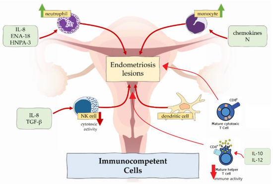
In endometriosis, number of macrophages is increased in eutopic endometrium [21,22] and peritoneal fluid [10] across all phases of menstrual cycle, but the cyclic changes are absent [23]. In contrast to higher density of macrophages, the phagocytic function is decreased because of the reduced expression of CD3, CD36 and annexin A2 [10,21,24]. It results in incomplete endometrial shedding, presence and survival of desquamated tissue in the peritoneal cavity [21]. Peritoneal macrophages release proinflammatory cytokines TNFα, IL-6, IL-8, IL-1β, which recruit neutrophils, provoke inflammation and support the development of endometrial lesions [3,10]. Macrophages also produce VEGF, which promotes angiogenesis in endometriosis [10].

Some authors noted predominance of M2 macrophage subtype in endometriotic lesions and peritoneal cavity [10,25]. This subtype classically promotes development of the tumours, e.g., colorectal cancer and osteosarcoma. M2 enhances nerve fibre growth, so excess of M2 macrophages could be related to severe pain, experienced in women with endometriosis [21,22].

In healthy endometrium, neutrophils are involved in endometrial repair and regulation of cyclic vascular proliferation. In endometriosis, neutrophil counts in peritoneal fluid are elevated. This could be attributable to the locally increased concentration of chemoattractants secreted by epithelial cells such as IL-8, epithelial neutrophil-activating peptide 8 (ENA-78) and human neutrophil peptides 1-3 (HNP1-3), which attract neutrophils to the peritoneal cavity [10,21].

According to the results in the mouse model, depleting of neutrophils with anti-Gr-1 antibody in the early stage of endometriosis significantly decreased the number of endometrial lesions [26]. In contrast, this antibody had no effect in advanced disease, which suggested that neutrophils did not take part in endometriosis progression, but only in induction [10]. However, neutrophils express cytokines, e.g., VEGF, IL-8 and C-X-C chemokine motif ligand 10 (CXCL10), which cause progression of the disease [10].

NK cells normally produce cytokines, which control tumour immunity and microbial infections. Regarding endometriosis, their cytotoxic function is suppressed by the IL-6, IL-15 and transforming growth factor β (TGF-β) [10,27]. Therefore, endometrial cells, which enter the peritoneal cavity, tend to stay there. However, the amount of the NK cells shows no differences in women with and without endometriosis.

Dendritic cells are responsible for antigen presentation to T cells and, therefore, are involved in immune responses in mucosal surfaces [22]. There are two types of dendritic cells—plasmocytoid dendritic cells and myeloid dendritic cells. Plasmocytoid dendritic cells function in recognition of viruses and produce interferons, while myeloid dendritic cells participate in T cell activation and are relevant to endometriosis. In healthy individuals, the amount of the dendritic cells increases to clear endometrial debris during menstruation. In ladies affected by endometriosis, the density of myeloid dendritic cells in endometrium is significantly reduced [28]. In the peritoneal cavity, numbers of dendritic cells are increased and may promote neuroangiogenesis, causing and enhancing pain sensation [22].

One of the important factors, which maintains the development of endometriosis, is imbalance between type 1 T lymphocytes (Th1) and type 2 T lymphocytes (Th2). These two types have different immune functions: Th1 lymphocytes produce cytokines and promote cellular responses, but Th2 lymphocytes influence differentiation of B lymphocytes and suppress cellular and humoral responses [3]. In endometriosis, Th2 lymphocytes represent the main population of T cells, allowing potentially harmful cells to escape from immune surveillance. On the other hand, the immune response of CD4+ Th1 lymphocytes in peritoneal fluid is suppressed due to an increased expression of IL-10 and IL-12 [29].

Moreover, the peripheral concentration of cytotoxic (CD8+) T cells and activated (HLA-DR) T cells in healthy women increases in luteal phase compared with the follicular phase of the menstrual cycle, but there are no such fluctuations of cytotoxic and activated T cells in patients with endometriosis [30].

Recently, the association between regulatory T cells (Tregs) and endometriosis was reported. The main function of regulatory T cells is the modulation of the immune system, maintaining tolerance to self-antigens and preventing autoimmune diseases [29]. In endometriosis patients, there is an increased amount of Tregs in the peritoneal fluid and decreased – in the peripheral blood. These changes can suppress local cellular immune response and facilitate autoimmune reactions [29].

4. Coelomic Metaplasia

In 1924 Robert Meyer proposed the theory of coelomic metaplasia. The idea is based on the embryogenesis of female reproductive tract: it develops from a pair of Müllerian ducts, which arise from coelomic epithelial cells of mesodermal origin [31]. The Mayer's theory assumes that the original coelomic membrane is able to undergo metaplasia and generate endometrial stroma and glands. Along with embryonic rest theory (see further, please), it is the most suitable explanation for cases of endometriosis in men, who have received high doses of oestrogen for prostatic carcinoma treatment. Coelomic metaplasia concept is also applicable to endometriosis in Rokitansky-Kuster-Hauser syndrome patients who do not have functioning endometrial tissue because of congenital aplasia of the uterus and the upper part of the vagina [9,31,32]. In both these clinical groups, endometriosis cannot be explained by Sampson’s implantation theory due to lack of eutopic endometrium.
Regarding the three common forms of endometriosis, coelomic metaplasia easily accounts for ovarian endometrioma. The mesothelium, which derives from the coelomic epithelium covering the ovary, has great metaplastic potential and can invaginate into the ovarian cortex [9,32]. These mesothelial inclusions could be transformed into endometriosis by metaplasia [9]. However, growth factors, which influence this phenomenon, should be identified. Thus, similarly to retrograde menstruation theory, the concept of coelomic metaplasia most likely represents a part of the whole puzzle. 

5. Embryonic Rest Theory

Embryonic rest theory is a variation on the metaplasia concepts. It states that remnants of embryonic cells of Wolffian or Müllerian duct origin may differentiate into endometriotic lesions [12,33]. In the coelomic metaplasia theory, the transformation capacity is ascribed only to mesothelium, but there is no such restriction in the embryonic rest theory [33]. In this concept, it is supposed that changes in cell differentiation or relocation of the Müllerian ducts during embryogenesis can maintain the spreading of embryonic cells – primordial endometrial cells [34]. Generally, these cells are located in the posterior pelvic floor and remain inactive until puberty, when the formation of endometriotic lesions is triggered via oestrogen stimulation [12]. Recently, as a proof of the embryonic rest theory, Signorile et al. published autopsy data of female fetuses, where ectopic endometrium was found in the posterior pelvic floor structures: Douglas pouch, recto-vaginal septum, rectal tube and posterior wall of uterus [12]. These places are frequently affected in the diagnosed endometriosis cases. This theory is suitable not only for endometriosis cases in women, but men too, because Wolffian ducts also may contain embryonic cells.

6. Endometrial Stem Cell Theory

Stem cells represent a minor fraction of multipotent cells with high replicative potential, having unlimited ability to renew themselves and produce more differentiated daughter cells [35]. There are several populations of somatic stem cells in endometrium, including epithelial, mesenchymal and mixed side population [6,36]. The main functions of these cells are remodelling, regeneration and homeostasis of the tissue. In endometrium, epithelial stem cells are found in the basal layer, and are responsible for regeneration of the functional layer during the proliferative phase, but mesenchymal stem cells are localized in the perivascular area of the basal and functional layers and are responsible for generation of functional stroma [6,12].
The migration of endometrial stem cells remains hypothetical. Some of the above-listed theories could be used to explain the mechanism. Firstly, endometrial stem cells are also found in menstrual blood [36]. This blood, containing stem cells can reach the peritoneal cavity via patient fallopian tubes as Sampson’s retrograde menstruation theory considers [12]. The second theoretical mechanism of the migration of stem cells to the ectopic sites is abnormal cell migration during organogenesis of the female reproductive tract, which is associated with aberrant expression of WNT and HOX genes [36]. The last mechanism is the ability of endometrial stem cells to enter the angiolymphatic space passively during menstruation and move around the circulation [6].
After the migration phase, stem cells adhere and start to form endometrial lesion. The stem cell potential of lesion formation has been proven by Cervelló et al. in 2011, when endometrial side population cells were implanted beneath the kidney capsule in immunocompromised NOD-SCID mice and this experiment resulted in endometriosis [37].
This theory is important because it can explain the pathogenesis of all three subtypes of endometriosis and its ectopic localization outside the abdominal cavity [6]. Nevertheless, additional factors, e.g., proinflammatory background, might be necessary to release endometrial stem cells from eutopic tissues.

7. Bone Marrow-Derived Stem Cell Recruitment Theory

This variant of stem cell theory is based on another source of stem cells – bone marrow. These cells are able to incorporate themselves into the endometrium to regenerate the tissue [36]. Several populations of cells take part in endometrial regeneration – mesenchymal, hematopoietic and endothelial progenitor cells [6].
The conception of theory is the following: bone marrow stem cells, which circulate via blood vessels, are settled in soft tissue instead of going to the endometrium, while reduced number of cells is recruited to eutopic endometrium [6,12]. Recent studies suggest that the CXCL12/CXCR4 axis is involved in recruiting bone marrow-derived stem cells, so the malfunction of this axis can cause the misplacement of stem cells [6].
The advantage of bone marrow-derived stem cell theory is its capability to explain extrapelvic endometriosis without the concept of “benign metastasis” which is contradictory to the fact that endometriosis itself is not a malignant tumour.

8. Hormonal Imbalance

In the healthy endometrium, progesterone and oestrogen signalling is strictly coordinated and menstrual cycle phase-dependent. This is important to maintain a normal menstrual cycle, embryo implantation and pregnancy [38]. Oestrogen induces epithelial growth during the proliferative phase, while progesterone inhibits the action of oestrogen and initiates the secretory phase, when stromal cells begin the decidualization [20,38]. The dysregulation of these two hormones – resistance to progesterone and oestrogen dominance [38,39] – leads to the development of endometriosis (Figure 2).
Figure 2. The effects of progesterone and oestrogen dysregulation on endometrium. Figure replicated from [38] under Creative Commons license, provided at https://creativecommons.org/licenses/by-nc/4.0/ (accessed on 17 January 2023). Changes made: Figure legend.
 
Endometriosis is occasionally considered an “oestrogen-dependant” disease because of epidemiologic correlations: endometriosis mostly affects women of reproductive age, and it can appear in postmenopausal ladies having high oestrogen levels or undergoing oestrogen-replacement therapy [40].

The main functions of oestrogen in healthy endometrium include stimulation of epithelial proliferation and induction of leukaemia inhibitory factor (LIF), an IL-6 family cytokine, which is important for successful embryo implantation and decidualization of the endometrium [38].

In endometriosis, studies report on higher levels of oestradiol – oestrogen steroid hormone – in menstrual blood and abnormal expression of enzymes involved in oestrogen metabolism, which can lead to increased oestrogen concentration and suppressed inactivation of oestrogen synthesis [40].

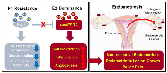
There are two oestrogen receptors – ERα and ERβ, which are coded by different genes: ESR1 and ESR2, respectively [38,39]. In endometriosis patients, expression of the receptors is changed – the ERα:ERβ ratio is significantly reduced due to high ERβ levels [41]. The main problem caused by abnormal expression of ERα is increased synthesis of inflammatory cytokines, prostaglandins, angiogenic and growth factors [20,29]. On the other hand, ERβ overexpression leads to inhibition of TNFα-induced apoptosis and also promotes the inflammation [27,41]. As a result, synthesized prostaglandins induce inflammation and prevent cell apoptosis; growth factors and angiogenic agents support the progression of the endometrial lesions, and inhibition of apoptosis promotes cell proliferation and lesion growth [20,27]. In addition, oestrogen is able to stimulate growth of peripheral nerve fibres by upregulating nerve growth factors (NGF) causing nociceptive pain [27].

The expression of the progesterone receptor (PGR) is induced by oestrogen action through its receptor ERα. PGR has two isoforms: PR-A and PR-B, expression of which increase during the proliferative phase and decrease after the ovulation [41]. Expressed PGR inhibits ERα expression, establishing a feedback system.

In endometriosis, as a result of low ERα:ERβ ratio and high oestrogen levels, progesterone resistance develops: PR-B is undetectable and PR-A levels are significantly lower than in the endometrium of healthy individuals [41]. Progesterone resistance manifests as a decreased responsiveness to progesterone of endometrial stromal cells [2].

Moreover, mutation of PGR gene causes sterility in mice due to reduced or absent ovulation, uterine hyperplasia, lack of decidualization of the endometrium and limited mammary gland development [38].

Due to the significant impact of oestrogen and progesterone on pathogenesis of endometriosis, several treatment approaches target the balance of these hormones. Current therapy options include combined oral contraceptives, progestins, gonadotropin-releasing hormone agonists, danazol and aromatase inhibitors. Other therapeutic pathways are under development, e.g., gonadotropin-releasing hormone antagonists, selective oestrogen receptor modulators and selective progesterone receptor modulators [3,38]. However, hormonal therapy is associated with adverse systemic effects including weight gain, fluid retention, acne, hot flashes, decreased libido, insomnia and vaginal dryness [3], which might decrease the compliance to long-term hormonal treatment.

9. Alterations in Epigenetic Regulation

In recent years, a growing body of evidence suggests that epigenetic changes have a certain role in the development of endometriosis. Epigenetic changes are defined as alterations of gene expression without any changes in DNA sequence. They are represented by the alterations in DNA methylation, histone acetylation, RNA transcription and chromatin remodelling [42]. The epigenome can be influenced by environmental factors, e.g., metabolism and nutritional deficiencies [39].

DNA methylation depends on DNA methyltransferases. Normally, the expression of these enzymes in endometrium is regulated by oestrogen and progesterone and varies depending on the cycle phase [42]. In endometriosis, hypermethylation of DNA of the local cells occurs due to increased expression of DNA methyltransferases DNMT1, DNMT3A and DNMT3B [39,43].

Human Homeobox A10 (HOXA10) genes are important for the endometrial changes throughout the normal menstrual cycle – they regulate endometrial growth, differentiation, and embryo implantation [39,44]. HOXA10 expression is regulated by oestrogen and progesterone [39]. In endometriosis, the expression of HOXA10 is decreased during the secretory phase, and, as the result, uterine receptivity is decreased and endometriosis-related infertility occurs [39,43]. Probably, HOXA10 gene expression is reduced due to hypermethylation of the HOXA10 gene promoter in the endometrial tissue [39,43].

Histone deacetylases are responsible for histone modulation and acetylation. In endometriosis, activity of histone deacetylases HDAC1 and HDAC2 is increased. It leads to the hypoacetylation of cyclins, which causes cell cycle induction and propagation [42,43].

10. Micro-RNAs

Micro-RNAs are short non-coding RNA molecules that regulate translation of mRNA post-transcriptionally by repression and mRNA degradation, acting as large-scale molecular switches [45]. According to recent findings, endometriosis is characterised by abnormal spectrum of micro-RNAs, further influencing the expression of the relevant target mRNAs [45]. Wide spectrum of micro-RNAs are involved in different steps of endometriosis. For example, miRNA-135a/b, regulating HOXA10, is upregulated in endometriosis and cause progesterone resistance [42,46]. MiR-199 is downregulated, so COX-2 translation is not suppressed, and it leads to pro-inflammatory milieu, characterised by active prostaglandin synthesis and elevated concentration of IL-8 [42,45]. MiRNA-96b is also downregulated, and it is the cause of increased proliferation in endometrial lesions [42]. MiR-126 increases VEGF and fibroblast growth factor (FGF) signalling in endothelial cells, resulting in neoangiogenesis and the subsequent maturation of the new vasculature [45]. MiRNA-223 also shows a significant impact on endometriosis. This micro-RNA is involved in signal transduction, regulation of transcription, cell growth and development, modulation of inflammation and tumorigenesis [47]. In 2022, Xue et al. found that miRNA-223 levels are decreased in eutopic and ectopic endometrial stromal cells in women with endometriosis [47]. They also proved that upregulation of miRNA-223 would lead to suppressed proliferation, invasion and migration of endometrial stromal cells and reversed epithelial-to-mesenchymal transition [47]. These findings highlight miRNA-223 as a potential new therapeutic target.
The other micro-RNA, which promotes the growth, proliferation and angiogenesis of ectopic stromal cells, is miRNA-21 [46,48]. In the serum of endometriosis patients, expression of miR-26b-5p and miR-215-5p is downregulated, but miR-6795-3p (Table 1) – upregulated [48]. These three micro-RNAs are involved in MAPK and PI3K-Akt molecular pathways. MAPK signalling is important in regulation of inflammation and the following cellular processes: differentiation, proliferation, stress response, metabolism and apoptosis [48]. PI3K-Akt pathway is also involved in the listed cellular reactions as well as in angiogenesis. These two molecular cascades are significant for development and progression of endometriosis, theoretically becoming future therapeutic targets.
Table 1. Micro-RNAs and their effects in endometriosis.

11. Carcinogenetic Pathways in Endometriosis

Although endometriosis is a benign disease, it still has a potential to transform into malignancy. This event is rather rare, affecting 1% of all endometriosis patients [39,45]. Endometriosis-related malignant change most frequently takes place in ovaries, and the predominant endometriosis-related ovarian tumours are ovarian endometrioid carcinoma and ovarian clear cell carcinoma, which are found in 76% of the relevant cases [39,49].
Recently, several carcinogenetic pathways have been reported for endometriosis-related malignant transformation. Uncontrolled cell division, infiltration of surrounding tissues, neoangiogenesis and escape of apoptosis can be caused by the demethylation of oncogenes and the hypermethylation of tumour suppressor genes [39]. This has been demonstrated in endometriosis. For example, the following events are involved in malignant change of the endometriosis: the hypermethylation of the human mutL homolog 1 (hMLH1) gene promoter which causes a decrease in DNA mismatch repair gene expression, the hypomethylation of long interspersed element-1 (LINE-1), inactivation of the tumour suppressor genes runt-related transcription factor 3 (RUNX3) gene and Ras-association domain family member 2 (RASSF2) gene by their promoter hypermethylation [39]. Also, in case of endometrioid cancer, activation of the KRAS oncogene and inactivation of the PTEN tumour suppressor gene occurs [44,49]. Loss of PTEN activity is supposed to be an early event in malignant transformation of endometriosis, and is related to the mutation of PTEN gene itself [45]. In addition, Anglesio et al. found that in deep infiltrating endometriosis there are somatic mutations in cancer driver genes ARID1A, PIK3CA, KRAS, and PPP2R1A [50].

12. External Environmental Factors

Environmental factors are suspected to have an impact on the pathogenesis of endometriosis. The main potentially harmful lifestyle factors include lack of physical activity, smoking, caffeine and alcohol intake, as well as diet. It is supposed that physical activity helps to reduce the risk of endometriosis, because it decreases menstrual flow and normalizes oestrogen balance [46]. Tobacco smoking increases the expression of pro-inflammatory mediators, disrupts synthesis of prostaglandin E2 and natural steroids [46]. Disrupted steroidogenesis leads to increased oestrogen and decreased progesterone synthesis [51]. Caffeine reduces production of sex-hormone binding globulin (SHBG) [46]. It is suggested that caffeine might have a protective potential, but the published data are conflicting. Kechagias et al. found a correlation between endometriosis and intense caffeine intake (>300 mg/day), admitting that it could be the risk factor for the disease [52]. Alcohol has the opposite effect on synthesis of hormones compared to caffeine. It has an impact on pituitary luteinizing hormone and activates the enzyme aromatase, resulting in increased oestrogen production and increased testosterone conversion to oestrogen [46,51]. Increased consumption of red meat is considered to have a negative effect on endometriosis development, probably because of high content of saturated fat [51,53].

Dioxins and polychlorinated biphenyls (PCBs) are organic pollutants produced by industrial processes. The most toxic environmental pollutant is called 2,3,7,8-tetrachlorodibenzo-p-dioxin (TCDD) [53]. PCB and TCDD may disrupt endocrine processes. In context of endometriosis, it was found that increased amount of PCB and TCDD have been accumulated in adipose tissue of patient with deep infiltrating endometriosis [51,54], but the mechanism of these changes remained uncertain.

Conclusions

In conclusion, the pathogenesis of endometriosis is complex. Retrograde menstruation, so-called benign metastasis, immune dysregulation, coelomic metaplasia, embryonic rest theory, recruitment of endometrial and/or bone marrow-derived stem cells, hormonal imbalance, alterations in epigenetic regulation and micro-RNA spectrum as well as influence of external environmental factors are among the proposed theories and research directions in endometriosis. There is no single theory which could explain all aspects of endometriosis. The future concept of endometriosis is likely to incorporate elements from all the listed pathogenetic theories. 

This entry is adapted from the peer-reviewed paper 10.3390/ijms24054254

This entry is offline, you can click here to edit this entry!
ScholarVision Creations