High-Density Lipoprotein Alterations in T2D and Obesity: History
Please note this is an old version of this entry, which may differ significantly from the current revision.
Contributor: , , , ,

Alterations affecting high-density lipoproteins (HDLs) are one of the various abnormalities observed in dyslipidemia in type 2 diabetes mellitus (T2DM) and obesity. Kinetic studies have demonstrated that the catabolism of HDL particles is accelerated. Both the size and the lipidome and proteome of HDL particles are significantly modified, which likely contributes to some of the functional defects of HDLs. Studies on cholesterol efflux capacity have yielded heterogeneous results, ranging from a defect to an improvement. HDLs are less able to inhibit the nuclear factor kappa-B (NF-κB) proinflammatory pathway, and subsequently, the adhesion of monocytes on endothelium and their recruitment into the subendothelial space. In addition, the antioxidative function of HDL particles is diminished, thus facilitating the deleterious effects of oxidized low-density lipoproteins on vasculature. Lastly, the HDL-induced activation of endothelial nitric oxide synthase is less effective in T2DM and metabolic syndrome, contributing to several HDL functional defects, such as an impaired capacity to promote vasodilatation and endothelium repair, and difficulty counteracting the production of reactive oxygen species and inflammation
  • high-density lipoprotein
  • type 2 diabetes
  • obesity
  • metabolic syndrome

1. HDL Size and Composition

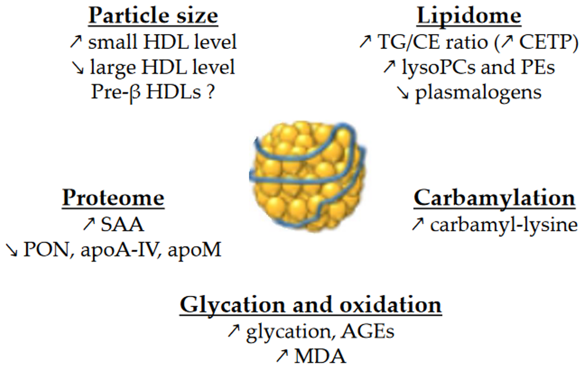
High-density lipoproteins (HDLs) are composed of apolipoproteins (mainly apoA-I), non-structural proteins, and lipids. They are a heterogeneous group of particles that vary in size as well as protein and lipid composition. Basically, HDLs are composed of a surface monolayer, made up of amphipathic phospholipids (PLs) and sphingolipids (SPLs) and unesterified cholesterol (UC) molecules, which surround the particle core, which consists of a mixture of hydrophobic TG and cholesteryl ester (CE) molecules. The alterations in HDL size and composition in contexts of insulin resistance are important because there is a close relationship between HDL size and composition and their functions. An overview of the changes in HDL size and composition is presented in Figure 1.
Figure 1. Main changes in the size and composition of HDLs in T2DM and MetS. ↗ and ↘ mean increase and decrease, respectively. AGEs, advanced glycation end-products; CE, cholesteryl esters; CETP, cholesteryl ester transfer protein; MDA, malondialdehyde; PCs, phosphatidylcholines; PEs, phosphatidylethanolamines; PON, paraoxonase; SAA, serum amyloid A; TG, triglycerides.

1.1. Particle Size

Size-based HDL nomenclature can be confusing because it depends on the technology used to separate HDL subclasses. In any case, HDLs are classified as very small, small, medium, large and very large particles. Classically, those particles are called HDL2a and HDL2b (large), HDL3a, HDL3b, and HDL3c (medium or small) and unlipidated apoA-I or pre-β (discoidal).
Overall, the size of HDLs is decreased in T2DM [1][2]. More precisely, nuclear magnetic resonance spectroscopy studies revealed that small HDL particles are more numerous in T2DM, contrary to large HDL particles [3][4][5][6]. It is also the case in insulin-resistant individuals without diabetes [6][7][8], and the intensity of changes actually increases with the degree of insulin resistance [6].

1.2. Lipidome

HDL particles are enriched in TGs in patients with T2DM [1][2][9][10][11], obesity [12], insulin resistance [10][13] or MetS [14]. The replacement of CE by TG molecules in HDLs affects the conformation of apoA-I [15], which could modulate binding to receptors and the functions of HDLs.

PLs and SPLs represent more than one third of the total mass of HDLs. They play a major role in HDL functions, either by binding to a specific receptor, such as sphingosine-1-phosphate (S1P) receptors or by modulating the physicochemical properties of HDLs. Lipidomic studies have revealed several changes in the HDL phosphosphingolipidome, although the results are quite heterogeneous. The expression of results in relation to either total HDL mass, total HDL lipids, or apoA-I levels makes it challenging to compare between studies. The HDL content in total PLs has been found to be either decreased in T2DM when the results are expressed in relation to total lipids [10], or normal when the results are expressed in relation to total mass or total lipids [1][11].

Taken together, phosphatidylcholines (PCs, the main PL class) and sphingomyelins (SMs) represent more than 80% of PLs/SPLs in HDLs. When the results are expressed in relation to apoA-I, the HDL content in PCs and SMs in T2DM has been found to be either normal or decreased [2][11]. When the results are expressed in relation to total HDL lipids, PCs and SMs have been found decreased in T2DM HDLs [10].
1.3. Proteome

Proteins form a major structural and functional component of HDL particles. The protein cargo of HDLs is comprised of over 100 proteins [16]. It should be noted that the HDL isolation method has a significant impact on HDL protein composition [17]. The HDL proteome has been extensively studied in diabetes, especially using mass spectrometry. Besides apoA-I, the kinetics of several HDL-proteins is perturbed in T2DM, as described hereafter. Reduced half-lives are observed for apoA-II, apoJ, apoA-IV, transthyretin, vitamin-D binding protein, and complement C3 [16]. A recent large proteomic study evaluated 182 proteins in isolated HDL fraction and found that HDLs from T2DM patients are enriched in 17 proteins and are deprived of 44 proteins compared to healthy individuals [5]. More precisely, HDLs were particularly enriched in pulmonary surfactant protein B and in serum amyloid A proteins (SAA1 and SAA2). In contrast, T2DM HDLs were deprived in apoA-IV, clusterin, paraoxonases (PON1 and PON3), apoD, apoE, apoF, apoM, apoC-II, and apoC-III [5]. The enrichment of HDLs in SAA in diabetes [5][18][19] is of particular interest due to its deleterious role in reverse cholesterol transport (RCT) [19][20].

1.4. Glycation and Oxidation

Chronic hyperglycemia in diabetes facilitates accelerated glycation, which is usually divided into early and late glycation. Thus, reducing sugar reacts with amino groups of proteins to form Schiff bases and then Amadori compounds (early glycation products). It may be followed by irreversible dehydration, condensation, and cross-linking reactions, resulting in a heterogeneous family of derivatives called advanced glycation end-products (AGEs), also known as late glycation products or glycoxidation products. Besides glucose itself, chronic hyperglycemia and oxidative stress in diabetes induces the production of dicarbonyls (glyoxal, methylglyoxal, 3-deoxyglucosone), which also react with proteins to yield AGEs [21]. Carboxymethyl-lysine (CML) is thought to be the most abundant AGE in vivo. Among the amino acids with nucleophilic residues prone to glycation, lysine residues are particularly abundant in apolipoproteins, and they are the preferred site of glycation.
The level of HDL glycation is significantly increased in T2DM, and seven glycation sites on lysine residues of apoA-I have been identified in T2DM subjects [22]. T2DM HDLs are enriched in methylglyoxal [23].
Beyond its role in the glycoxidation of HDL proteins, oxidative stress also leads to the peroxidation of lipids in HDLs. The proinflammatory enzyme myeloperoxidase (MPO) represents a major source of reactive-oxygen species (ROS) in atherosclerotic lesions. MPO-derived oxidants modify lipids and lead to the formation of dicarbonyls, such as malondialdehyde (MDA). The increased oxidative stress in subjects with T2DM and/or obesity is a consequence of several abnormalities, including hyperglycemia, insulin resistance, inflammation, and dyslipidemia [24]. Thus, not surprisingly, T2DM patients have increased concentrations of oxidized HDLs [25] and apoA-I [26], and their HDLs are enriched in MDA [27] and oxidized fatty acids derived from arachidonic and linoleic acids [28].

1.5. Carbamylation

Carbamylation (carbamoylation stricto sensu) is another post-translational modification affecting lipoproteins in diabetes. It is an irreversible non-enzymatic process mediated by isocyanate, and it is characterized by the binding of a carbamoyl moiety (-CONH2) to lysine, resulting in carbamyl-lysine. It originates from MPO within atherosclerotic lesions or from urea or smoking. Plasma MPO levels are increased in T2DM [29][30], and patients with T2DM exhibit higher levels of carbamylated HDLs even without renal impairment [31][32].

2. HDL/apoA-I Kinetics

Numerous in vivo kinetic studies demonstrate that low HDL-C levels in T2DM and obesity are due to an accelerated catabolism of HDL particles [13][14][33][34][35]. The fractional catabolic rate (FCR) of HDL-apoA-I is increased, resulting in shorter plasma residence times for HDL particles [9][33][36]. Obese patients at an early stage of insulin resistance (i.e., without impaired glucose tolerance) already have an accelerated HDL-apoA-I catabolism [37].
Studies have found the production rate (PR) of HDL-apoA-I to be either elevated [9][13][34][36] or normal [14][37] in insulin-resistant individuals. The PR of HDL-cholesterol has recently been reported to be higher in patients with T2DM. In any case, the increase in HDL-apoA-I PR is usually smaller than the increase in the FCR, which explains the decreased concentrations of HDL-apoA-I. Interestingly, chronic endogenous hyperinsulinemia without insulin resistance (patients with insulinoma) does not induce an increase in the HDL-apoA-I PR [13]. This suggests that hyperinsulinemia per se is not responsible for the increased PR of HDL-apoA-I that is sometimes reported in patients with insulin resistance.
Glycation and glycoxidation may play a role in the accelerated catabolism of HDLs, although there is reportedly no correlation between HDL-apoA-I FCR and HbA1c [35]. Thus, the turnover of glycated apoA-I is almost three times faster than its non-glycated counterpart [36]. In addition, the methylglyoxal modification of HDLs reduces plasma half-life in vivo [23]. Yet considerable evidence suggests that the kinetic properties of HDLs are linked to TG metabolism. For instance, the HDL-apoA-I FCR in obese individuals is associated with triglyceridemia [38][39], VLDL-apoB PR [38], VLDL1-TG FCR and PR [39], and also with the HDL-TG/CE ratio [37]. In T2DM, the HDL-apoA-I FCR is also positively associated with plasma TG [35] and with the TG content in HDLs [14][35]. HDLs enriched in TGs are hydrolyzed by hepatic lipase, leading to lipid-poor HDLs. Such HDLs are thermodynamically unstable and exhibit structural modifications in apoA-I, which facilitates both its dissociation from HDL particles [40] and its renal glomerular filtration [41].

3. HDL Functions

3.1. Reverse Cholesterol Transport

The best-known function of HDLs is their major role in RCT, which enables the removal of cholesterol from lipid-laden macrophages and artery walls. To sum up, HDLs promote cholesterol efflux from macrophage foam cells in atherosclerotic plaques either specifically by interacting with the transporters ABCA1 and ABCG1, or by aqueous diffusion, through a process facilitated by the scavenger receptor B1 (SR-B1). ABCA1 mediates cholesterol efflux preferentially to lipid-poor apoA-I and small dense HDLs, whereas ABCG1 transports cholesterol to the more mature lipidated HDL particles. Free cholesterol is then esterified by LCAT, and this esterification is important for maintaining the dynamics of cholesterol efflux. Cholesterol is ultimately cleared by the liver, either directly by selective uptake through SR-B1 or by a more recently discovered pathway mediated by F1-ATPase and P2Y13 receptor, or indirectly after CETP-mediated transfer to apoB-containing lipoproteins, which are then internalized by the LDL receptor. In addition to this classical hepatobiliary pathway, cholesterol can be also eliminated by a transintestinal pathway called transintestinal cholesterol efflux (TICE) [42].
Cholesterol efflux is thus the first step in the atheroprotective RCT pathway, and the cholesterol efflux capacity (CEC) of HDL particles is a crucial determinant of cholesterol clearance from lipid-laden macrophages. Over the past few years, studies have demonstrated that the CEC of HDLs is more strongly and inversely associated with incident cardiovascular events than the circulating HDL-C level itself [43][44].
Some authors reported an increased CEC in patients with T2DM [45] and in diabetic patients with hypertriglyceridemia [46]. ABCA1-dependent efflux was also increased using apoB-depleted serum from T2DM patients with hypertriglyceridemia compared to T2DM patients without hypertriglyceridemia [47]. On the other hand, CEC in T2DM patients was unmodified using fibroblasts and whole plasma [46][48][49], human THP-1 macrophages, and apoB-depleted serum [11], or when using THP-1 cells and HDLs isolated by dextran sulfate precipitation [28]. Lastly, CEC was decreased in T2DM patients using adipocytes and LpA-I (i.e., HDL particles containing apoA-I but not apoA-II) [50], Fu5AH hepatoma cells and whole plasma/serum [19][51][52][53][54][55], mouse peritoneal macrophages and isolated HDL3 [56], murine RAW264.7 macrophages and apoB-depleted serum [36], and also THP-1 macrophages and isolated HDLs [1][57]. Similarly, it was recently reported that small HDL particles and apoB-depleted serum from patients with T2DM both have impaired ABCA1-dependent CEC using baby hamster kidney (BHK) cells [36][58]. However, medium and large HDL particles had a similar capacity to promote ABCA1-specific CEC in T2DM patients compared to control individuals in the study [58]. Otherwise, all three sizes of HDL particles from T2DM subjects had similar ABCG1-dependent CEC compared to controls [58].
Changes in lipid composition may also affect HDL CEC. In particular, the replacement of CE by TG molecules in HDLs affects the conformation of apoA-I [15] and could therefore modulate binding to receptors. In addition, the literature suggests that the content of HDLs in total PLs [59][60] and in SMs [61] modulates CEC, but, as mentioned above, the changes in these parameters are very heterogeneous across studies. Cardner et al. recently reported that CEC of apoB-depleted serum is mainly driven by apoA-I level in diabetic individuals [5].

3.2. Anti-Inflammatory Properties

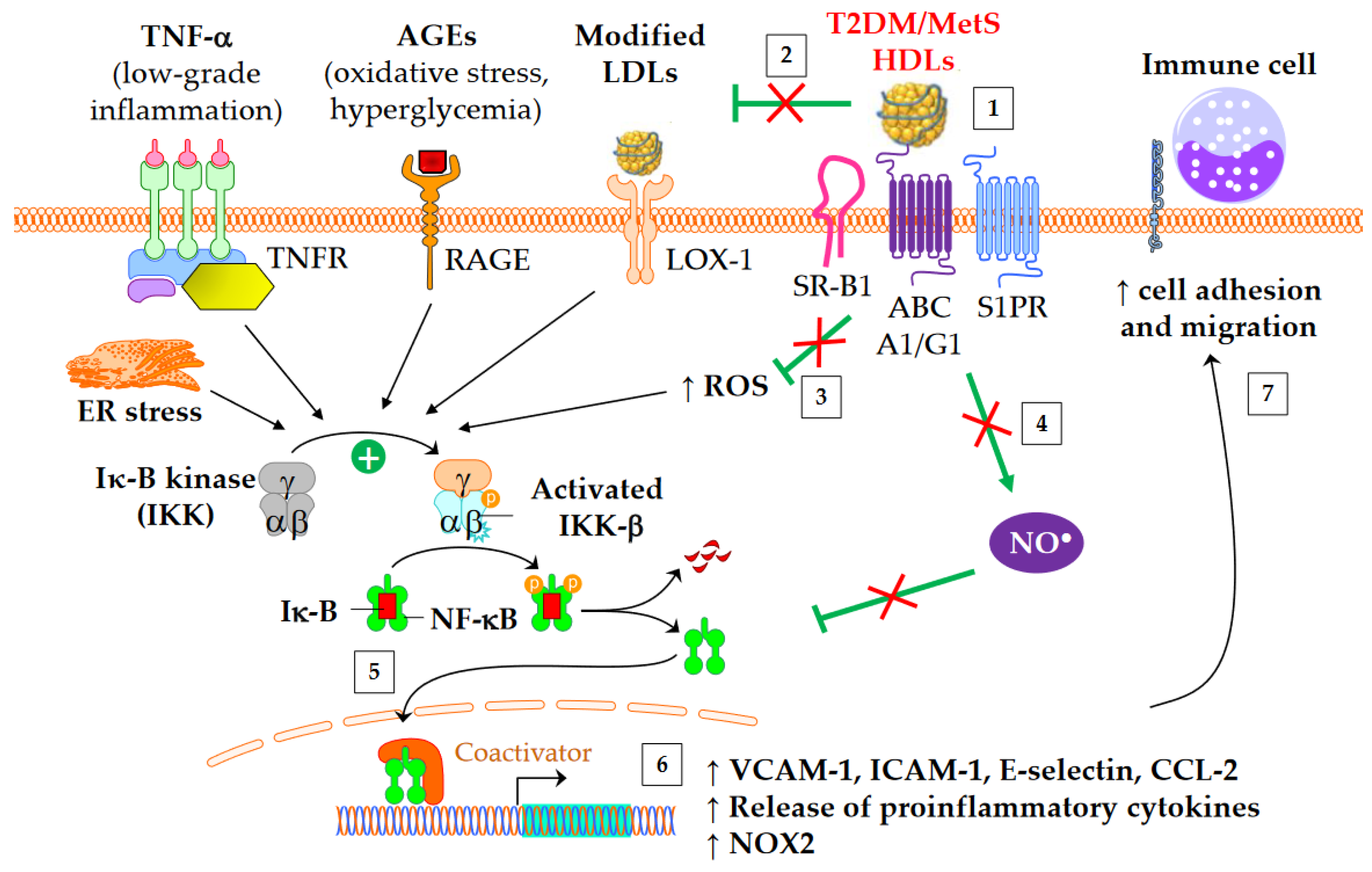
Both diabetes and obesity are associated with low-grade inflammation, which substantially contributes to endothelial dysfunction and atherosclerosis. As shown in Figure 2, HDL particles exert an anti-inflammatory function by downregulating the expression of molecules involved in the recruitment of immune cells into the subendothelial space. These molecules include chemokine CCL-2 [62], vascular cell adhesion molecule (VCAM)-1, intracellular adhesion molecule (ICAM)-1, and selectin-E [11]. In addition, HDLs inhibit the release of inflammatory cytokines, such as TNF-α and IL-1β. The HDL anti-inflammatory function seems of particular relevance for CV outcomes, since it predicts new cardiac events in patients with myocardial infarction, independently of HDL-C [63]. Moreover, an inverse association between the anti-inflammatory capacity of HDLs and incident CV events was recently observed in a study of individuals from the general population cohort, independently of both HDL-C and CEC [64].
Figure 2. Anti-inflammatory functions of HDLs in T2DM and MetS. ↑ means increase. The inflammatory NF-κB pathway is triggered by several mediators, including TNF-α, advanced glycation end-products (AGEs), modified LDLs, reactive oxygen species (ROS), and endoplasmic reticulum (ER) stress. This leads to an increased gene expression of adhesion molecules, proinflammatory cytokines, and NADPH oxidase (NOX2). Green arrows represent the functions of healthy HDLs. (1) The binding of HDLs to receptors is modified by conformational changes in HDL particles in insulin resistant conditions. The depletion in S1P of MetS HDLs likely decreases the binding to S1P receptors (S1PR). (2) HDL-mediated protection of LDLs against oxidation is affected, (3) in particular due to the loss of capacity of HDLs to dampen ROS production. This promotes the NF-κB activation triggered by the recognition of oxidized LDLs by scavenger receptors in vasculature, particularly by LOX-1 (i.e., SR-E1) and SR-A1. (4) The activation of endothelial NO synthase by HDLs is reduced (see 4.3) and subsequently inhibits nitrosylation of NF-κB. (5) Ultimately, HDLs are less able to inhibit the translocation of NF-κB into the nucleus, and, (6) afterwards, the gene expression of adhesion molecules and proinflammatory cytokines. (7) This facilitates the recruitment of immune cells into the subendothelial space.
HDLs from T2DM patients are less able to inhibit the migration of monocytes towards endothelial cells [32][65]. Interestingly, this loss of function correlates with plasma SAA [65] and carbamylated HDL levels [32]. The fact that in vitro carbamylation of HDLs reproduces the loss of capacity to inhibit the migration of monocytes reinforces the potential role of carbamylated HDLs [32]. Although HbA1c or glucose levels do not correlate with this loss of HDL function [65], some evidence suggests that glycoxidative changes in HDLs may play a role. Thus, the ex vivo treatment of plasma with L-4F, an apoA-I mimetic peptide able to bind oxidized lipids with a higher affinity than apoA-I itself [66], restores the anti-inflammatory function of HDLs [65]
The activation of the classical IKK/IκB-α/NF-κB pathway plays a crucial role in the expression of adhesion molecules and in the release of proinflammatory cytokines into cardiovascular tissue. HDLs/apoA-I inhibit the NF-κB pathway via several overlapped mechanisms following interaction with SR-B1, S1PR, ABCA1, and ABCG1: cholesterol efflux, endothelial nitric oxide synthase (eNOS) activation [67] and subsequent S-nitrosylation [68], upregulation of 3β-hydroxysteroid-δ24 reductase [69][70] and heme oxygenase-1 [70], and inhibition of the toll-like receptors TLR4 [71] and TLR2 [72]. HDLs or apoA-I ultimately prevent NF-κB p65 subunit translocation to the nucleus and DNA binding [71], thus inhibiting the transcription of adhesion molecules, CCL-2, proinflammatory cytokines, and also NADPH-oxidase (NOX2) genes. It has been shown that HDLs isolated from T2DM patients are unable to suppress the activating phosphorylation of the NF-κB p65 subunit in endothelial cells [73]. Glycated apoA-I partially loses its ability to inhibit cytosolic IκB-α phosphorylation and NF-κB p65 subunit translocation to the nucleus [22].

3.3. Antioxidative Properties

T2DM and obesity are known to be associated with increased oxidative stress, which is closely linked to low-grade inflammation. In particular, oxidative stress in artery walls is responsible for the formation of oxidized LDLs (oxLDLs), rendering them more atherogenic. There are several mechanisms through which HDLs present in the intima can protect LDLs against oxidation [74]. HDLs directly protect LDLs from oxidation induced by one-electron oxidants (free radicals), and they are also able to remove oxidized lipids from LDLs. These activities can decrease local concentrations of oxLDLs. The antioxidative potential of HDL particles originates both from the activities of their proteins and from lipid components. Different HDL-associated apolipoproteins, lipid transfer proteins, and enzymes have been shown to contribute to the antioxidative capacity of HDLs.

3.4. Nitric Oxide Production

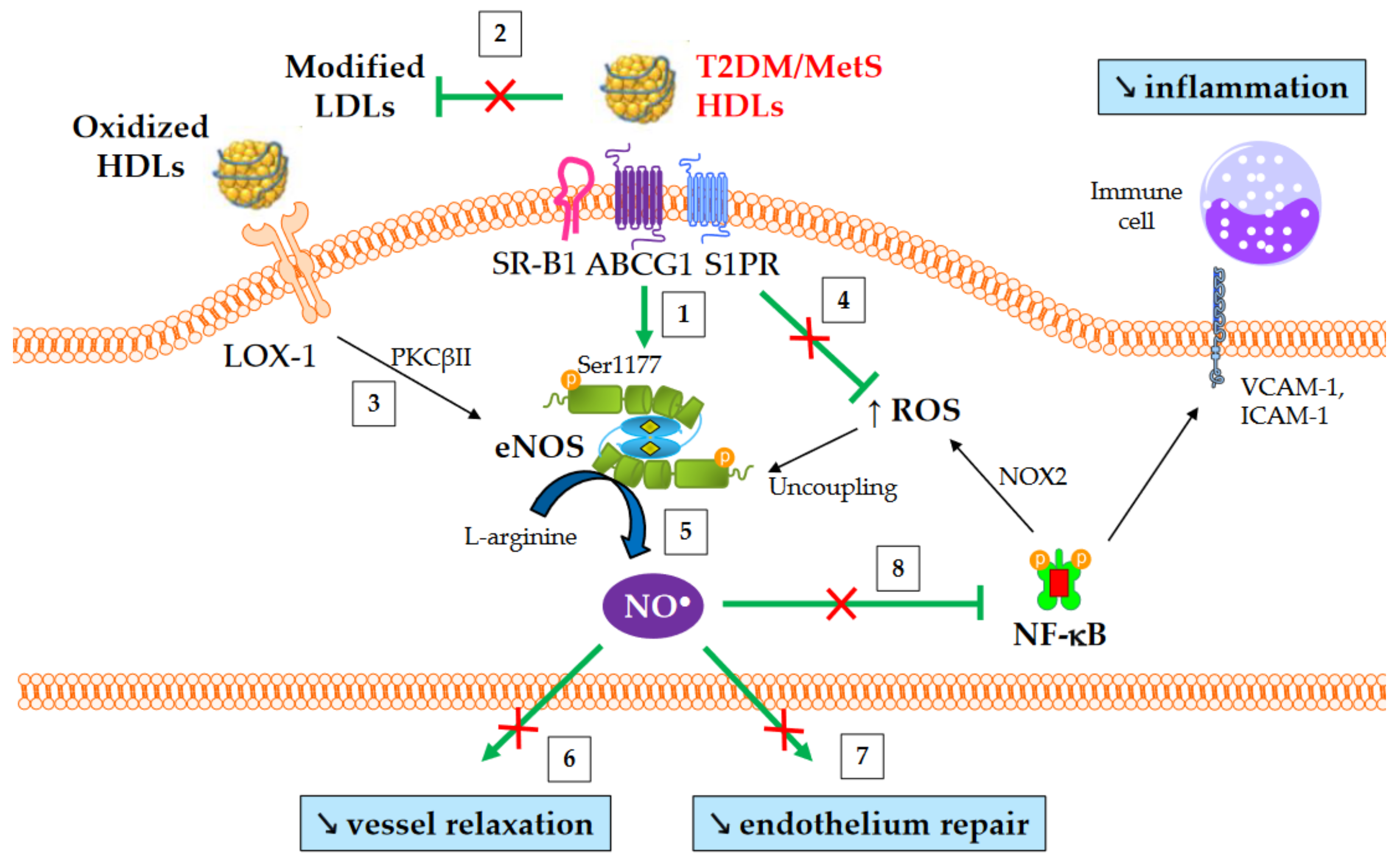
The production of nitric oxide (NO) is important for normal endothelial function and protects against endothelial dysfunction, an early hallmark of atherosclerosis. HDLs improve the bioavailability of NO in vasculature mainly by inducing the activating-phosphorylation of eNOS at serine 1177, which then promotes NO synthesis. NO production contributes to a number HDL’s beneficial effects, such as vasorelaxation, inhibition of NF-κB pathway, and endothelium repair [75]. Several mechanisms are involved in eNOS activation mediated by HDLs. These include the binding of HDL-apoA-I to SR-B1 [76] and ABCG1 [77], the binding of HDL-S1P to S1PR1/3 receptors [78], the activation of mitogen-activated protein (MAP) kinases [79], the inhibition of protein kinase C (PKC) βII [67], cholesterol efflux facilitating the dissociation of eNOS from caveolin-1 [77], and, lastly, the suppression of ROS production which preserves from eNOS uncoupling.
An overview of NO-mediated HDL functions in T2DM and MetS is presented in Figure 3. HDLs from T2DM patients have been demonstrated to be less able to induce the activating-phosphorylation of eNOS at serine 1177 [73], NO production [27], and vessel relaxation [27]. Interestingly, eNOS phosphorylation and activity are even affected in obese patients in the absence of diabetes [80].
Figure 3. NO-mediated HDL functions in T2DM and MetS. ↑ and ↘ mean increase and decrease, respectively. Green arrows represent the functions of healthy HDLs. (1) HDLs from T2DM and non-diabetic MetS individuals are less able to induce Akt-dependent eNOS phosphorylation at Ser1177. (2) HDL-mediated protection of LDLs against oxidation is affected, thus facilitating eNOS inhibition after the binding of modified LDLs to LOX-1. (3) Oxidized HDLs are also able to bind to LOX-1 receptor, leading to PKCβII activation and subsequently to eNOS inhibition. (4) The loss of capacity of HDLs to dampen ROS production promotes eNOS uncoupling. (5) Ultimately, HDL-mediated NO production is reduced, (6) affecting the relaxation of vascular smooth muscle cells and (7) endothelium repair. (8) Reduced NO synthesis diminishes the inhibition of nitrosylation of NF-κB, as well as NOX2-mediated ROS production and the recruitment of immune cells into the subendothelial space.

3.5. Antiapoptotic Properties and Endothelium Repair

The integrity of endothelial cells is crucial for vascular homeostasis, and endothelial cell death triggers vascular damage and promotes inflammation and endothelial dysfunction. HDL particles can inhibit apoptosis in endothelial cells, thus preserving endothelium integrity [81][82][83].
The antiapoptotic activity of small HDL particles is reduced in MetS individuals [84][85], and is closely associated with altered physicochemical properties, such as core CE depletion and TG enrichment in apoA-I-poor HDL3c [84].
The depletion of HDLs in plasmalogens in T2DM and MetS may also play a role, since enrichment of rHDLs with plasmalogens enhances their antiapoptotic activity [81]. Changes in the HDL-apoM/S1P axis are also likely to be involved considering its important role in HDL antiapoptotic activity [82][86].

4. Antidiabetic Properties

Emerging data suggest that HDL particles could actually contribute to the development of diabetes. Firstly, HDL-C levels are inversely associated with T2DM development in epidemiological studies [87][88][89], and this metric has been included in scores for T2DM risk [89][90]. In addition, Mendelian randomization studies showed that HDL-C elevation is associated with a lower risk of developing T2DM [91][92]. Moreover, both HDL particle size and the concentration of large HDL particles were inversely associated with incident T2DM in the general population [4]. Moreover, CETP inhibitors, which increased HDL-C concentration of 29 to 132% in large interventional studies, reduced the risk of new-onset diabetes by 16% on average [93].
Many of the antidiabetic mechanisms induced by HDLs or apoA-I have now been identified [94]. Infusions of rHDLs and apoA-I stimulate insulin secretion and reduce plasma glucose concentrations in obese mice and in T2DM patients [95][96]. From a mechanistic point of view, apoA-I enhances the expression of key enzymes involved in insulin maturation in β-cells [97]. In addition, HDLs protect β-cells from apoptosis induced by endoplasmic reticulum stressors [98].

This entry is adapted from the peer-reviewed paper 10.3390/metabo13020253

References

  1. Apro, J.; Tietge, U.J.F.; Dikkers, A.; Parini, P.; Angelin, B.; Rudling, M. Impaired Cholesterol Efflux Capacity of High-Density Lipoprotein Isolated from Interstitial Fluid in Type 2 Diabetes Mellitus-Brief Report. Arterioscler. Thromb. Vasc. Biol. 2016, 36, 787–791.
  2. Ståhlman, M.; Fagerberg, B.; Adiels, M.; Ekroos, K.; Chapman, J.M.; Kontush, A.; Borén, J. Dyslipidemia, but Not Hyperglycemia and Insulin Resistance, Is Associated with Marked Alterations in the HDL Lipidome in Type 2 Diabetic Subjects in the DIWA Cohort: Impact on Small HDL Particles. Biochim. Biophys. Acta 2013, 1831, 1609–1617.
  3. Mora, S.; Otvos, J.D.; Rosenson, R.S.; Pradhan, A.; Buring, J.E.; Ridker, P.M. Lipoprotein Particle Size and Concentration by Nuclear Magnetic Resonance and Incident Type 2 Diabetes in Women. Diabetes 2010, 59, 1153–1160.
  4. Sokooti, S.; Flores-Guerrero, J.L.; Kieneker, L.M.; Heerspink, H.J.L.; Connelly, M.A.; Bakker, S.J.L.; Dullaart, R.P.F. HDL Particle Subspecies and Their Association with Incident Type 2 Diabetes: The PREVEND Study. J. Clin. Endocrinol. Metab. 2021, 106, 1761–1772.
  5. Cardner, M.; Yalcinkaya, M.; Goetze, S.; Luca, E.; Balaz, M.; Hunjadi, M.; Hartung, J.; Shemet, A.; Kränkel, N.; Radosavljevic, S.; et al. Structure-Function Relationships of HDL in Diabetes and Coronary Heart Disease. JCI Insight 2020, 5, e131491.
  6. Davidson, W.S.; Heink, A.; Sexmith, H.; Dolan, L.M.; Gordon, S.M.; Otvos, J.D.; Melchior, J.T.; Elder, D.A.; Khoury, J.; Geh, E.; et al. Obesity Is Associated with an Altered HDL Subspecies Profile among Adolescents with Metabolic Disease. J. Lipid Res. 2017, 58, 1916–1923.
  7. Garvey, W.T.; Kwon, S.; Zheng, D.; Shaughnessy, S.; Wallace, P.; Hutto, A.; Pugh, K.; Jenkins, A.J.; Klein, R.L.; Liao, Y. Effects of Insulin Resistance and Type 2 Diabetes on Lipoprotein Subclass Particle Size and Concentration Determined by Nuclear Magnetic Resonance. Diabetes 2003, 52, 453–462.
  8. Festa, A.; Williams, K.; Hanley, A.J.G.; Otvos, J.D.; Goff, D.C.; Wagenknecht, L.E.; Haffner, S.M. Nuclear Magnetic Resonance Lipoprotein Abnormalities in Prediabetic Subjects in the Insulin Resistance Atherosclerosis Study. Circulation 2005, 111, 3465–3472.
  9. Vergès, B.; Florentin, E.; Baillot-Rudoni, S.; Petit, J.-M.; Brindisi, M.C.; Pais de Barros, J.-P.; Lagrost, L.; Gambert, P.; Duvillard, L. Rosuvastatin 20 Mg Restores Normal HDL-ApoA-I Kinetics in Type 2 Diabetes. J. Lipid Res. 2009, 50, 1209–1215.
  10. Kostara, C.E.; Karakitsou, K.S.; Florentin, M.; Bairaktari, E.T.; Tsimihodimos, V. Progressive, Qualitative, and Quantitative Alterations in HDL Lipidome from Healthy Subjects to Patients with Prediabetes and Type 2 Diabetes. Metabolites 2022, 12, 683.
  11. Denimal, D.; Benanaya, S.; Monier, S.; Simoneau, I.; Pais de Barros, J.-P.; Le Goff, W.; Bouillet, B.; Vergès, B.; Duvillard, L. Normal HDL Cholesterol Efflux and Anti-Inflammatory Capacities in Type 2 Diabetes despite Lipidomic Abnormalities. J. Clin. Endocrinol. Metab. 2022, 107, e3816–e3823.
  12. Mocciaro, G.; D’Amore, S.; Jenkins, B.; Kay, R.; Murgia, A.; Herrera-Marcos, L.V.; Neun, S.; Sowton, A.P.; Hall, Z.; Palma-Duran, S.A.; et al. Lipidomic Approaches to Study HDL Metabolism in Patients with Central Obesity Diagnosed with Metabolic Syndrome. IJMS 2022, 23, 6786.
  13. Duvillard, L.; Florentin, E.; Pont, F.; Petit, J.-M.; Baillot-Rudoni, S.; Penfornis, A.; Vergès, B. Chronic Hyperinsulinemia Does Not Increase the Production Rate of High-Density Lipoprotein Apolipoprotein AI: Evidence from a Kinetic Study in Patients with Insulinoma. Arterioscler. Thromb. Vasc. Biol. 2013, 33, 2460–2465.
  14. Vergès, B.; Petit, J.M.; Duvillard, L.; Dautin, G.; Florentin, E.; Galland, F.; Gambert, P. Adiponectin Is an Important Determinant of ApoA-I Catabolism. Arterioscler. Thromb. Vasc. Biol. 2006, 26, 1364–1369.
  15. Curtiss, L.K.; Bonnet, D.J.; Rye, K.A. The Conformation of Apolipoprotein A-I in High-Density Lipoproteins Is Influenced by Core Lipid Composition and Particle Size: A Surface Plasmon Resonance Study. Biochemistry 2000, 39, 5712–5721.
  16. HDL Proteome Watch Page. Available online: https://homepages.uc.edu/~davidswm/HDLproteome.html (accessed on 25 November 2022).
  17. Holzer, M.; Ljubojevic-Holzer, S.; Souza Junior, D.R.; Stadler, J.T.; Rani, A.; Scharnagl, H.; Ronsein, G.E.; Marsche, G. HDL Isolated by Immunoaffinity, Ultracentrifugation, or Precipitation Is Compositionally and Functionally Distinct. J. Lipid Res. 2022, 63, 100307.
  18. Zhao, D.; Yang, L.-Y.; Wang, X.-H.; Yuan, S.-S.; Yu, C.-G.; Wang, Z.-W.; Lang, J.-N.; Feng, Y.-M. Different Relationship between ANGPTL3 and HDL Components in Female Non-Diabetic Subjects and Type-2 Diabetic Patients. Cardiovasc. Diabetol. 2016, 15, 132.
  19. Tsun, J.G.S.; Shiu, S.W.M.; Wong, Y.; Yung, S.; Chan, T.M.; Tan, K.C.B. Impact of Serum Amyloid A on Cellular Cholesterol Efflux to Serum in Type 2 Diabetes Mellitus. Atherosclerosis 2013, 231, 405–410.
  20. Annema, W.; Nijstad, N.; Tölle, M.; de Boer, J.F.; Buijs, R.V.C.; Heeringa, P.; van der Giet, M.; Tietge, U.J.F. Myeloperoxidase and Serum Amyloid A Contribute to Impaired in Vivo Reverse Cholesterol Transport during the Acute Phase Response but Not Group IIA Secretory Phospholipase A(2). J. Lipid Res. 2010, 51, 743–754.
  21. Brownlee, M. Biochemistry and Molecular Cell Biology of Diabetic Complications. Nature 2001, 414, 813–820.
  22. Liu, D.; Ji, L.; Zhao, M.; Wang, Y.; Guo, Y.; Li, L.; Zhang, D.; Xu, L.; Pan, B.; Su, J.; et al. Lysine Glycation of Apolipoprotein A-I Impairs Its Anti-Inflammatory Function in Type 2 Diabetes Mellitus. J. Mol. Cell. Cardiol. 2018, 122, 47–57.
  23. Godfrey, L.; Yamada-Fowler, N.; Smith, J.; Thornalley, P.J.; Rabbani, N. Arginine-Directed Glycation and Decreased HDL Plasma Concentration and Functionality. Nutr. Diabetes 2014, 4, e134.
  24. Aroor, A.R.; DeMarco, V.G. Oxidative Stress and Obesity: The Chicken or the Egg? Diabetes 2014, 63, 2216–2218.
  25. Marin, M.T.; Dasari, P.S.; Tryggestad, J.B.; Aston, C.E.; Teague, A.M.; Short, K.R. Oxidized HDL and LDL in Adolescents with Type 2 Diabetes Compared to Normal Weight and Obese Peers. J. Diabetes Complicat. 2015, 29, 679–685.
  26. Sartore, G.; Seraglia, R.; Burlina, S.; Bolis, A.; Marin, R.; Manzato, E.; Ragazzi, E.; Traldi, P.; Lapolla, A. High-Density Lipoprotein Oxidation in Type 2 Diabetic Patients and Young Patients with Premature Myocardial Infarction. Nutr. Metab. Cardiovasc. Dis. 2015, 25, 418–425.
  27. Sorrentino, S.A.; Besler, C.; Rohrer, L.; Meyer, M.; Heinrich, K.; Bahlmann, F.H.; Mueller, M.; Horváth, T.; Doerries, C.; Heinemann, M.; et al. Endothelial-Vasoprotective Effects of High-Density Lipoprotein Are Impaired in Patients with Type 2 Diabetes Mellitus but Are Improved after Extended-Release Niacin Therapy. Circulation 2010, 121, 110–122.
  28. Feng, J.; Wang, Y.; Li, W.; Zhao, Y.; Liu, Y.; Yao, X.; Liu, S.; Yu, P.; Li, R. High Levels of Oxidized Fatty Acids in HDL Impair the Antioxidant Function of HDL in Patients with Diabetes. Front. Endocrinol. 2022, 13, 993193.
  29. Wiersma, J.J.; Meuwese, M.C.; van Miert, J.N.I.; Kastelein, A.; Tijssen, J.G.P.; Piek, J.J.; Trip, M.D. Diabetes Mellitus Type 2 Is Associated with Higher Levels of Myeloperoxidase. Med. Sci. Monit. 2008, 14, CR406–CR410.
  30. Shiu, S.W.M.; Xiao, S.-M.; Wong, Y.; Chow, W.-S.; Lam, K.S.L.; Tan, K.C.B. Carbamylation of LDL and Its Relationship with Myeloperoxidase in Type 2 Diabetes Mellitus. Clin. Sci. 2013, 126, 175–181.
  31. Tan, K.C.B.; Cheung, C.-L.; Lee, A.C.H.; Lam, J.K.Y.; Wong, Y.; Shiu, S.W.M. Carbamylated Lipoproteins and Progression of Diabetic Kidney Disease. Clin. J. Am. Soc. Nephrol. 2020, 15, 359–366.
  32. Chen, Z.; Ding, S.; Wang, Y.P.; Chen, L.; Mao, J.Y.; Yang, Y.; Sun, J.T.; Yang, K. Association of Carbamylated High-Density Lipoprotein with Coronary Artery Disease in Type 2 Diabetes Mellitus: Carbamylated High-Density Lipoprotein of Patients Promotes Monocyte Adhesion. J. Transl. Med. 2020, 18, 460.
  33. Kheniser, K.G.; Osme, A.; Kim, C.; Ilchenko, S.; Kasumov, T.; Kashyap, S.R. Temporal Dynamics of High-Density Lipoprotein Proteome in Diet-Controlled Subjects with Type 2 Diabetes. Biomolecules 2020, 10, 520.
  34. Duvillard, L.; Pont, F.; Florentin, E.; Gambert, P.; Vergès, B. Inefficiency of Insulin Therapy to Correct Apolipoprotein A-I Metabolic Abnormalities in Non-Insulin-Dependent Diabetes Mellitus. Atherosclerosis 2000, 152, 229–237.
  35. Frénais, R.; Ouguerram, K.; Maugeais, C.; Mahot, P.; Maugère, P.; Krempf, M.; Magot, T. High Density Lipoprotein Apolipoprotein AI Kinetics in NIDDM: A Stable Isotope Study. Diabetologia 1997, 40, 578–583.
  36. Kashyap, S.R.; Osme, A.; Ilchenko, S.; Golizeh, M.; Lee, K.; Wang, S.; Bena, J.; Previs, S.F.; Smith, J.D.; Kasumov, T. Glycation Reduces the Stability of ApoAI and Increases HDL Dysfunction in Diet-Controlled Type 2 Diabetes. J. Clin. Endocrinol. Metab. 2018, 103, 388–396.
  37. Pont, F.; Duvillard, L.; Florentin, E.; Gambert, P.; Vergès, B. High-Density Lipoprotein Apolipoprotein A-I Kinetics in Obese Insulin Resistant Patients. An in Vivo Stable Isotope Study. Int. J. Obes. Relat. Metab. Disord. 2002, 26, 1151–1158.
  38. Chan, D.C.; Barrett, P.H.R.; Ooi, E.M.M.; Ji, J.; Chan, D.T.; Watts, G.F. Very Low Density Lipoprotein Metabolism and Plasma Adiponectin as Predictors of High-Density Lipoprotein Apolipoprotein A-I Kinetics in Obese and Nonobese Men. J. Clin. Endocrinol. Metab. 2009, 94, 989–997.
  39. Vergès, B.; Adiels, M.; Boren, J.; Barrett, P.H.; Watts, G.F.; Chan, D.; Duvillard, L.; Söderlund, S.; Matikainen, N.; Kahri, J.; et al. Interrelationships between the Kinetics of VLDL Subspecies and HDL Catabolism in Abdominal Obesity: A Multicenter Tracer Kinetic Study. J. Clin. Endocrinol. Metab. 2014, 99, 4281–4290.
  40. Sparks, D.L.; Davidson, W.S.; Lund-Katz, S.; Phillips, M.C. Effects of the Neutral Lipid Content of High Density Lipoprotein on Apolipoprotein A-I Structure and Particle Stability. J. Biol. Chem. 1995, 270, 26910–26917.
  41. Horowitz, B.S.; Goldberg, I.J.; Merab, J.; Vanni, T.M.; Ramakrishnan, R.; Ginsberg, H.N. Increased Plasma and Renal Clearance of an Exchangeable Pool of Apolipoprotein A-I in Subjects with Low Levels of High Density Lipoprotein Cholesterol. J. Clin. Investig. 1993, 91, 1743–1752.
  42. Garçon, D.; Berger, J.-M.; Cariou, B.; Le May, C. Transintestinal Cholesterol Excretion in Health and Disease. Curr. Atheroscler. Rep. 2022, 24, 153–160.
  43. Rohatgi, A.; Khera, A.; Berry, J.D.; Givens, E.G.; Ayers, C.R.; Wedin, K.E.; Neeland, I.J.; Yuhanna, I.S.; Rader, D.R.; de Lemos, J.A.; et al. HDL Cholesterol Efflux Capacity and Incident Cardiovascular Events. N. Engl. J. Med. 2014, 371, 2383–2393.
  44. Ebtehaj, S.; Gruppen, E.G.; Bakker, S.J.L.; Dullaart, R.P.F.; Tietge, U.J.F. HDL (High-Density Lipoprotein) Cholesterol Efflux Capacity Is Associated with Incident Cardiovascular Disease in the General Population. Arterioscler. Thromb. Vasc. Biol. 2019, 39, 1874–1883.
  45. Low, H.; Hoang, A.; Forbes, J.; Thomas, M.; Lyons, J.G.; Nestel, P.; Bach, L.A.; Sviridov, D. Advanced Glycation End-Products (AGEs) and Functionality of Reverse Cholesterol Transport in Patients with Type 2 Diabetes and in Mouse Models. Diabetologia 2012, 55, 2513–2521.
  46. De Vries, R.; Groen, A.K.; Perton, F.G.; Dallinga-Thie, G.M.; van Wijland, M.J.A.; Dikkeschei, L.D.; Wolffenbuttel, B.H.R.; van Tol, A.; Dullaart, R.P.F. Increased Cholesterol Efflux from Cultured Fibroblasts to Plasma from Hypertriglyceridemic Type 2 Diabetic Patients: Roles of Pre β-HDL, Phospholipid Transfer Protein and Cholesterol Esterification. Atherosclerosis 2008, 196, 733–741.
  47. Yassine, H.N.; Belopolskaya, A.; Schall, C.; Stump, C.S.; Lau, S.S.; Reaven, P.D. Enhanced Cholesterol Efflux to HDL through the ABCA1 Transporter in Hypertriglyceridemia of Type 2 Diabetes. Metabolism 2014, 63, 727–734.
  48. Dullaart, R.P.F.; Pagano, S.; Perton, F.G.; Vuilleumier, N. Antibodies Against the C-Terminus of ApoA-1 Are Inversely Associated with Cholesterol Efflux Capacity and HDL Metabolism in Subjects with and without Type 2 Diabetes Mellitus. Int. J. Mol. Sci. 2019, 20, 732.
  49. Plomgaard, P.; Dullaart, R.P.F.; de Vries, R.; Groen, A.K.; Dahlbäck, B.; Nielsen, L.B. Apolipoprotein M Predicts Pre-Beta-HDL Formation: Studies in Type 2 Diabetic and Nondiabetic Subjects. J. Intern. Med. 2009, 266, 258–267.
  50. Cavallero, E.; Brites, F.; Delfly, B.; Nicolaïew, N.; Decossin, C.; De Geitere, C.; Fruchart, J.C.; Wikinski, R.; Jacotot, B.; Castro, G. Abnormal Reverse Cholesterol Transport in Controlled Type II Diabetic Patients. Studies on Fasting and Postprandial LpA-I Particles. Arterioscler. Thromb. Vasc. Biol. 1995, 15, 2130–2135.
  51. Shiu, S.W.; Zhou, H.; Wong, Y.; Tan, K.C. Endothelial Lipase and Reverse Cholesterol Transport in Type 2 Diabetes Mellitus. J. Diabetes Investig. 2010, 1, 111–116.
  52. Shiu, S.W.; Wong, Y.; Tan, K.C. Pre-Β1 HDL in Type 2 Diabetes Mellitus. Atherosclerosis 2017, 263, 24–28.
  53. Zhou, H.; Tan, K.C.B.; Shiu, S.W.M.; Wong, Y. Cellular Cholesterol Efflux to Serum Is Impaired in Diabetic Nephropathy. Diabetes Metab. Res. Rev. 2008, 24, 617–623.
  54. Syvänne, M.; Castro, G.; Dengremont, C.; De Geitere, C.; Jauhiainen, M.; Ehnholm, C.; Michelagnoli, S.; Franceschini, G.; Kahri, J.; Taskinen, M.R. Cholesterol Efflux from Fu5AH Hepatoma Cells Induced by Plasma of Subjects with or without Coronary Artery Disease and Non-Insulin-Dependent Diabetes: Importance of LpA-I:A-II Particles and Phospholipid Transfer Protein. Atherosclerosis 1996, 127, 245–253.
  55. Zhou, H.; Shiu, S.W.; Wong, Y.; Tan, K.C. Impaired Serum Capacity to Induce Cholesterol Efflux Is Associated with Endothelial Dysfunction in Type 2 Diabetes Mellitus. Diabetes Vasc. Dis. Res. 2009, 6, 238–243.
  56. Passarelli, M.; Shimabukuro, A.F.; Catanozi, S.; Nakandakare, E.R.; Rocha, J.C.; Carrilho, A.J.; Quintão, E.C. Diminished Rate of Mouse Peritoneal Macrophage Cholesterol Efflux Is Not Related to the Degree of HDL Glycation in Diabetes Mellitus. Clin. Chim. Acta 2000, 301, 119–134.
  57. Murakami, H.; Tanabe, J.; Tamasawa, N.; Matsumura, K.; Yamashita, M.; Matsuki, K.; Murakami, H.; Matsui, J.; Suda, T. Reduction of Paraoxonase-1 Activity May Contribute the Qualitative Impairment of HDL Particles in Patients with Type 2 Diabetes. Diabetes Res. Clin. Pract. 2013, 99, 30–38.
  58. He, Y.; Ronsein, G.E.; Tang, C.; Jarvik, G.P.; Davidson, W.S.; Kothari, V.; Song, H.D.; Segrest, J.P.; Bornfeldt, K.E.; Heinecke, J.W. Diabetes Impairs Cellular Cholesterol Efflux From ABCA1 to Small HDL Particles. Circ. Res. 2020, 127, 1198–1210.
  59. Agarwala, A.P.; Rodrigues, A.; Risman, M.; McCoy, M.; Trindade, K.; Qu, L.; Cuchel, M.; Billheimer, J.; Rader, D.J. High-Density Lipoprotein (HDL) Phospholipid Content and Cholesterol Efflux Capacity Are Reduced in Patients With Very High HDL Cholesterol and Coronary Disease. Arterioscler. Thromb. Vasc. Biol. 2015, 35, 1515–1519.
  60. Yancey, P.G.; Kawashiri, M.; Moore, R.; Glick, J.M.; Williams, D.L.; Connelly, M.A.; Rader, D.J.; Rothblat, G.H. In Vivo Modulation of HDL Phospholipid Has Opposing Effects on SR-BI- and ABCA1-Mediated Cholesterol Efflux. J. Lipid Res. 2004, 45, 337–346.
  61. Subbaiah, P.V.; Gesquiere, L.R.; Wang, K. Regulation of the Selective Uptake of Cholesteryl Esters from High Density Lipoproteins by Sphingomyelin. J. Lipid Res. 2005, 46, 2699–2705.
  62. Tölle, M.; Pawlak, A.; Schuchardt, M.; Kawamura, A.; Tietge, U.J.; Lorkowski, S.; Keul, P.; Assmann, G.; Chun, J.; Levkau, B.; et al. HDL-Associated Lysosphingolipids Inhibit NAD(P)H Oxidase-Dependent Monocyte Chemoattractant Protein-1 Production. Arterioscler. Thromb. Vasc. Biol. 2008, 28, 1542–1548.
  63. Dullaart, R.P.F.; Annema, W.; Tio, R.A.; Tietge, U.J.F. The HDL Anti-Inflammatory Function Is Impaired in Myocardial Infarction and May Predict New Cardiac Events Independent of HDL Cholesterol. Clin. Chim. Acta 2014, 433, 34–38.
  64. Jia, C.; Anderson, J.L.C.; Gruppen, E.G.; Lei, Y.; Bakker, S.J.L.; Dullaart, R.P.F.; Tietge, U.J.F. High-Density Lipoprotein Anti-Inflammatory Capacity and Incident Cardiovascular Events. Circulation 2021, 143, 1935–1945.
  65. Morgantini, C.; Natali, A.; Boldrini, B.; Imaizumi, S.; Navab, M.; Fogelman, A.M.; Ferrannini, E.; Reddy, S.T. Anti-Inflammatory and Antioxidant Properties of HDLs Are Impaired in Type 2 Diabetes. Diabetes 2011, 60, 2617–2623.
  66. Van Lenten, B.J.; Wagner, A.C.; Jung, C.-L.; Ruchala, P.; Waring, A.J.; Lehrer, R.I.; Watson, A.D.; Hama, S.; Navab, M.; Anantharamaiah, G.M.; et al. Anti-Inflammatory ApoA-I-Mimetic Peptides Bind Oxidized Lipids with Much Higher Affinity than Human ApoA-I. J. Lipid Res. 2008, 49, 2302–2311.
  67. Besler, C.; Heinrich, K.; Rohrer, L.; Doerries, C.; Riwanto, M.; Shih, D.M.; Chroni, A.; Yonekawa, K.; Stein, S.; Schaefer, N.; et al. Mechanisms Underlying Adverse Effects of HDL on ENOS-Activating Pathways in Patients with Coronary Artery Disease. J. Clin. Investig. 2011, 121, 2693–2708.
  68. Marshall, H.E.; Stamler, J.S. Inhibition of NF-Kappa B by S-Nitrosylation. Biochemistry 2001, 40, 1688–1693.
  69. McGrath, K.C.Y.; Li, X.H.; Puranik, R.; Liong, E.C.; Tan, J.T.M.; Dy, V.M.; DiBartolo, B.A.; Barter, P.J.; Rye, K.A.; Heather, A.K. Role of 3beta-Hydroxysteroid-Delta 24 Reductase in Mediating Antiinflammatory Effects of High-Density Lipoproteins in Endothelial Cells. Arterioscler. Thromb. Vasc. Biol. 2009, 29, 877–882.
  70. Wu, B.J.; Chen, K.; Shrestha, S.; Ong, K.L.; Barter, P.J.; Rye, K.-A. High-Density Lipoproteins Inhibit Vascular Endothelial Inflammation by Increasing 3β-Hydroxysteroid-Δ24 Reductase Expression and Inducing Heme Oxygenase-1. Circ. Res. 2013, 112, 278–288.
  71. Cheng, A.M.; Handa, P.; Tateya, S.; Schwartz, J.; Tang, C.; Mitra, P.; Oram, J.F.; Chait, A.; Kim, F. Apolipoprotein A-I Attenuates Palmitate-Mediated NF-ΚB Activation by Reducing Toll-like Receptor-4 Recruitment into Lipid Rafts. PLoS ONE 2012, 7, e33917.
  72. Dueñas, A.I.; Aceves, M.; Fernández-Pisonero, I.; Gómez, C.; Orduña, A.; Crespo, M.S.; García-Rodríguez, C. Selective Attenuation of Toll-like Receptor 2 Signalling May Explain the Atheroprotective Effect of Sphingosine 1-Phosphate. Cardiovasc. Res. 2008, 79, 537–544.
  73. Vaisar, T.; Couzens, E.; Hwang, A.; Russell, M.; Barlow, C.E.; DeFina, L.F.; Hoofnagle, A.N.; Kim, F. Type 2 Diabetes Is Associated with Loss of HDL Endothelium Protective Functions. PLoS ONE 2018, 13, e0192616.
  74. Brites, F.; Martin, M.; Guillas, I.; Kontush, A. Antioxidative Activity of High-Density Lipoprotein (HDL): Mechanistic Insights into Potential Clinical Benefit. BBA Clin. 2017, 8, 66–77.
  75. Iwakiri, Y. S-Nitrosylation of Proteins: A New Insight into Endothelial Cell Function Regulated by ENOS-Derived NO. Nitric. Oxide 2011, 25, 95–101.
  76. Yuhanna, I.S.; Zhu, Y.; Cox, B.E.; Hahner, L.D.; Osborne-Lawrence, S.; Lu, P.; Marcel, Y.L.; Anderson, R.G.; Mendelsohn, M.E.; Hobbs, H.H.; et al. High-Density Lipoprotein Binding to Scavenger Receptor-BI Activates Endothelial Nitric Oxide Synthase. Nat. Med. 2001, 7, 853–857.
  77. Terasaka, N.; Westerterp, M.; Koetsveld, J.; Fernández-Hernando, C.; Yvan-Charvet, L.; Wang, N.; Sessa, W.C.; Tall, A.R. ATP-Binding Cassette Transporter G1 and High-Density Lipoprotein Promote Endothelial NO Synthesis through a Decrease in the Interaction of Caveolin-1 and Endothelial NO Synthase. Arterioscler. Thromb. Vasc. Biol. 2010, 30, 2219–2225.
  78. Nofer, J.-R.; van der Giet, M.; Tölle, M.; Wolinska, I.; von Wnuck Lipinski, K.; Baba, H.A.; Tietge, U.J.; Gödecke, A.; Ishii, I.; Kleuser, B.; et al. HDL Induces NO-Dependent Vasorelaxation via the Lysophospholipid Receptor S1P3. J. Clin. Investig. 2004, 113, 569–581.
  79. Mineo, C.; Yuhanna, I.S.; Quon, M.J.; Shaul, P.W. High Density Lipoprotein-Induced Endothelial Nitric-Oxide Synthase Activation Is Mediated by Akt and MAP Kinases. J. Biol. Chem. 2003, 278, 9142–9149.
  80. Denimal, D.; Monier, S.; Brindisi, M.-C.; Petit, J.-M.; Bouillet, B.; Nguyen, A.; Demizieux, L.; Simoneau, I.; Pais de Barros, J.-P.; Vergès, B.; et al. Impairment of the Ability of HDL from Patients with Metabolic Syndrome but without Diabetes Mellitus to Activate ENOS: Correction by S1P Enrichment. Arterioscler. Thromb. Vasc. Biol. 2017, 37, 804–811.
  81. Sutter, I.; Velagapudi, S.; Othman, A.; Riwanto, M.; Manz, J.; Rohrer, L.; Rentsch, K.; Hornemann, T.; Landmesser, U.; von Eckardstein, A. Plasmalogens of High-Density Lipoproteins (HDL) Are Associated with Coronary Artery Disease and Anti-Apoptotic Activity of HDL. Atherosclerosis 2015, 241, 539–546.
  82. Ruiz, M.; Okada, H.; Dahlbäck, B. HDL-Associated ApoM Is Anti-Apoptotic by Delivering Sphingosine 1-Phosphate to S1P1 & S1P3 Receptors on Vascular Endothelium. Lipids Health Dis. 2017, 16, 36.
  83. Nofer, J.R.; Levkau, B.; Wolinska, I.; Junker, R.; Fobker, M.; von Eckardstein, A.; Seedorf, U.; Assmann, G. Suppression of Endothelial Cell Apoptosis by High Density Lipoproteins (HDL) and HDL-Associated Lysosphingolipids. J. Biol. Chem. 2001, 276, 34480–34485.
  84. De Souza, J.A.; Vindis, C.; Hansel, B.; Nègre-Salvayre, A.; Therond, P.; Serrano, C.V.; Chantepie, S.; Salvayre, R.; Bruckert, E.; Chapman, M.J.; et al. Metabolic Syndrome Features Small, Apolipoprotein A-I-Poor, Triglyceride-Rich HDL3 Particles with Defective Anti-Apoptotic Activity. Atherosclerosis 2008, 197, 84–94.
  85. Sang, H.; Yao, S.; Zhang, L.; Li, X.; Yang, N.; Zhao, J.; Zhao, L.; Si, Y.; Zhang, Y.; Lv, X.; et al. Walk-Run Training Improves the Anti-Inflammation Properties of High-Density Lipoprotein in Patients with Metabolic Syndrome. J. Clin. Endocrinol. Metab. 2015, 100, 870–879.
  86. Kontush, A.; Therond, P.; Zerrad, A.; Couturier, M.; Négre-Salvayre, A.; de Souza, J.A.; Chantepie, S.; Chapman, M.J. Preferential Sphingosine-1-Phosphate Enrichment and Sphingomyelin Depletion Are Key Features of Small Dense HDL3 Particles: Relevance to Antiapoptotic and Antioxidative Activities. Arterioscler. Thromb. Vasc. Biol. 2007, 27, 1843–1849.
  87. Seo, M.H.; Bae, J.C.; Park, S.E.; Rhee, E.J.; Park, C.Y.; Oh, K.W.; Park, S.W.; Kim, S.W.; Lee, W.-Y. Association of Lipid and Lipoprotein Profiles with Future Development of Type 2 Diabetes in Nondiabetic Korean Subjects: A 4-Year Retrospective, Longitudinal Study. J. Clin. Endocrinol. Metab. 2011, 96, E2050–E2054.
  88. Abbasi, A.; Corpeleijn, E.; Gansevoort, R.T.; Gans, R.O.B.; Hillege, H.L.; Stolk, R.P.; Navis, G.; Bakker, S.J.L.; Dullaart, R.P.F. Role of HDL Cholesterol and Estimates of HDL Particle Composition in Future Development of Type 2 Diabetes in the General Population: The PREVEND Study. J. Clin. Endocrinol. Metab. 2013, 98, E1352–E1359.
  89. Wilson, P.W.F.; Meigs, J.B.; Sullivan, L.; Fox, C.S.; Nathan, D.M.; D’Agostino, R.B. Prediction of Incident Diabetes Mellitus in Middle-Aged Adults: The Framingham Offspring Study. Arch. Intern. Med. 2007, 167, 1068–1074.
  90. Hu, P.L.; Koh, Y.L.E.; Tan, N.C. The Utility of Diabetes Risk Score Items as Predictors of Incident Type 2 Diabetes in Asian Populations: An Evidence-Based Review. Diabetes Res. Clin. Pract. 2016, 122, 179–189.
  91. White, J.; Swerdlow, D.I.; Preiss, D.; Fairhurst-Hunter, Z.; Keating, B.J.; Asselbergs, F.W.; Sattar, N.; Humphries, S.E.; Hingorani, A.D.; Holmes, M.V. Association of Lipid Fractions with Risks for Coronary Artery Disease and Diabetes. JAMA Cardiol. 2016, 1, 692–699.
  92. Fall, T.; Xie, W.; Poon, W.; Yaghootkar, H.; Mägi, R.; Knowles, J.W.; Lyssenko, V.; Weedon, M.; Frayling, T.M.; the GENESIS Consortium; et al. Using Genetic Variants to Assess the Relationship Between Circulating Lipids and Type 2 Diabetes. Diabetes 2015, 64, 2676–2684.
  93. Dangas, K.; Navar, A.-M.; Kastelein, J.J.P. The Effect of CETP Inhibitors on New-Onset Diabetes: A Systematic Review and Meta-Analysis. Eur. Heart J. Cardiovasc. Pharmacother. 2022, 8, 622–632.
  94. Cochran, B.J.; Ong, K.-L.; Manandhar, B.; Rye, K.-A. High Density Lipoproteins and Diabetes. Cells 2021, 10, 850.
  95. Stenkula, K.G.; Lindahl, M.; Petrlova, J.; Dalla-Riva, J.; Göransson, O.; Cushman, S.W.; Krupinska, E.; Jones, H.A.; Lagerstedt, J.O. Single Injections of ApoA-I Acutely Improve in Vivo Glucose Tolerance in Insulin-Resistant Mice. Diabetologia 2014, 57, 797–800.
  96. Drew, B.G.; Duffy, S.J.; Formosa, M.F.; Natoli, A.K.; Henstridge, D.C.; Penfold, S.A.; Thomas, W.G.; Mukhamedova, N.; de Courten, B.; Forbes, J.M.; et al. High-Density Lipoprotein Modulates Glucose Metabolism in Patients with Type 2 Diabetes Mellitus. Circulation 2009, 119, 2103–2111.
  97. Nilsson, O.; Del Giudice, R.; Nagao, M.; Grönberg, C.; Eliasson, L.; Lagerstedt, J.O. Apolipoprotein A-I Primes Beta Cells to Increase Glucose Stimulated Insulin Secretion. Biochim. Biophys. Acta Mol. Basis Dis. 2020, 1866, 165613.
  98. Pétremand, J.; Puyal, J.; Chatton, J.-Y.; Duprez, J.; Allagnat, F.; Frias, M.; James, R.W.; Waeber, G.; Jonas, J.-C.; Widmann, C. HDLs Protect Pancreatic β-Cells against ER Stress by Restoring Protein Folding and Trafficking. Diabetes 2012, 61, 1100–1111.
More
This entry is offline, you can click here to edit this entry!
ScholarVision Creations