Peripheral Muscle Fatigue: History
Please note this is an old version of this entry, which may differ significantly from the current revision.

Muscle fatigue is defined as a decrease in maximal force or power generated in response to contractile activity, and it is a risk factor for the development of musculoskeletal injuries. One of the many stressors imposed on skeletal muscle through exercise is the increased production of reactive oxygen species (ROS) and reactive nitrogen species (RNS), which intensifies as a function of exercise intensity and duration. The progressive reduction in muscle fibers’ ability to generate force originates at different levels of the motor system and can be categorized into two types, namely, central and peripheral fatigue. Peripheral mechanisms of fatigue refer to activity-induced mechanical failure through processes at or distal to neuromuscular junctions, so they can be attributed to neuromuscular transmission and excitation–contraction coupling.

  • muscle fatigue
  • exercise
  • oxidative stress
  • antioxidants

1. Peripheral Muscle Fatigue

The progressive reduction in muscle fibers’ ability to generate force originates at different levels of the motor system and can be categorized into two types, namely, central and peripheral fatigue [1]. Peripheral mechanisms of fatigue refer to activity-induced mechanical failure through processes at or distal to neuromuscular junctions, so they can be attributed to neuromuscular transmission and excitation–contraction coupling. Additionally, muscle bioenergetics, which can be understood as changes in energy demand, the accumulation of metabolites, or the depletion of fuels, provides control signals for the regulation of muscle fatigue. From a biochemical point of view, metabolic acidosis (a drop of pH up to 6.2–6.5 during maximal contractions) is related to the function of each ATP-producing system, including phosphagen, glycolysis, and mitochondrial respiration, and the consequent accumulation of inorganic phosphates (Pi), lactate and H+. In humans, metabolic acidosis was shown to impair muscle’s ability to sustain submaximal force, but it did not exert an inhibitory effect on maximal isometric-force output [2]. H+ contributes to fatigue by affecting ATP generation, calcium (Ca2+) release from the sarcoplasmic reticulum, and depressing actin affinity to myosin, thus reducing the number of cross-bridges and inhibiting muscle’s ability to produce force and motion [3]. Physical exercise is also a physiological factor that impacts the oxidant–antioxidant balance through the enhanced generation of ROS (reactive oxygen species)/RNS. Under normal circumstances, cells maintain redox homeostasis by matching the intensity of generating ROS/RNS (reactive nitrogen species) with the activity of oxidant scavengers, which may be overwhelmed due to exercise. Traditionally, ROS/RNS were associated with skeletal muscle fatigue development and damage; however, the role of ROS/RNS in muscle performance is not as straightforward as initially thought. Contrary to massive ROS/RNS synthesis, low-to-moderate concentrations were generally thought to be engaged in adaptations to physical effort [4], which was based on the concept of hormesis. The latest data based on the implementation of blood flow restriction (BFR) training protocols, however, challenged this hypothesis. BFR involves a controlled form of vascular occlusion proximal to muscle, typically using a pneumatic cuff, combined with aerobic or resistance training. During the successive periods of cuff deflation, where blood flow was sustained at a level higher than at rest and convective O2 transport in the muscle was maximal, ROS/RNS production was amplified [5][6]. The direct measurement of free radical generation and redox markers confirmed systemic increases in ROS following BFR exercise sessions [7][8]. Such a profound rise in ROS/RNS, associated with local reactive hyperemia, might contribute to increases in muscle mass, strength, and performance [9].

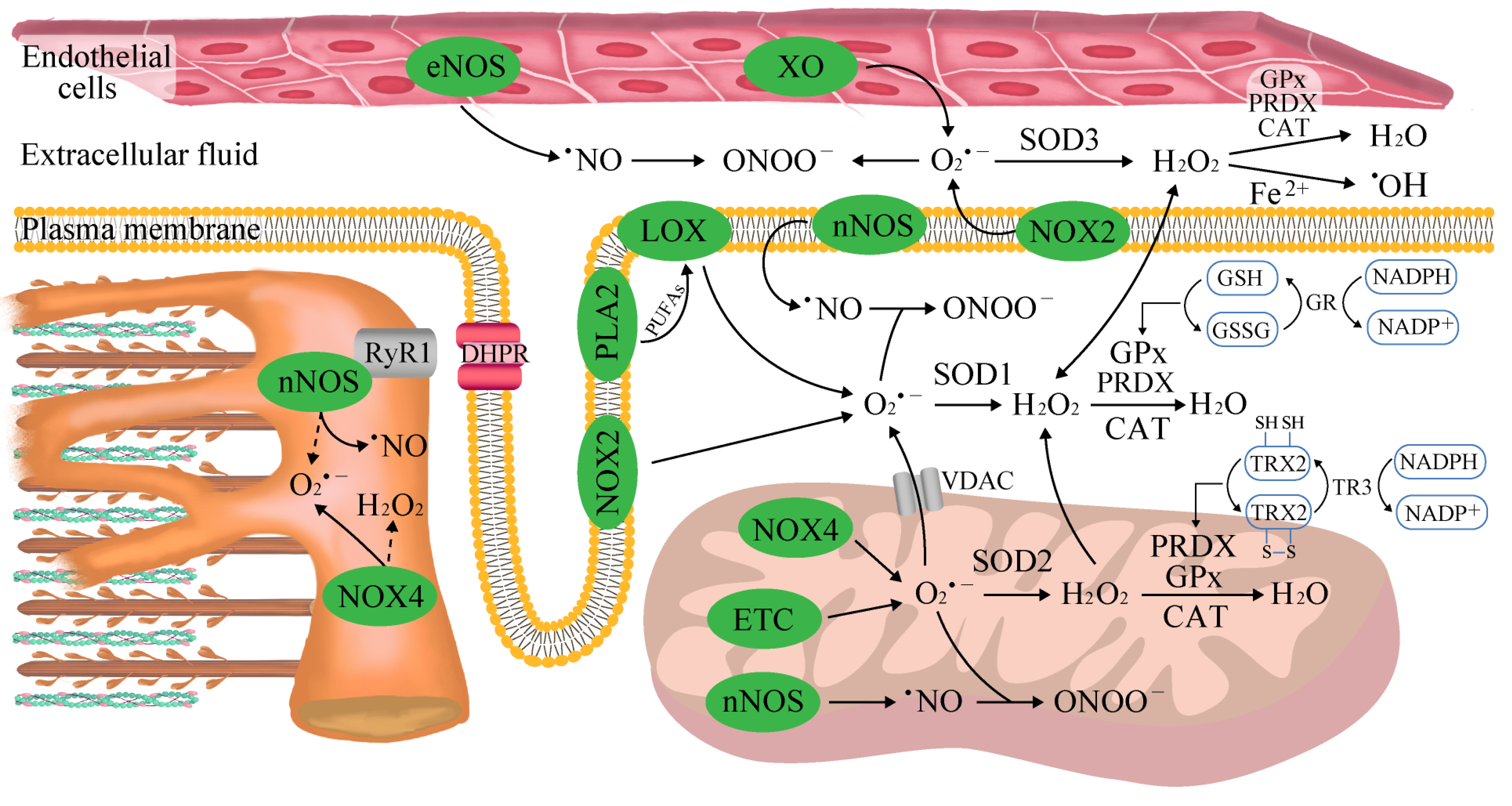
2. The Sources of Reactive Oxygen and Nitrogen Species (ROS/RNS) in Skeletal Muscle

Skeletal muscle is a heterogeneous tissue composed of three major fiber types that possess certain unique metabolic and contractile features. Specifically, slow-twitch oxidative fibers (type I) have a higher oxidative capacity and a higher fatigue threshold that enables them to support sustained aerobic activity. In contrast, fast-twitch glycolytic fibers (type IIx) exhibit a lower oxidative capacity and enzymatic profile that favor anaerobic metabolism, slower calcium kinetics, faster shortening velocities, and the ability to generate more power than slow-twitch fibers. Fast-twitch oxidative–glycolytic fibers (type IIa) exert higher twitch speeds than type I fibers but are less fatigue-resistant. Such a broad range of capabilities has emerged through the selection of a characteristic molecular profile designated to achieve a particular contractile phenotype for each fiber type. Specifically, myosin heavy chain (MHC) isoforms dictate the ATPase activity of muscle and underly the above classification. The MHC I isoform is characterized by a high ATP hydrolysis power, such that a lower maximum shortening velocity and higher economy of ATP usage are developed in MHC I muscle compared with fast MHC IIa and IIx fibers [10]. Furthermore, MHC-I-rich fibers exhibit a lower maximum Ca2+-activated force but a higher Ca2+ sensitivity compared with MHC-II-rich fibers [11]. At rest, a certain amount of ROS/RNS is continuously produced in muscle as an important component of normal cell signaling in all fiber types, although the magnitude of this process is the highest in glycolytic fibers [12]. Free radicals consist of atoms or molecules with an unpaired electron in their outer shell, which makes them unstable and highly reactive. This group includes superoxides (O2•−), oxygen radicals (O2••), hydroxyl radicals (OH), alkoxy radicals (RO), peroxyl radicals (ROO), nitric oxide (NO), and nitrogen dioxide (NO2). Non-radical derivatives include hydrogen peroxide (H2O2), hypochlorous acid (HOCl), ozone (O3), singlet oxygen (1O2), nitrous acid (HNO2), nitrosyl cations (NO+), nitroxyl anions (NO), and peroxynitrite (ONOO), and they can easily lead to free radical reactions in living organisms [12].
Davies et al. [13] were the first to report that free radicals are elevated in contracting rat muscles in 1982. In 1978, Dillard et al. observed an increased content of expired pentane, an index of lipid peroxidation, after 60 min of cycle ergometer exercise at 50% VO2 max intensity, in humans [14]. The first direct evidence for intramuscular free radical accumulation following exercise in humans was provided by Bailey et al. in 2007 with the use of electron paramagnetic spectroscopy [15]. The rate of ROS/RNS synthesis differs with respect to both the intensity and duration of exercise, which together constitute exercise volume. In general, short-term, low-intensity aerobic exercise (<40% VO2max) generates relatively less ROS than moderate-intensity exercise (65–75% VO2max). However, it needs to be highlighted that this association was concluded based on the net activity of antioxidant systems and the level of oxidative damage to macromolecules (e.g., increased DNA damage, protein oxidation and lipid peroxidation) in both the blood and active skeletal muscles, not the direct measurement of ROS/RNS content. The assessment of radical expression faces difficulties due to the high reactivity and short half-life of ROS/RNS [16][17]. Furthermore, a higher frequency of exercise (five times per week) was found to more efficiently reduce oxidative stress based on malondialdehyde content and improved mitochondrial oxidation capacity compared with exercise performed three times per week [18]. Other aspects determining the rate of ROS/RNS generation include fluctuations in blood flow and local oxygenation. It was shown that isometric muscle contractions at 60% of maximal voluntary contraction (MVC) occlude blood flow, presumably at the level of lower-order arterioles or the capillary bed. The extent of flow restriction is lower at both 30% and 100% of MVC because of the insufficient tension created and shorter duration of time when contraction can be sustained, respectively. The successive reperfusion period (muscle relaxation) was found to enable oxygen delivery, although it was not linearly dependent on the degree of blood perfusion during sustained isometric contractions and accounted for ~42%, ~22%, and ~22% at 30%, 60%, and 100% of MVC, respectively [19]. Regained oxygenation could precede the production of ROS/RNS and provide time for muscle regeneration and adaptation to exercise. To support this notion, the employment of BFR training protocols in human studies coincided with several improvements, including upregulated glucose extraction, Na+/K+-pump expression, and K+ homeostasis, that delay fatigue [6][20]. Moreover, increased muscle temperatures lead to higher levels of ROS during contractions [21]. Whenever ROS production is managed with a biological system’s ability to readily detoxify ROS, exercise appears to not promote oxidative stress. However, this imbalance occurs as a result of the excess level of ROS/RNS and overridden antioxidant system capacity. It is well-established that acute bouts of endurance exercise in untrained humans and animals result in increases in biomarkers of oxidative stress (e.g., increased protein oxidation and lipid peroxidation) in both blood and active skeletal muscles [22].
The main sources of muscular ROS during exercise remain uncertain due to the difficulties in measuring and quantifying real-time contraction-stimulated ROS production. These difficulties are caused by radicals’ relatively short half-life, instantaneous removal by defense mechanisms, and continuous generation in tissues other than skeletal muscle. Under normal physiological conditions, only 0.1–0.2% of the O2 consumed by mitochondria is converted into O2•−, and that value depends on both metabolic status and the oxidative substrate used [23]. Mitochondria can generate ROS from at least 11 different sites, and conventionally, complexes I (site IQ) and III (site IIIQo) within the respiratory electron transport chain, as well as monoamine oxidases activity, are thought to be the dominant contributors of mitochondrial O2•− and H2O2 during exercise [24]. Results regarding both permeabilized fibers and isolated mitochondria showed that fast-glycolytic muscles emit higher H2O2 levels than slow-oxidative fibers, most likely as a consequence of a considerably lower mitochondrial ROS/RNS scavenging capacity [25]. Most importantly, under ex vivo conditions resembling mild and aerobic exercise in skeletal muscles, the overall rate of mitochondrial O2•− and H2O2 production was found to account for only 15% of that at rest. This was shown to be related to a transition to so-called state 3 respiration (ADP-stimulated oxidative phosphorylation) associated with a lower protonmotive force and the oxidation of ubiquinone pool, while site IF (with low-capacity to O2•− production) was shown to be the major ROS contributor [26]. Moreover, the 66 kD isoform of spontaneous human combustion (shc) adaptor proteins (p66Shc) translocates to the mitochondrial matrix upon exercise, wherein it oxidizes cytochrome c to form H2O2. p66shc-induced mitochondrial ROS synthesis was shown to be protective in conditions of high levels of cell stress, such as during exercise. This was evident in p66shc−/− mice, which were slightly less fatigued during downhill running than control animals. The knockdown of p66shc, however, did not affect skeletal muscle structure and function at rest [27].
NADPH oxidase (NOX) enzymes, rather than mitochondria, appear to be the predominant contributors of ROS in skeletal muscle. Of the NOX family, NOX1, NOX2 (producing O2•−), as well as NOX4 and DUOX1/2 (producing H2O2), have been reported to increase during both high-intensity exercise [28] and moderate-intensity exercise [29]. In the presence of ADP and Fe3+, the enzymes transfer electrons from NADPH to molecular oxygen to produce O2•− and then H2O2 [30], and the inhibition of NOX enzymes blocks both basal and stretch/contraction-stimulated skeletal muscle ROS production [31]. The results of a study examining the loss of NOX2 in both acute and repeated exercise implied potential cross-talk between different sites of ROS production in skeletal muscle responses to exercise [32]. It is also likely that NOX2 (present in membrane-enriched protein fractions) and NOX4 (mitochondria) mediate specific signaling pathways via ROS production in different subcellular microdomains. Matrix O2•− is possibly more important for stress resistance than intermembrane space ROS [33]. On the other hand, NOX4 is activated following decreases in mitochondrial ATP levels and is involved in the adjustment of glucose and fatty acid oxidation to exercise [34].
Concurrently, human skeletal muscles contain approximately 16 different phospholipase A2 (PLA2) isoforms, both calcium-dependent and calcium-independent, capable of stimulating ROS generation in muscles under rest [35] and exercise conditions [36]. Augmented PLA2 activity (in mitochondria and the cytosol) and arachidonic acid release (i.e., a substrate for several ROS-generating enzyme systems including lipoxygenases) during exercise can stimulate NOX [37][38].
XO has been recognized as contributing to O2•− generation in the extracellular space following muscle contraction [39] due to its high expression in endothelial cells and activation in response to shear stresses applied to skeletal muscle cells during exercise [23]. XO is most strongly stimulated under conditions of exhaustive exercise when blood flow does not meet requirements. An increase in XO activity is associated with an accelerated conversion of ATP into AMP and eventually into hypoxanthine. XO then catalyzes the conversion of hypoxanthine into xanthine and xanthine into uric acid [23][40].
Skeletal muscles constitutively express neuronal and endothelial nitric oxide synthases (nNOS and eNOS, respectively), which generate NO from L-arginine, NADPH and O2. Approximately 75% of nNOS is associated with submembrane scaffolds that are part of the dystrophin glycoprotein complex, and the remainder is detected in the mitochondria and the sarcoplasmic reticulum [41]. Both neuronal and endothelial isoforms become activated by increases in free cytosolic Ca2+ concentration during contractile activities [42]. The produced NO is an uncharged and freely diffusible molecule, and it may exert effects over distances exceeding 100 μm [43]. NO undergoes complex interactions with ROS, including direct quenching by O2•−, to generate the toxic oxidant ONOO that damages proteins, lipids, and DNA [44]. Even at high physiological concentrations of superoxide dismutase (SOD), the rate of ONOO synthesis is 6 times faster than the rate at which SOD creates H2O2 from O2•− [45].

3. Aerobic Exercise

Despite a 1–3-fold increase in O2•− during strenuous exercise, mitochondria only account for a small portion of the ROS generated as a consequence of lowered mitochondrial NADH/NAD+ ratios and consequently reduced complex-I-dependent ROS production [30]. Studies based on both human and animal models have shown that the major pool of ROS in strenuous exercises is produced by the action of NOX2 and NOX4 [46]. Importantly, specific mitochondrial phenotypes exist in slow- and fast-twitch fibers in line with distinct patterns of pro-oxidative enzymes [28]. In this case, some specificity in NOX2 and NOX4 responses to exercise was shown between different types of muscle fibers. Particularly, initial NOX activity was significantly higher in the slow-twitch oxidative soleus muscle compared with the fast-twitch oxidative–glycolytic red gastrocnemius and the fast-twitch glycolytic red gastrocnemius [28]. Other biological systems more strongly contributing to exercise-induced ROS production include XO activated by the breakdown of ATP to support repetitive contractions [47].

4. Anaerobic Exercise

In 2000, ROS/RNS production during and after anaerobic exercise was observed based on a higher lipid hydroperoxide content in the blood following isometric exercise [48], and direct increases in blood free radical levels were later observed in male individuals performing the Wingate test (a 30-s full-strength pedaling exercise) [49]. Further insight into the underlying mechanisms revealed that the main site of ROS/RNS production in the fast-twitch fibers was a cytosolic compartment. These results were also supported by the fact that during short-term anaerobic exercises, only 0.15% of O2•− was found to be produced in the mitochondria [50]. Some studies have shown that high-intensity exercise is also coupled with the activation of NOX2 and NOX4 located in the sarcolemma, transverse tubules, and sarcoplasmic reticulum of muscle cells [32][51]. The accelerated ATP degradation and accumulation of downstream metabolites lead to the activation of XO [50], which is directly correlated with lactic acid levels [52]. However, there were no reported differences in the levels of ROS generation between eccentric contractions that damage the muscle and isometric contractions that do not cause injury [53]. Additionally, increased real-time NO production was visualized in skeletal muscle isolated from mice using a fluorescent NO probe, 4-amino-5-methylamino-2′,7′-difluorofluorescein diacetate (DAF-FM DA). Five minutes of electrically stimulated contractions led to a marked increase in NO production in fast-twitch fibers, which rapidly normalized by 15 min post-stimulation [54]. The abrupt nature of RNS changes in skeletal muscle highlights the importance of real-time monitoring systems for the direct detection of ROS/RNS production throughout exercise protocols.

5. Endogenous Mechanisms of Reactive Oxygen and Nitrogen Species Detoxification

Skeletal muscle is a highly plastic tissue, and under most exercise conditions, oxidative balance is preserved within physiological range via the upregulation of ROS scavenging, hence minimizing the potential for oxidative damage. The intensity and duration of physical exercises are critical in this respect. When exercise is being performed on a regular basis, even 5–10 consecutive days of moderate-intensity exercise markedly increase both the oxidative and antioxidant capacity of skeletal muscle fibers, which upregulates fatigue resistance (Figure 1) [55][56]. Similarly, long-term (12 weeks) endurance exercise training augments antioxidant enzyme activities in the muscle and reduces contraction-induced oxidative stress [57]. Increases in the antioxidant enzyme activities in plasma were detected after both maximal and submaximal exercise periods in untrained men [58].
Figure 1. Overview of the major oxidant and antioxidant systems in skeletal muscle. Mitochondrial respiration shifts from state 4 to active state 3 when muscle contractions start, which is characterized by a lower superoxide (O2•−) production than at rest. NADPH oxidase (NOX) enzymes become activated in response to sarcolemma depolarization, ATP release, and fiber stretching associated with exercise. NOX2 resides in the plasma membrane and transverse tubules, and it is the main source for cytosolic O2•− generation in contracting muscle. NOX4 was reported to localize in the sarcoplasmic reticulum and mitochondrial intermembrane space. Xanthine oxidase (XO) is abundant in endothelial cells and activates via increases in shear stresses applied to skeletal muscle and ATP hydrolysis. The release of polyunsaturated fatty acids (PUFAs) from plasma membrane is stimulated by phospholipase A2 (PLA2), and the released free PUFAs are subsequently oxidized by lipoxygenase (LOX). The formed O2•− is detoxified with the assistance of superoxide dismutase (SOD), catalase (CAT), glutathione peroxidase (GPx), peroxiredoxin (PRDX) or thioredoxin (TRX). The PRDX-mediated reduction in H2O2 uses TRX as the electron donor. In the course of the reaction, one sulfhydryl group of the cysteine residues in PRDX is oxidized and a disulfide bridge is formed. Then, the disulfide bridge is reduced by TRX at the expense of NADPH oxidation. Abbreviations: DHPR, dihydropyridine receptor; ETC, mitochondrial electron transport chain; GR, glutathione reductase; RyR1, ryanodine receptor 1; TR3, thioredoxin reductase 3; VDAC, voltage-dependent anion channel.
The multifaced antioxidant aperture consists of the three main strategies. First, numerous low-molecular-weight molecules able to scavenge ROS co-exist in the extracellular space and within cells. This group includes glutathione (GSH), uric acid, lipoic acid uric acid, bilirubin, vitamin E, and vitamin C. Second, some enzymatic antioxidants act by converting ROS into less reactive molecules, including copper–zinc superoxide dismutase (SOD1), manganese superoxide dismutase (SOD2), extracellular superoxide dismutase (SOD3), catalase, and glutathione peroxidase (GPX). A final antioxidant mechanism is based on the binding of pro-oxidant transition metals (e.g., iron and copper) via metal-binding proteins. The chelating molecules preclude these transition metals from participating in ROS formation [16].
A distinct phenotypical predominance of exercise-induced antioxidants holds true for SOD, which is most noticeably upregulated in active skeletal muscles composed of highly oxidative fibers (e.g., type I and type IIa) [28][59]. The importance of SOD1 to preserve muscle function has been confirmed by studies employing SOD-deprived mice (Sod1−/−). Sod1−/− animals were found to exhibit a characteristic phenotype with peripheral nerve integrity and denervated motor end plates, which resulted in fiber loss and muscle atrophy [60] and was linked to elevated oxidative damage in DNA, proteins, and lipids, as well as an increase in proteolytic activity [60]. Compared with whole-body Sod1−/− strain, muscle-restricted SOD1 deficiency was found to be insufficient to reproduce the accelerated neuromuscular degenerative phenotype, partially through the compensative upregulation of other antioxidative pathways, and these animals maintained levels of muscle mass similar to respective control mice. Nevertheless, both systemic and skeletal muscle-specific SOD1 knockout affected muscle structure, with the relocation of nuclei towards the cell center, hence reflecting continuing cycles of degeneration and regeneration [61]. Similarly, muscle Sod2−/− mice showed centralized nuclei in their muscle fibers and the selective loss of respiratory enzyme activities, including complex I and complex II (succinate dehydrogenase), as well as reduced ATP levels in their skeletal muscles. A single dose of the EUK-8 antioxidant significantly improved exercise activity in mice [62], which demonstrated the critical contribution of the O2•− generated in mitochondria in the progression of deficits in muscle structure and force generation and the development of exercise intolerance.

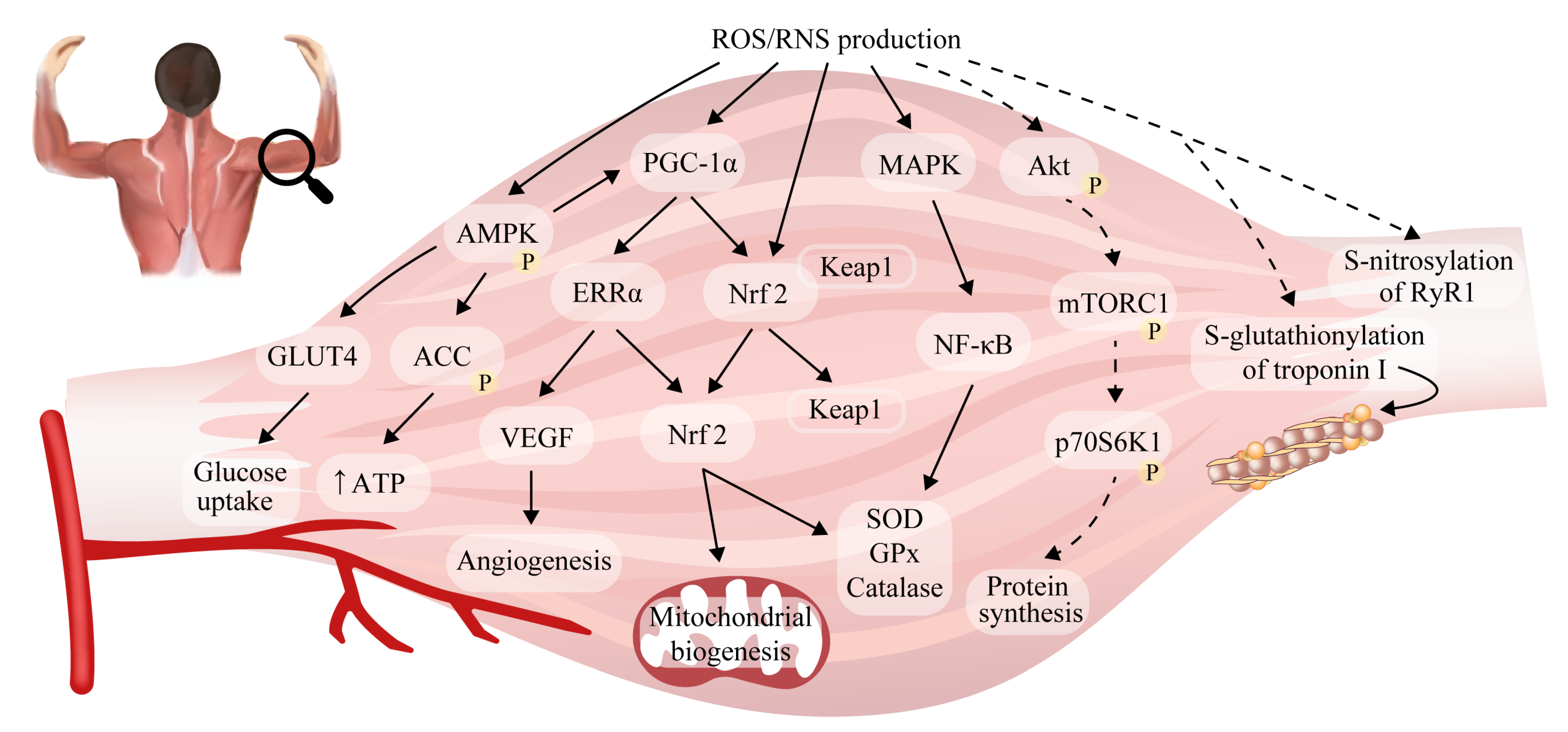
6. Reactive Oxygen/Nitrogen Species and Adaptations to Exercise in Skeletal Muscle

Subjects involved in interval training, which is characterized by short periods of high-intensity exercise interspersed with periods of recovery, develop adaptive mechanisms related to not only the renewal of glycogen stores in skeletal muscle but also higher levels of mitochondrial content and the effectiveness of oxidative damage repair systems. Additionally, the subjects produce lower levels of ROS at a given intensity of exercise compared with less-trained individuals [63]. Several important pathways have been proposed in mediating these physiological adaptations to training (Figure 2), although the extent of muscle reprogramming depends on workload and exercise volume [64]. Although some human studies have shown that exercise intensity is the most important variable determining the stimulation of mitochondrial function [65], other research has suggested that exercise training volume is more significant [66].
Figure 2. Redox-regulated signaling pathways in muscle adaptations to exercise. During endurance exercise (solid arrows), ROS/RNS participate in AMPK activation to promote glucose uptake, ATP generation, vascularization, mitochondrial biogenesis, and antioxidant defense (via the peroxisome-proliferator activated co-activator 1α (PGC-1α) pathway). Dashed arrows indicate mechanisms that dominate in response to resistance exercise in fast-twitch fibers. ROS/RNS trigger post-translational modifications in ryanodine receptor (RyR1) and troponin I to increase ongoing force production. The long-term effect is the stimulation of mTORC1-p70S6K1 signaling to orchestrate overload-induced muscle hypertrophy. Abbreviations: ACC, acetyl-CoA carboxylase; AMPK, AMP-activated protein kinase; ERRα, estrogen-related receptor α; Keap1, Kelch-like ECH-associated protein 1; MAPK, mitogen-activated protein kinase; mTORC1 (mammalian target of rapamycin (mTOR)), mammalian target of rapamycin 1; NF-κB, nuclear factor-kappa B; Nrf2, nuclear factor erythroid 2-related factor 2; p70S6K1, p70 ribosomal protein subunit 6 kinase 1; RyR1, ryanodine receptor 1; VEGF, vascular endothelial growth factor.

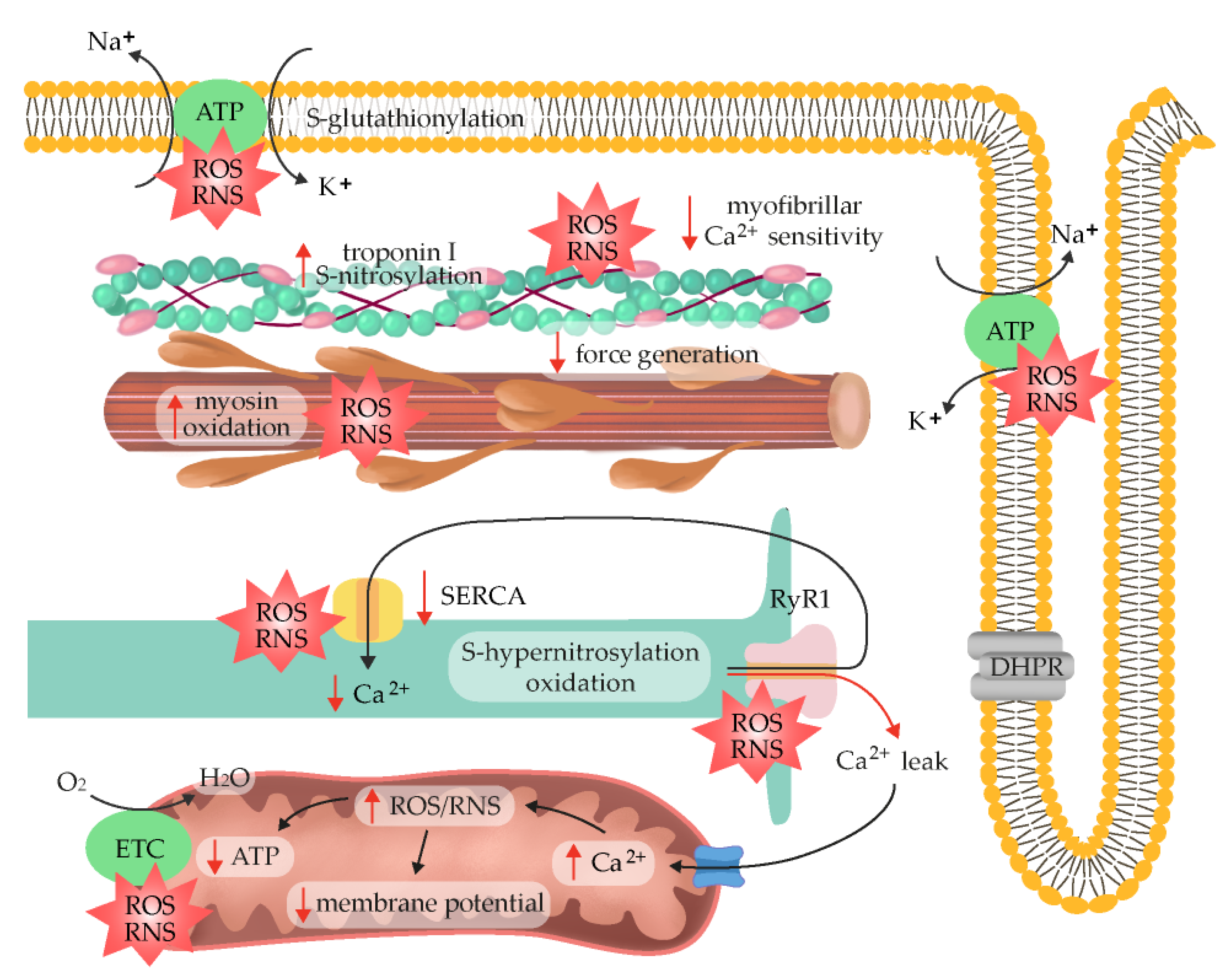
7. ROS/RNS as Fatigue Mediators

Contrary to an intermittent regular exercise protocol, wherein exercise-induced ROS production is mostly overcompensated for by an upregulated antioxidant defense, exhaustive exercise or chronic exposure to ROS can exceed the antioxidant barrier and lead to oxidative stress and damage to muscle fibers [22][63]. The first discovery that radicals contribute to muscle fatigue in animals was reported in 1990 [67], and since then, several ROS-triggered mechanisms have been shown to limit muscle performance. The inhibitory effects of O2•− and H2O2 [68], NO [69], and ONOO [70] on maximum force production have been shown to be most pronounced in type II fibers. This could be attributed to variations in endogenous ROS/RNS scavenging capabilities; in fact, the substantially higher antioxidative potential of slow-twitch fibers was confirmed in several studies. For instance, an approximately 5-fold higher GSH level was noticed in slow-twitch muscle compared with fast-twitch muscle [25][28][71].
One of the potential mechanisms that contributes to fatigue is a decrease in membrane excitability, which is most pronounced deep within the transverse tubular system [72]. According to research on mechanically skinned rat muscle fibers undergoing fatiguing stimulation, the decreased excitability of transverse tubules is caused by the S-glutathionylation of Na+/K+-ATPase in response to the excessive production of ROS, which might be accelerated by contraction-induced ATP decline and preclude the restoration of the Na+/K+ balance with repeated contractions [73]. Some basal level of the inhibitory S-glutathionylation of the Na+/K+-pump β-subunit that intensified during intense exercise, which coincided with fatigue, was also noticed in biopsies of human vastus lateralis muscles [74]. Experimental evidence to support these data was provided following antioxidant N-acetylcysteine (NAC) administration in humans. More specifically, NAC supplementation in humans markedly alleviated the percentage decrease in maximal Na+/K+-pump activity caused by submaximal fatiguing exercise, most likely via the preservation of muscle GSH and cysteine levels, which can prevent the oxidation of SH groups on the Na+/K+-pump [75], suggesting that ROS/RNS may jeopardize the potential transmission of cellular action. However, another study did not reproduce action potential failure due to H2O2 exposure in an animal model employing isolated intact mouse flexor digitorum brevis fibers [4]. Similarly, slow-twitch soleus mouse muscle did not exhibit impaired excitability during fatiguing stimulation [76].
The described data indicate several target points through which ROS/RNS can impede skeletal muscle force generation (Figure 3). Collectively, the dual consequences of ROS/RNS in muscle performance can be described with a bell-shaped dose–response curve, where a low dose of activity facilitates adaptation and excessive exercise is harmful. Moderate exercise appears to extend the levels of tolerable ROS due to higher antioxidant enzyme activity, thus increasing physiological functioning [77]. However, data from diabetic individuals challenged this assumption, showing that both high (exercise-induced) and low (antioxidant intervention) ROS concentrations can trigger beneficial responses as long as they do not override the redox balance threshold range [78]. Currently, there is no unambiguous protocol or biomarker that can differentiate moderate and excessive exercise and predict either healthful effects or reduced muscle performance.
Figure 3. Mechanisms underlying muscle fatigue development in response to high intramuscular ROS/RNS concentration. Increased ROS/RNS levels can influence muscle force production through: (1) enhanced extracellular K+ and reduced membrane excitability, (2) the oxidation/nitrosylation of RyR1 and sarco(endo)plasmic reticulum Ca2+ ATPase (SERCA) to affect Ca2+ release from the sarcoplasmic reticulum and clearance in skeletal muscle, (3) changes in Ca2+-sensitivity and cross-bridge kinetics, (4) mitochondrial Ca2+ overload and mitochondrial ROS emission, and (5) lowered ATP production.

8. Exogenous Antioxidants

The mechanistic findings presented above provide a basis for nutritional supplementation strategies or pharmacological manipulations in order to prevent fatigue development. Exogenous antioxidant delivery modifies multiple aspects of skeletal muscle signaling during and immediately following a single bout of exercise, and this can include the modulation of mitochondrial biogenesis, glucose uptake, force production and cell excitability. It has become common practice for athletes and health-conscious, physically active individuals to chronically supplement their normal diets with high doses of antioxidants including vitamins C and E, coenzyme Q10 (CoQ10), α-lipoic acid, and NAC [79]. However, at present, there is not enough evidence to support a role for antioxidants supplementation in preventing the cumulative effects on skeletal muscle caused by radicals during exercise, and this practice may actually hamper certain adaptations to exercise training [46].
Among the most well-known and prevalent antioxidants, vitamins can be easily obtained through natural foods such as vegetables and fruits [80]. The potential prophylactic effects of antioxidant vitamins have been evaluated in an abundance of animal and human studies. He et al. showed that short-term concomitant vitamin C and E administration not only attenuated levels of creatine kinase (a muscle damage biomarker) and muscle soreness but also enhanced muscle protection following a second bout of aerobic exercise [50]. Likewise, Fogarty et al. reported that exhaustive exercise-induced lipid peroxidation and DNA damage can be mitigated by both the short- and long-term consumption of watercress, which is rich in lipid-soluble antioxidants, such as α-tocopherol, β-carotene, and xanthophyll [17]. Despite the beneficial effects mentioned above, a thorough understanding of the application of vitamin and antioxidant supplements such as the optimal dosage, duration, and administration method is necessary to avoid unfavorable effects. Several studies have indicated that antioxidant supplements fail to protect against damaging effects of oxidative stress such as exercise-induced lipid peroxidation and inflammation, both of which hamper muscle recovery. Specifically, prolonged antioxidant ingestion can disrupt endogenous antioxidant levels and impede exercise-induced adaptation, thereby blunting the body’s defense against oxidative stress [81]. Studies on vitamin E have reported the acceleration of oxidative stress in exercising participants following the stimulation of lipid peroxidation and inflammatory process [82][83]. To monitor the effects of ascorbic acid supplementation on post-exercise recovery, muscle strength and redox status were measured 14 days after downhill running. Participants in both groups experienced delayed-onset muscle soreness (DOMS), but those receiving vitamin C also showed delayed muscle recovery [84]. Most importantly, the daily ingestion of vitamin C and vitamin E was found to almost completely blunt mitochondrial adaptive responses by abolishing the changes in PGC-1α gene expression and increases in insulin sensitivity elicited by physical exercise [85][86]. Soon after, Gomez-Cabrera et al. reported that 8 weeks of vitamin C supplementation prevented training-induced mitochondrial biogenesis by suppressing the expression of SOD and GPx [79]. One research showed that consuming large dosages of vitamin C (500 mg kg−1) for 14 days inhibited the hypertrophy of overworked muscles in Wistar rats. This event was accompanied by reductions in the ROS-regulated p70S6K and ERK1/2 (regulators of skeletal muscle hypertrophy), although the level of oxidative stress markers was similar between groups. Most likely, the long gap between tissue sampling and the last vitamin C delivery affected the measurements, indicating the transient nature of antioxidant impacts on redox balance [87]. In research by Theodorou et al., males performed eccentric exercise twice a week for four weeks. Concomitantly, they were given vitamins E and C (400 IU and 1000 mg per day, respectively) for 11 weeks, but the high antioxidant doses were unable to improve the antioxidant status (GSH and catalase) of blood and skeletal muscle during and after exercise. The vitamins likely hindered any effect of antioxidant supplementation, so these supplements had no impact on muscular function or post-exercise recovery [88]. On the other hand, an oral antioxidant cocktail of vitamins C, E, and α-lipoic acid (αLA) attenuated circulating free radicals during exercise but impaired exercise-induced brachial artery vasodilation [89]. Following 6 weeks of eccentric leg exercise training, the effects of the administration of αLA and oral vitamins C and E on resting blood pressure and brachial artery vasodilation were assessed via the flow-mediated dilation evoked by both post-cuff occlusion hyperemia and during progressive handgrip exercise. Antioxidants limited free radical concentration, negated training-induced improvements in resting and exercising arterial blood pressure, and significantly decreased flow-mediated vasodilation [90]. Similar findings in isolated rat arterioles confirmed the critical roles of ROS and RNS in flow-induced vasodilation [91], and antioxidants were found to prevent that effect.
αLA can provide effective protection against oxidative stress via a thiol/disulfide exchange mechanism. The compound is unique among antioxidants because it retains powerful antioxidant abilities in its oxidized (αLA) and reduced (dihydrolipoic acid, DHLA) forms. Multiple mechanisms are implicated in αLA’s and DHLA’s antioxidant power, including direct ROS/RNS quenching actions, metal-chelating properties, and the regeneration of endogenous antioxidants such as GSH, vitamin C, and vitamin E [92]. Exercise training and αLA have synergistic effects on improving skeletal muscle glucose transport activity, whole-body glucose tolerance, and lipid profile [93][94]. Athletes supplemented with αLA at a dose of 1200 mg/d for 10 days before exercise showed reduced levels of inflammatory cytokines because of changes in thiol redox status [95]. αLA can have positive effects on maintaining muscle force and reducing muscle damage and inflammation by downregulating the expression of redox-sensitive proinflammatory cytokines, such as TNF-α, Il-6, and inducible NOS. These positive outcomes were associated with training, though not when acute resistance exercise was performed [96]. In other research, no differences in force generation between control and αLA groups were noticed in isolated fibers [97]. Because strategies employing αLA in the prevention of muscle fatigue are in their infancy, there is insufficient evidence to support chronic antioxidant delivery.

This entry is adapted from the peer-reviewed paper 10.3390/antiox12020501

References

  1. Suzuki, K.; Tominaga, T.; Ruhee, R.T.; Ma, S. Characterization and Modulation of Systemic Inflammatory Response to Exhaustive Exercise in Relation to Oxidative Stress. Antioxidants 2020, 9, 401.
  2. Sahlin, K.; Ren, J.M. Relationship of Contraction Capacity to Metabolic Changes during Recovery from a Fatiguing Contraction. J. Appl. Physiol. 1989, 67, 648–654.
  3. Woodward, M.; Debold, E.P. Acidosis and Phosphate Directly Reduce Myosin’s Force-Generating Capacity through Distinct Molecular Mechanisms. Front. Physiol. 2018, 9, 862.
  4. Andrade, F.H.; Reid, M.B.; Westerblad, H. Contractile Response to Low Peroxide Concentrations: Myofibrillar Calcium Sensitivity as a Likely Target for Redox-modulation of Skeletal Muscle Function. FASEB J. 2001, 15, 309–311.
  5. Christiansen, D.; Eibye, K.; Hostrup, M.; Bangsbo, J. Training with Blood Flow Restriction Increases Femoral Artery Diameter and Thigh Oxygen Delivery during Knee-Extensor Exercise in Recreationally Trained Men. J. Physiol. 2020, 598, 2337–2353.
  6. Christiansen, D. Molecular Stressors Underlying Exercise Training-Induced Improvements in K + Regulation during Exercise and Na +, K + -ATPase Adaptation in Human Skeletal Muscle. Acta Physiol. 2019, 225, e13196.
  7. Centner, C.; Zdzieblik, D.; Dressler, P.; Fink, B.; Gollhofer, A.; König, D. Acute Effects of Blood Flow Restriction on Exercise-Induced Free Radical Production in Young and Healthy Subjects. Free Radic. Res. 2018, 52, 446–454.
  8. Zuo, L.; Clanton, T.L. Reactive Oxygen Species Formation in the Transition to Hypoxia in Skeletal Muscle. Am. J. Physiol.-Cell Physiol. 2005, 289, 207–216.
  9. Korkmaz, E.; Dönmez, G.; Uzuner, K.; Babayeva, N.; Torgutalp, Ş.Ş.; Özçakar, L. Effects of Blood Flow Restriction Training on Muscle Strength and Architecture. J. Strength Cond. Res. 2022, 36, 1396–1403.
  10. Anderson, E.J.; Neufer, P.D. Type II Skeletal Myofibers Possess Unique Properties That Potentiate Mitochondrial H2O2 Generation. Am. J. Physiol.-Cell Physiol. 2006, 290, C844–C851.
  11. Gejl, K.D.; Hvid, L.G.; Andersson, E.P.; Jensen, R.; Holmberg, H.C.; Ørtenblad, N. Contractile Properties of MHC I and II Fibers From Highly Trained Arm and Leg Muscles of Cross-Country Skiers. Front. Physiol. 2021, 12, 855.
  12. Phaniendra, A.; Jestadi, D.B.; Periyasamy, L. Free Radicals: Properties, Sources, Targets, and Their Implication in Various Diseases. Indian J. Clin. Biochem. 2015, 30, 11–26.
  13. Davies, K.J.A.; Quintanilha, A.T.; Brooks, G.A.; Packer, L. Free Radicals and Tissue Damage Produced by Exercise. Biochem. Biophys. Res. Commun. 1982, 107, 1198–1205.
  14. Dillard, C.J.; Litov, R.E.; Savin, W.M.; Dumelin, E.E.; Tappel, A.L. Effects of Exercise, Vitamin E, and Ozone on Pulmonary Function and Lipid Peroxidation. J. Appl. Physiol. Respir. Environ. Exerc. Physiol. 1978, 45, 927–932.
  15. Bailey, D.A.; Lawrenson, L.; McEneny, J.; Young, I.S.; James, P.E.; Jackson, S.K.; Henry, R.R.; Mathieu-Costello, O.; McCord, J.M.; Richardson, R.S. Electron Paramagnetic Spectroscopic Evidence of Exercise-Induced Free Radical Accumulation in Human Skeletal Muscle. Free Radic. Res. 2007, 41, 182–190.
  16. Powers, S.K.; Deminice, R.; Ozdemir, M.; Yoshihara, T.; Bomkamp, M.P.; Hyatt, H. Exercise-Induced Oxidative Stress: Friend or Foe? J. Sport Health Sci. 2020, 9, 415–425.
  17. Fogarty, M.C.; Hughes, C.M.; Burke, G.; Brown, J.C.; Trinick, T.R.; Duly, E.; Bailey, D.M.; Davison, G.W. Exercise-Induced Lipid Peroxidation: Implications for Deoxyribonucleic Acid Damage and Systemic Free Radical Generation. Environ. Mol. Mutagen. 2011, 52, 35–42.
  18. Silva, L.A.; Tromm, C.B.; Doyenart, R.; Thirupathi, A.; Silveira, P.C.L.; Pinho, R.A. Effects of Different Frequencies of Physical Training on Electron Transport Chain and Oxidative Damage in Healthy Mice. Motriz. Rev. Educ. Fis. 2018, 24, 101804.
  19. McNeil, C.J.; Allen, M.D.; Olympico, E.; Shoemaker, J.K.; Rice, C.L. Blood Flow and Muscle Oxygenation during Low, Moderate, and Maximal Sustained Isometric Contractions. Am. J. Physiol.-Regul. Integr. Comp. Physiol. 2015, 309, R475–R481.
  20. Christiansen, D.; Eibye, K.H.; Hostrup, M.; Bangsbo, J. Blood Flow-Restricted Training Enhances Thigh Glucose Uptake during Exercise and Muscle Antioxidant Function in Humans. Metabolism 2019, 98, 1–15.
  21. Van Der Poel, C.; Edwards, J.N.; MacDonald, W.A.; Stephenson, D.G. Effect of Temperature-Induced Reactive Oxygen Species Production on Excitation-Contraction Coupling in Mammalian Skeletal Muscle. Clin. Exp. Pharmacol. Physiol. 2008, 35, 1482–1487.
  22. Supruniuk, E.; Maciejczyk, M.; Zalewska, A.; Górski, J.; Chabowski, A. Blood Profile of Cytokines, Chemokines, Growth Factors, and Redox Biomarkers in Response to Different Protocols of Treadmill Running in Rats. Int. J. Mol. Sci. 2020, 21, 8071.
  23. Bouviere, J.; Fortunato, R.S.; Dupuy, C.; Werneck-De-castro, J.P.; Carvalho, D.P.; Louzada, R.A. Exercise-Stimulated Ros Sensitive Signaling Pathways in Skeletal Muscle. Antioxidants 2021, 10, 537.
  24. Menazza, S.; Blaauw, B.; Tiepolo, T.; Toniolo, L.; Braghetta, P.; Spolaore, B.; Reggiani, C.; di Lisa, F.; Bonaldo, P.; Canton, M. Oxidative Stress by Monoamine Oxidases Is Causally Involved in Myofiber Damage in Muscular Dystrophy. Hum. Mol. Genet. 2010, 19, 4207–4215.
  25. Picard, M.; Hepple, R.T.; Burelle, Y. Mitochondrial Functional Specialization in Glycolytic and Oxidative Muscle Fibers: Tailoring the Organelle for Optimal Function. Am. J. Physiol.-Cell Physiol. 2012, 302, 629–641.
  26. Goncalves, R.L.S.; Quinlan, C.L.; Perevoshchikova, I.V.; Hey-Mogensen, M.; Brand, M.D. Sites of Superoxide and Hydrogen Peroxide Production by Muscle Mitochondria Assessed Ex Vivo under Conditions Mimicking Rest and Exercise. J. Biol. Chem. 2015, 290, 209–227.
  27. Granatiero, V.; Gherardi, G.; Vianello, M.; Salerno, E.; Zecchini, E.; Toniolo, L.; Pallafacchina, G.; Murgia, M.; Blaauw, B.; Rizzuto, R.; et al. Role of P66shc in Skeletal Muscle Function. Sci. Rep. 2017, 7, 6283.
  28. Alves, J.O.; Pereira, L.M.; Monteiro, I.C.C.D.R.; Dos Santos, L.H.P.; Ferraz, A.S.M.; Loureiro, A.C.C.; Lima, C.C.; Leal-Cardoso, J.H.; Carvalho, D.P.; Fortunato, R.S.; et al. Strenuous Acute Exercise Induces Slow and Fast Twitch-Dependent NADPH Oxidase Expression in Rat Skeletal Muscle. Antioxidants 2020, 9, 57.
  29. Henríquez-Olguin, C.; Knudsen, J.R.; Raun, S.H.; Li, Z.; Dalbram, E.; Treebak, J.T.; Sylow, L.; Holmdahl, R.; Richter, E.A.; Jaimovich, E.; et al. Cytosolic ROS Production by NADPH Oxidase 2 Regulates Muscle Glucose Uptake during Exercise. Nat. Commun. 2019, 10, 4623.
  30. Sakellariou, G.K.; Vasilaki, A.; Palomero, J.; Kayani, A.; Zibrik, L.; McArdle, A.; Jackson, M.J. Studies of Mitochondrial and Nonmitochondrial Sources Implicate Nicotinamide Adenine Dinucleotide Phosphate Oxidase(s) in the Increased Skeletal Muscle Superoxide Generation That Occurs during Contractile Activity. Antioxid. Redox Signal. 2013, 18, 603–621.
  31. Javeshghani, D.; Magder, S.A.; Barreiro, E.; Quinn, M.T.; Hussain, S.N.A. Molecular Characterization of a Superoxide-Generating NAD(P)H Oxidase in the Ventilatory Muscles. Am. J. Respir. Crit. Care Med. 2002, 165, 412–418.
  32. Henríquez-Olguín, C.; Renani, L.B.; Arab-Ceschia, L.; Raun, S.H.; Bhatia, A.; Li, Z.; Knudsen, J.R.; Holmdahl, R.; Jensen, T.E. Adaptations to High-Intensity Interval Training in Skeletal Muscle Require NADPH Oxidase 2. Redox Biol. 2019, 24, 101188.
  33. Wojtovich, A.P.; Berry, B.J.; Galkin, A. Redox Signaling through Compartmentalization of Reactive Oxygen Species: Implications for Health and Disease. Antioxid. Redox Signal. 2019, 31, 591–593.
  34. Specht, K.S.; Kant, S.; Addington, A.K.; McMillan, R.P.; Hulver, M.W.; Learnard, H.; Campbell, M.; Donnelly, S.R.; Caliz, A.D.; Pei, Y.; et al. Nox4 Mediates Skeletal Muscle Metabolic Responses to Exercise. Mol. Metab. 2021, 45, 101160.
  35. Gong, M.C.; Arbogast, S.; Guo, Z.; Mathenia, J.; Su, W.; Reid, M.B. Calcium-Independent Phospholipase A2 Modulates Cytosolic Oxidant Activity and Contractile Function in Murine Skeletal Muscle Cells. J. Appl. Physiol. 2006, 100, 399–405.
  36. Nethery, D.; Stofan, D.; Callahan, L.; DiMarco, A.; Supinski, G. Formation of Reactive Oxygen Species by the Contracting Diaphragm Is PLA2 Dependent. J. Appl. Physiol. 1999, 87, 792–800.
  37. Zhao, X.; Bey, E.A.; Wientjes, F.B.; Cathcart, M.K. Cytosolic Phospholipase A2 (CPLA2) Regulation of Human Monocyte NADPH Oxidase Activity: CPLA2 Affects Translocation but Not Phosphorylation of P67phox and P47phox. J. Biol. Chem. 2002, 277, 25385–25392.
  38. Zuo, L.; Christofi, F.L.; Wright, V.P.; Bao, S.; Clanton, T.L. Lipoxygenase-Dependent Superoxide Release in Skeletal Muscle. J. Appl. Physiol. 2004, 97, 661–668.
  39. Gomez-Cabrera, M.C.; Borrás, C.; Pallardo, F.V.; Sastre, J.; Ji, L.L.; Viña, J. Decreasing Xanthine Oxidase-Mediated Oxidative Stress Prevents Useful Cellular Adaptations to Exercise in Rats. J. Physiol. 2005, 567, 113–120.
  40. Sutkowy, P.; Wróblewska, J.; Wróblewski, M.; Nuszkiewicz, J.; Modrzejewska, M.; Woźniak, A. The Impact of Exercise on Redox Equilibrium in Cardiovascular Diseases. J. Clin. Med. 2022, 11, 4833.
  41. Tidball, J.G.; Wehling-Henricks, M. Nitric Oxide Synthase Deficiency and the Pathophysiology of Muscular Dystrophy. J. Physiol. 2014, 592, 4627–4638.
  42. Joyner, M.J.; Coyle, E.F. Endurance Exercise Performance: The Physiology of Champions. J. Physiol. 2008, 586, 35–44.
  43. Cheng, A.J.; Yamada, T.; Rassier, D.E.; Andersson, D.C.; Westerblad, H.; Lanner, J.T. Reactive Oxygen/Nitrogen Species and Contractile Function in Skeletal Muscle during Fatigue and Recovery. J. Physiol. 2016, 594, 5149–5160.
  44. Kawamura, T.; Muraoka, I. Exercise-Induced Oxidative Stress and the Effects of Antioxidant Intake from a Physiological Viewpoint. Antioxidants 2018, 7, 119.
  45. Beckman, J.S.; Koppenol, W.H. Nitric Oxide, Superoxide, and Peroxynitrite: The Good, the Bad, and the Ugly. Am. J. Physiol.-Cell Physiol. 1996, 271, C1424–C1437.
  46. Powers, S.K.; Jackson, M.J. Exercise-Induced Oxidative Stress: Cellular Mechanisms and Impact on Muscle Force Production. Physiol. Rev. 2008, 88, 1243–1276.
  47. Wiecek, M.; Maciejczyk, M.; Szymura, J.; Kantorowicz, M.; Szygula, Z. Impact of Single Anaerobic Exercise on Delayed Activation of Endothelial Xanthine Oxidase in Men and Women. Redox Rep. 2017, 22, 367–376.
  48. Alessio, H.M.; Hagerman, A.E.; Fulkerson, B.K.; Ambrose, J.; Rice, R.E.; Wiley, R.L. Generation of Reactive Oxygen Species after Exhaustive Aerobic and Isometric Exercise. Med. Sci. Sport. Exerc. 2000, 32, 1576–1581.
  49. Groussard, C.; Rannou-Bekono, F.; Machefer, G.; Chevanne, M.; Vincent, S.; Sergent, O.; Cillard, J.; Gratas-Delamarche, A. Changes in Blood Lipid Peroxidation Markers and Antioxidants after a Single Sprint Anaerobic Exercise. Eur. J. Appl. Physiol. 2003, 89, 14–20.
  50. He, F.; Li, J.; Liu, Z.; Chuang, C.C.; Yang, W.; Zuo, L. Redox Mechanism of Reactive Oxygen Species in Exercise. Front. Physiol. 2016, 7, e0185993.
  51. Morales-Alamo, D.; Calbet, J.A.L. Free Radicals and Sprint Exercise in Humans. Free Radic. Res. 2014, 48, 30–42.
  52. Radak, Z.; Asano, K.; Inoue, M.; Kizaki, T.; Oh-Ishi, S.; Suzuki, K.; Taniguchi, N.; Ohno, H. Superoxide Dismutase Derivative Reduces Oxidative Damage in Skeletal Muscle of Rats during Exhaustive Exercise. J. Appl. Physiol. 1995, 79, 129–135.
  53. Sloboda, D.D.; Brooks, S.V. Reactive Oxygen Species Generation Is Not Different during Isometric and Lengthening Contractions of Mouse Muscle. Am. J. Physiol.-Regul. Integr. Comp. Physiol. 2013, 305, R832.
  54. Pye, D.; Palomero, J.; Kabayo, T.; Jackson, M.J. Real-Time Measurement of Nitric Oxide in Single Mature Mouse Skeletal Muscle Fibres during Contractions. J. Physiol. 2007, 581, 309–318.
  55. Steinbacher, P.; Eckl, P. Impact of Oxidative Stress on Exercising Skeletal Muscle. Biomolecules 2015, 5, 356–377.
  56. Vincent, H.K.; Powers, S.K.; Stewart, D.J.; Demirel, H.A.; Shanely, R.A.; Naito, H. Short-Term Exercise Training Improves Diaphragm Antioxidant Capacity and Endurance. Eur. J. Appl. Physiol. Occup. Physiol. 2000, 81, 67–74.
  57. Vincent, H.K.; Powers, S.K.; Demirel, H.A.; Coombes, J.S.; Naito, H. Exercise Training Protects against Contraction-Induced Lipid Peroxidation in the Diaphragm. Eur. J. Appl. Physiol. Occup. Physiol. 1999, 79, 268–273.
  58. García, J.J.; Berzosa, C.; Cebrián, I.; Fuentes-Broto, L.; Gómez-Trullén, E.; Piedrafita, E.; Martínez-Ballarín, E.; López-Pingarrn, L.; Reiter, R.J. Acute Exercise Increases Plasma Total Antioxidant Status and Antioxidant Enzyme Activities in Untrained Men. J. Biomed. Biotechnol. 2011, 2011, 540458.
  59. Ferraro, E.; Giammarioli, A.M.; Chiandotto, S.; Spoletini, I.; Rosano, G. Exercise-Induced Skeletal Muscle Remodeling and Metabolic Adaptation: Redox Signaling and Role of Autophagy. Antioxid. Redox Signal. 2014, 21, 154–176.
  60. Larkin, L.M.; Davis, C.S.; Sims-Robinson, C.; Kostrominova, T.Y.; van Remmen, H.; Richardson, A.; Feldman, E.L.; Brooks, S.V. Skeletal Muscle Weakness Due to Deficiency of CuZn-Superoxide Dismutase Is Associated with Loss of Functional Innervation. Am. J. Physiol.-Regul. Integr. Comp. Physiol. 2011, 301, R1400–R1407.
  61. Sakellariou, G.K.; McDonagh, B.; Porter, H.; Giakoumaki, I.I.; Earl, K.E.; Nye, G.A.; Vasilaki, A.; Brooks, S.V.; Richardson, A.; Van Remmen, H.; et al. Comparison of Whole Body SOD1 Knockout with Muscle-Specific SOD1 Knockout Mice Reveals a Role for Nerve Redox Signaling in Regulation of Degenerative Pathways in Skeletal Muscle. Antioxid. Redox Signal. 2018, 28, 275–295.
  62. Kuwahara, H.; Horie, T.; Ishikawa, S.; Tsuda, C.; Kawakami, S.; Noda, Y.; Kaneko, T.; Tahara, S.; Tachibana, T.; Okabe, M.; et al. Oxidative Stress in Skeletal Muscle Causes Severe Disturbance of Exercise Activity without Muscle Atrophy. Free Radic. Biol. Med. 2010, 48, 1252–1262.
  63. Radak, Z.; Zhao, Z.; Koltai, E.; Ohno, H.; Atalay, M. Oxygen Consumption and Usage during Physical Exercise: The Balance between Oxidative Stress and ROS-Dependent Adaptive Signaling. Antioxid. Redox Signal. 2013, 18, 1208–1246.
  64. Pengam, M.; Amérand, A.; Simon, B.; Guernec, A.; Inizan, M.; Moisan, C. How Do Exercise Training Variables Stimulate Processes Related to Mitochondrial Biogenesis in Slow and Fast Trout Muscle Fibres? Exp. Physiol. 2021, 106, 938–957.
  65. MacInnis, M.J.; Gibala, M.J. Physiological Adaptations to Interval Training and the Role of Exercise Intensity. J. Physiol. 2017, 595, 2915.
  66. Bishop, D.J.; Botella, J.; Granata, C. CrossTalk Opposing View: Exercise Training Volume Is More Important than Training Intensity to Promote Increases in Mitochondrial Content. J. Physiol. 2019, 597, 4115–4118.
  67. Novelli, G.P.; Bracciotti, G.; Falsini, S. Spin-Trappers and Vitamin E Prolong Endurance to Muscle Fatigue in Mice. Free Radic. Biol. Med. 1990, 8, 9–13.
  68. Lamb, G.D.; Posterino, G.S. Effects of Oxidation and Reduction on Contractile Function in Skeletal Muscle Fibres of the Rat. J. Physiol. 2003, 546, 149–163.
  69. Nogueira, L.; Figueiredo-Freitas, C.; Casimiro-Lopes, G.; Magdesian, M.H.; Assreuy, J.; Sorenson, M.M. Myosin Is Reversibly Inhibited by S-Nitrosylation. Biochem. J. 2009, 424, 221–231.
  70. Dutka, T.L.; Mollica, J.P.; Lamb, G.D. Differential Effects of Peroxynitrite on Contractile Protein Properties in Fast- and Slow-Twitch Skeletal Muscle Fibers of Rat. J. Appl. Physiol. 2011, 110, 705–716.
  71. Ji, L.L.; Fu, R.; Mitchell, E.W. Glutathione and Antioxidant Enzymes in Skeletal Muscle: Effects of Fiber Type and Exercise Intensity. J. Appl. Physiol. 1992, 73, 1854–1859.
  72. Fauler, M.; Jurkat-Rott, K.; Lehmann-Horn, F. Membrane Excitability and Excitation-Contraction Uncoupling in Muscle Fatigue. Neuromuscul. Disord. 2012, 22, S162–S167.
  73. Watanabe, D.; Wada, M.; Hogan, M.; Hepple, R.; Watanabe, D.; Wada, M. Fatigue-Induced Change in T-System Excitability and Its Major Cause in Rat Fast-Twitch Skeletal Muscle In Vivo. J. Physiol. 2020, 598, 5195–5211.
  74. Moon, Y.; Balke, J.E.; Madorma, D.; Siegel, M.P.; Knowels, G.; Brouckaert, P.; Buys, E.S.; Marcinek, D.J.; Percival, J.M. Nitric Oxide Regulates Skeletal Muscle Fatigue, Fiber Type, Microtubule Organization, and Mitochondrial ATP Synthesis Efficiency Through CGMP-Dependent Mechanisms. Antioxid. Redox Signal. 2017, 26, 966–985.
  75. McKenna, M.J.; Medved, I.; Goodman, C.A.; Brown, M.J.; Bjorksten, A.R.; Murphy, K.T.; Petersen, A.C.; Sostaric, S.; Gong, X. N-Acetylcysteine Attenuates the Decline in Muscle Na+, K+-Pump Activity and Delays Fatigue during Prolonged Exercise in Humans. J. Physiol. 2006, 576, 279–288.
  76. Place, N.; Yamada, T.; Zhang, S.J.; Westerblad, H.; Bruton, J.D. High Temperature Does Not Alter Fatigability in Intact Mouse Skeletal Muscle Fibres. J. Physiol. 2009, 587, 4717–4724.
  77. Radak, Z.; Ishihara, K.; Tekus, E.; Varga, C.; Posa, A.; Balogh, L.; Boldogh, I.; Koltai, E. Exercise, Oxidants, and Antioxidants Change the Shape of the Bell-Shaped Hormesis Curve. Redox Biol. 2017, 12, 285–290.
  78. Wu, M.; Zhao, A.; Yan, X.; Gao, H.; Zhang, C.; Liu, X.; Luo, Q.; Xie, F.; Liu, S.; Shi, D. Hepatic AMPK Signaling Activation in Response to Dynamic REDOX Balance Is a Biomarker of Exercise to Improve Blood Glucose Control. eLife 2022, 11, e79939.
  79. Gomez-Cabrera, M.C.; Domenech, E.; Viña, J. Moderate Exercise Is an Antioxidant: Upregulation of Antioxidant Genes by Training. Free Radic. Biol. Med. 2008, 44, 126–131.
  80. Trapp, D.; Knez, W.; Sinclair, W. Could a Vegetarian Diet Reduce Exercise-Induced Oxidative Stress? A Review of the Literature. J. Sport. Sci. 2010, 28, 1261–1268.
  81. Peternelj, T.T.; Coombes, J.S. Antioxidant Supplementation during Exercise Training: Beneficial or Detrimental? Sport. Med. 2011, 41, 1043–1069.
  82. Nieman, D.C.; Henson, D.A.; McAnulty, S.R.; McAnulty, L.S.; Morrow, J.D.; Ahmed, A.; Heward, C.B. Vitamin E and Immunity after the Kona Triathlon World Championship. Med. Sci. Sport. Exerc. 2004, 36, 1328–1335.
  83. Sacheck, J.M.; Milbury, P.E.; Cannon, J.G.; Roubenoff, R.; Blumberg, J.B. Effect of Vitamin E and Eccentric Exercise on Selected Biomarkers of Oxidative Stress in Young and Elderly Men. Free Radic. Biol. Med. 2003, 34, 1575–1588.
  84. Close, G.L.; Ashton, T.; Cable, T.; Doran, D.; Holloway, C.; McArdle, F.; MacLaren, D.P.M. Ascorbic Acid Supplementation Does Not Attenuate Post-Exercise Muscle Soreness Following Muscle-Damaging Exercise but May Delay the Recovery Process. Br. J. Nutr. 2006, 95, 976–981.
  85. Ristow, M.; Zarse, K.; Oberbach, A.; Klöting, N.; Birringer, M.; Kiehntopf, M.; Stumvoll, M.; Kahn, C.R.; Blüher, M. Antioxidants Prevent Health-Promoting Effects of Physical Exercise in Humans. Proc. Natl. Acad. Sci. USA 2009, 106, 8665–8670.
  86. Paulsen, G.; Cumming, K.T.; Holden, G.; Hallén, J.; Rønnestad, B.R.; Sveen, O.; Skaug, A.; Paur, I.; Bastani, N.E.; Østgaard, H.N.; et al. Vitamin C and E Supplementation Hampers Cellular Adaptation to Endurance Training in Humans: A Double-Blind, Randomised, Controlled Trial. J. Physiol. 2014, 592, 1887–1901.
  87. Makanae, Y.; Kawada, S.; Sasaki, K.; Nakazato, K.; Ishii, N. Vitamin C Administration Attenuates Overload-Induced Skeletal Muscle Hypertrophy in Rats. Acta Physiol. 2013, 208, 57–65.
  88. Theodorou, A.A.; Nikolaidis, M.G.; Paschalis, V.; Koutsias, S.; Panayiotou, G.; Fatouros, I.G.; Koutedakis, Y.; Jamurtas, A.Z. No Effect of Antioxidant Supplementation on Muscle Performance and Blood Redox Status Adaptations to Eccentric Training. Am. J. Clin. Nutr. 2011, 93, 1373–1383.
  89. Richardson, R.S.; Donato, A.J.; Uberoi, A.; Wray, D.W.; Lawrenson, L.; Nishiyama, S.; Bailey, D.M. Exercise-Induced Brachial Artery Vasodilation: Role of Free Radicals. Am. J. Physiol.-Heart Circ. Physiol. 2007, 292, H1516–H1522.
  90. Wray, D.W.; Uberoi, A.; Lawrenson, L.; Bailey, D.M.; Richardson, R.S. Oral Antioxidants and Cardiovascular Health in the Exercise-Trained and Untrained Elderly: A Radically Different Outcome. Clin. Sci. 2009, 116, 433–441.
  91. Sindler, A.L.; Delp, M.D.; Reyes, R.; Wu, G.; Muller-Delp, J.M. Effects of Ageing and Exercise Training on ENOS Uncoupling in Skeletal Muscle Resistance Arterioles. J. Physiol. 2009, 587, 3885–3897.
  92. Rochette, L.; Ghibu, S.; Richard, C.; Zeller, M.; Cottin, Y.; Vergely, C. Direct and Indirect Antioxidant Properties of α-Lipoic Acid and Therapeutic Potential. Mol. Nutr. Food Res. 2013, 57, 114–125.
  93. Henriksen, E.J. Exercise Training and the Antioxidant α-Lipoic Acid in the Treatment of Insulin Resistance and Type 2 Diabetes. Free Radic. Biol. Med. 2006, 40, 3–12.
  94. Maciejczyk, M.; Żebrowska, E.; Nesterowicz, M.; Supruniuk, E.; Choromańska, B.; Chabowski, A.; Żendzian-Piotrowska, M.; Zalewska, A. α-Lipoic Acid Reduces Ceramide Synthesis and Neuroinflammation in the Hypothalamus of Insulin-Resistant Rats, While in the Cerebral Cortex Diminishes the β-Amyloid Accumulation. J. Inflamm. Res. 2022, 15, 2295–2312.
  95. Pingitore, A.; Lima, G.P.P.; Mastorci, F.; Quinones, A.; Iervasi, G.; Vassalle, C. Exercise and Oxidative Stress: Potential Effects of Antioxidant Dietary Strategies in Sports. Nutrition 2015, 31, 916–922.
  96. Isenmann, E.; Trittel, L.; Diel, P. The Effects of Alpha Lipoic Acid on Muscle Strength Recovery after a Single and a Short-Term Chronic Supplementation—A Study in Healthy Well-Trained Individuals after Intensive Resistance and Endurance Training. J. Int. Soc. Sport. Nutr. 2020, 17, 1–13.
  97. Coombes, J.S.; Powers, S.K.; Rowell, B.; Hamilton, K.L.; Dodd, S.L.; Shanely, R.A.; Sen, C.K.; Packer, L. Effects of Vitamin E and α-Lipoic Acid on Skeletal Muscle Contractile Properties. J. Appl. Physiol. 2001, 90, 1424–1430.
More
This entry is offline, you can click here to edit this entry!
Video Production Service