Your browser does not fully support modern features. Please upgrade for a smoother experience.
Please note this is an old version of this entry, which may differ significantly from the current revision.
Subjects:
Physics, Applied
Due to the wide application of wearable electronic devices in daily life, research into flexible electronics has become very attractive. Various polymer-based sensors have emerged with great sensing performance and excellent extensibility. It is well known that different structural designs each confer their own unique, great impacts on the properties of materials. For polymer-based pressure/strain sensors, different structural designs determine different response-sensing mechanisms, thus showing their unique advantages and characteristics.
- polymer-based sensors
- pressure/strain sensors
- topography optimization
1. Pyramid Microstructure
Pyramid structures have been employed in pressure sensors because of their unique triangular structure. This special microstructure is usually accompanied by high sensitivity under low pressure, which is imparted by the sharp tip [93,94,95,96,97].
Figure 1a shows a mechanistic schematic diagram of the classic pyramidal microstructure pressure sensor [98]. When pressure is concentrated on the upper surface of the sensor, R1 and R2 will increase with the deformation of the soft surface substrate. As presented, the surface resistance (R1) and bottom resistance (R2) are several orders of magnitude smaller than the total resistance (Rc) of the sensor (greater than 1G ohm). Relative to the overall resistance of the sensor, R1 and R2 can be ignored. When the sensor surface is subjected to an external force, Rc will decrease with the increase in pressure due to the increasing contact area. Then, the small deformation of external load is converted into the change in the current signal under constant bias voltage. However, pyramidal microstructure sensors with high sensitivity show a very narrow pressure-sensing range.
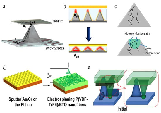
Figure 1. (a) Response mechanism of a SWCNT-coated polymer pyramidal pressure sensor [98]. (b) Mechanism of structural change of a transparent linear pressure sensor under pressure [99]. (c) Schematic of the conductive PDMS/CNT micropyramids, showing more conductive paths under stress [100]. (d) Schematic of fabrication process of electrospun fibers combined film with nanofiber mat [101]. (e) A pair of designed pyramids with dome-like tops touching the bottom film.
To solve this problem, Park et al. [99] improved the pyramidal structure to increase the effective contact area, thus obtaining high linearity in a wide pressure range, as shown in Figure 1b. The capacitance change of the device is determined by the distance between the electrodes coated on the surface and the top of the pyramid and the contact area between the electrodes and the pyramid structure. Moreover, with the increase in pressure, the distance between the two electrodes and the contact area between the electrode and the pyramid structure increase linearly, which promotes the linear range of the capacitor. Moreover, Li etc. [100] modified the internal structure and further increased the number of conductive paths, resulting in a decrease in volume resistivity. Figure 1c shows that when stress is applied, the stress concentrated at the tip of the pyramid reduces the distance between the internal conductive networks of PDMS/CNT composites. As the pyramid’s height decreases, the contact area increases significantly, which leads to a decrease in contact resistance, thus increasing the device’s current. Subsequently, Zhang etc. [101] combined a nanofiber mat and a micropyramid array by electrospinning to increase their pressure sensitivity and greatly decrease their detection limit, as shown in Figure 1d. They developed a pressure sensor featuring micropyramid arrays (HD-μPA) and an active piezoelectric material composed of a poly(vinylidenefluoride-co-trifluoroethylene) [P(VDF-TrFE)]/barium titanate (BTO) nanofiber-based mat with silver nanowires (AgNWs) assembled as connected film electrodes. Consequently, the sensitivity was improved by about 1.7 times that of a flat substrate sensor because of the stress concentration on the top and the unbalance of the elastic modulus between the polymer-based nanofiber mat. At the same time, the pressure detection limit of the structural pressure sensor can be as low as 0.6 Pa, which shows great potential with respect to wearable applications.
Furthermore, Liu et al. [102] designed a flexible pressure sensor with a uniquely engineered pyramid wall grid microstructure (PWGM) on polydimethylsiloxane (PDMS) film, as shown in Figure 1e. The square pyramid with a dome-shaped head and a large top area and the reinforcing wall connecting all the pyramids endow the sensor with a series of resistance changes; consequently, first the pyramid and then the various walls contact and deform under vertical pressure. Meanwhile, the interconnection between the pyramid and the wall improves the crushing pressure of the PWGM PDMS film, and its new delamination deformation mechanism makes it highly sensitive and quickly responsive and endows it with long-term stability. The pressure sensor shows excellent sensitivity and mechanical durability of 383,665.9, 269,662.9, 48,689.1, and 1266.8 kPa−1 in the pressure ranges 0–1.6, 1.6–6, 6.1–11, and 11–56 kPa, respectively, thus showcasing its potential applications in wearable health-monitoring electronics. In conclusion, a single-row pyramid structure presents high sensitivity under micro pressure due to its special triangular pyramid structure.
2. Pillar Microstructure
In order to pursue both high sensitivity performance as well as a large and stable linear-sensing recognition area, researchers have designed pressure sensors with a pillar-shaped microstructure. By varying the pillar’s geometry, a pressure sensor with a desirable pressure-sensing region can be obtained. The micropillar array is applied to the pressure sensor with the help of the air gap between the pillar structure and the underlying substrate, and its response and relaxation time scales can reach milliseconds [103]. The current passing through the device will depend on the contact resistance between the covered micro-pillar and the substrate film and the resistance of the substrate film between two adjacent micropillars. By changing these two resistance parameters, pressure sensors with different sensitivities in different pressure areas can be designed [104].
Figure 2a shows a typical pressure sensor device composed of Au micropillar arrays and a deformable PPy/PDMS substrate film [6]. One micropillar-structured pressure sensor achieved high sensitivity in the low-pressure range by controlling the change in the contact resistance between the Au-coated micropillar surface and the yielding PPy film. Figure 2a shows a typical pressure sensor device composed of Au micropillar arrays and a deformable PPy/PDMS substrate film. The bottom of the figure shows a top view and a cross section of a regular and uniform pillar array. With a device composed of similar structures, Ha et al. [105] replaced the electrode with an improved Ag nanowire sticker to stabilize the electrical performance of pressure sensor on a deformable substrate under tension, as shown in Figure 2b. The polyaniline nanofibers are collected on a PET film as the bottom layer, and the gold-coated PDMS micropillars are used as the top layer.
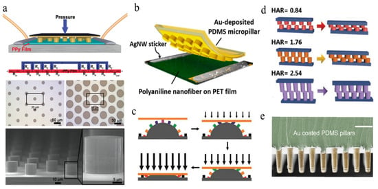
Figure 2. Schematic of micropillar array-based pressure sensor. (a) Detailed structure of pressure sensor based on micropillar array includes the PPy/PDMS substrate, a gold-covered micropillar array, and the optical images of photolithographically fabricated patterns [6]. (b) Primary design of the pressure sensor composed of PDMS micropillars and polyaniline nanofiber film [105]. (c) Schematic of in situ structural variation of hybrid structure under increased pressure load [106]. (d) Comparison of pillar-based pressure sensor with different ratio of height to diameter under pressure [107]. (e) SEM image of Au-coated PDMS micropillars. Scale bar: 50 μm [108].
More recently, different structural designs have been developed in order to improve sensitivity, such as that developed by Zhou et al. [106], which demonstrated a hybrid structure of interlocked micropillars and mesodomes (shown in Figure 2c). The increased number of micropillars brought about by a structural design featuring interlocked assembly not only increased the surface contact area but also caused larger pressure-induced deformation induced by the interlocking micropillars and mesodomes. Therefore, in terms of sensitivity and detectable pressure range, compared with the traditional structure based on single-fold domes, this hybrid structure shows excellent sensing performance.
To modify the micropillar structure, Hu et al. [107] discussed the influence of aspect ratios on micropillar structures by comparing the sensitivity of pressure sensors with three different height-to-diameter ratios, as shown in Figure 2d. It can be seen that the sensor with a greater height-to-diameter ratio shows higher sensitivity in the low-pressure range. Furthermore, Fang et al. [108] reported an Au-coated PDMS micropillar sensor with high-aspect-ratio microstructures, which can sense touch and detect weak physiological signals, even fingertip pulses. Figure 2e shows the skin–electrode mechanosensing structure of micropillars with a length (L) to radius (R) aspect ratio of 6. Unlike other sandwich structures, the skin–electrode mechanosensing structure consists of two electrodes, and the skin is designed to utilize ion transport in biological systems. Due to its ionic and electronic properties, the structure presents low noise but high signal strength when touched. Further mechanical analysis reveals that the instability of high-aspect ratio microstructures plays a critical role in sensing.
3. Microdome
Another form of microdome-shaped geometry has been developed to obtain a stable pressure sensor without experiencing fatigue during multiple cycles. Compared with pyramid and pillar structures where the pressure is concentrated at the tip, because a microdome’s geometry can expand the external tension more uniformly, this microstructure offers a stabler structure under pressure [109,110,111] and leads to a wider linear range and cyclic stability [112]. The linear range can be further widened by adjusting the microstructure of the sensing layer. Due to the special microdome shape of the elastomer, the contact area with the conductive electrode increases under external pressure, resulting in a significant reduction in the tunneling resistance or contact resistance. Although the sensors assembled with this structure show excellent sensing performance, they still suffer from problems such as the imbalance between high sensitivity and a wide linear range, which requires further optimization of the structure and composition [113,114].
To improve its sensitivity, Ko et al. used [115] a microdome structure design of interlocked microdome arrays, which display extreme resistance-switching behavior, including significant improvements in sensitivity and response/relaxation times. This massive improvement is achieved by the interlocking of the microdome arrays, which leads to an increase in the tunneling resistance at the contact point and the tunneling piezoresistance in the flexible film, as shown in Figure 3a. Later, Lee et al. [116] filled urchin-shaped metal nanoparticles into a polyurethane particle array to prepare a highly sensitive pressure sensor (71.37 kPa−1) with high optical transmittance (77.7% at 550 nm), as shown in Figure 3b. The excellent sensing performance of the transparent piezoresistive pressure sensor can be attributed to the effective quantum tunneling effect caused by the stress concentration at the small contact point and the deformation in the contact area.
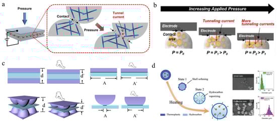
Figure 3. Schematic of microdome array-based pressure sensor. (a) Schematic of the working principle for interlocked microdome arrays with conductive nanofiber inside. Under pressure, the deformation of the micro dome increases the contact area and tunneling current of nanofibers [115]. (b) Schematic of microdomes composed of metal nanoparticles under pressure [116]. (c) Schematic diagram showing the difference in distance and contact area between film and microdome array under pressure [117]. (d) Thermal expansion of microdome with irregular structure under applied temperature [118].
Recently, Hu et al. [117] assembled a capacitive pressure sensor by adding a dielectric layer of polyvinylidene fluoride (PVDF) between two microdome-shaped conductive soft layers. For comparison, the internal stresses of the devices with flat and microdome-shaped conductive layers under pressure are shown in Figure 3c. As the contact area of the device with a flat conductive layer hardly changes under pressure, the relative capacitance of the device is only determined by the change in the dielectric layer’s thickness. In contrast, the microstructure in the devices with microdome-shaped conductive layers will deform significantly under pressure, leading to a change in the thickness and contact area of the dielectric layer. Hence, a noticeable improvement in sensitivity was observed in the microdome device. To modify the surface morphology of microdome arrays, Cho et al. [118] fabricated a 3D microstructure elastomer for flexible pressure sensors via the internal popping of microspheres. Each microsphere possesses a core–shell structure, consisting of thermoplastic resin as the shell and liquid hydrocarbon as the core. Figure 3d (left) shows the volume expansion process of the microspheres from state 1 to state 2 and, finally, the “expansion state” under a specific temperature. For a comparison of the volume change, Figure 3d (right) shows the SEM images and particle diameter distribution before and after the expansion.
4. Porous Sponge Microstructure
The structural characteristics of porous materials are similar to spongy and loose microstructures, which make them easier to deform under low pressure; thus, these materials exhibit a low detection limit and a larger deformation detection range. In addition, porous materials are very suitable for wearable flexible electronic devices due to their ultra-high illumination and good permeability. Researchers used an internal porous microstructure as a conductive or dielectric layer to fabricate lightweight pressure sensors. At the same time, the unique pressure detection function of porous microstructures in a large size range and in different planes enables them to be applied to detection in 3D space [119,120,121]. According to the requirements of different flexible wearable devices, such as light weight, low detection limit, and other characteristics, researchers have developed various porous microstructures with low density and high compressibility [122].
Due to the unique interconnective network of a porous sponge, electrons can move very quickly through the 3D network of the seamless interconnections of the conductive network, thus imparting very high electrical conductivity to the structure. It is very important to build a highly connected conductive filler network in the insulating matrix to improve the conductivity of the composite [123]. A typical fabrication process of a porous sponge-microstructured sensor is shown in Figure 4a [124]. This figure shows multilayer graphene grown on a nickel foam template by the chemical vapor deposition (CVD) method. Then, nickel foam coated with graphene sheets was immersed into prepared PDMS. The composite sample was subsequently cured in a hot plate at 100 °C, and the nickel skeleton was removed using hydrochloric acid. A composite of a graphene skeleton surrounded by PMDS was fabricated by removing the redundant PDMS. Moreover, the composite displayed good bending, torsional, and stretching properties.
Recently, some conductive organic composite polymers have replaced the PDMS-based 3D frame structure, and the assembled sensors show good pressure-sensing abilities [125,126,127,128]. A conductive sponge copolymer composed of poly (3,4-ethylenedioxythiophene): poly (styrene sulfonate) (PEDOT: PSS) was constructed, and it presented a stable piezoresistive response under a compression strain of up to 80% [129]. Figure 4b shows a photograph and SEM image of the porous sponge sample as well as the structural changes during compression. Under compression force, the conduction mechanism is called the negative piezoresistive effect, which forms more conductive paths in the conductive elastomer composite or sponge, resulting in the lower resistance of the sensor. Due to the pores in the composite’s pressure and strain sensors, it can realize the strain-sensing characteristics of pressure and tension at the same time and shows a wide pressure-sensing range. The sponge reveals its robust porous morphology, high mechanical compressibility, high sensitivity, and stable compress−release cycles over 1000 cycles.
More recently, with the development of 1D nano/micro particles or 2D materials such as Fe2O3 particles, reduced graphene oxide, carbon nanofibers, MXene, etc. [130,131,132,133,134,135,136], Li et al. [137] successfully combined MXene with aerogel to increase its conductivity in order to assemble an interdigital sensor system, as shown in Figure 4c. The 3D porous MXene aerogel was composed of MXene nanosheets and cellulose nanofibers, which provide enough conductive channels with and without an external force. Finally, the 3D MXene aerogel was encapsulated in a PU layer to integrate a pressure-sensing device with self-healing properties. In addition, the MXene aerogel pressure sensor shows excellent response sensitivity of 306 kPa−1 with a wide pressure detection range from 2.3 Pa to 87.3 kPa and a fast response time of 35 ms. Moreover, it shows remarkable stability over 2000 cycles and confers self-healing characteristics to the system, constituting properties that can be applied in a variety of wearable sensor devices.
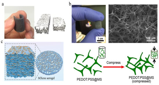
Figure 4. Schematic of pressure sensor based on porous composite material. (a) Photograph of a bendable porous composite sensor with GPN-PDMS and Schematic of the porous structure [124]. (b) Photograph of PEDOT:PSS-coated melamine sponge, SEM image of the porous sponge, and schematic of structural change under compression [129]. (c) Schematic of 3D porous sensor composed of MXene aerogel [137].
5. Bio-Inspired Microstructure
Biomimetic materials are widely used in multi-functional flexible electronic materials [138,139]. In 2014, for the first time, Choi et al. [31] made use of nanoscale crack connections inspired by the geometry of a spider’s slit organs to prepare sensors with ultra-high sensitivity (gauge factor exceeds 2000) when detecting small vibrations (0–2% strain range). Figure 5a shows a schematic of the disconnection–reconnection mechanism of the sensor with ultra-high mechanical sensitivity and a zipper-like nanoscale crack connection under strain. Spiders use the slit organs on their leg joints to detect external vibrations and transmit signals to the nervous system.
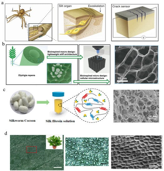
Figure 5. Schematic illustrations and images of bio-inspired pressure sensors. (a) Schematic of a spider’s sensory system and an ultra-mechanosensitive pressure sensor based on spider-inspired nanoscale crack junctions [31]. (b) Schematic of Elytrigia repens and 3D-printed hierarchical structure. SEM image of the hierarchical structure [140]. (c) Schematic illustration of the fabrication of the silk fibroin-based hydrogel. SEM image of interconnected porous architecture (right) [141]. (d) Optical images of an aureum leaf and SEM image of a replicated microstructured PDMS film. Scale bar: 1000 μm (left), 100 μm (middle), and 100 μm (right) [142].
Later, Jiang et al. [140] reported a bionic hierarchical graphene material (BHGM) that exhibits ultra-high elasticity and stability when the compressive strain reaches 95%. The BHGM was constructed by imitating the structure of Elytrigia repens and is similar to an ice-crystal-induced cellular microstructure, as shown in Figure 5b (left). Elytrigia repens shows remarkable mechanical properties and light weight due to its gradually formed hierarchical structure and macro hollow structure and micro cellular structure. Researchers utilized an ink-based 3D printing strategy to prepare 3D multi-layer porously structured BHGMs (Figure 5b, right). The imitation of Elytrigia repens’s structure also grants the BHGM remarkably low weight and extremely high stiffness and elasticity.
Other structures such as silk fibroin-based hydrogels exhibit remarkable extensibility and compressibility, thus enabling them to be assembled into strain/pressure sensors with a wide range from 2% to 600% and good stability over multiple cycles [141]. Figure 5c shows the fabrication procedures of the composite hydrogel composed of silk fibroin, polyacrylamide, graphene oxide, and poly(3,4-ethylenedioxythiophene):poly(styrenesulfonate). The SEM images display the interconnected porous structure of the composite hydrogel, which leads to its excellent elastic and mechanical stability, thus allowing it to be manufactured into any shape.
Recently, researchers have used elastic PDMS to replicate the rough surface of Epipremnum aureum leaves and successfully prepared this bio-inspired microstructure [142,143]. Not only does this pressure sensor with a bio-inspired hybrid porous surface show an excellent sensitivity of 83.9 kPa−1 and excellent stability (>28,000 cycles), but it is also more attractive due to its ultra-low detection limit of less than 0.5 Pa, as shown Figure 5d. The optical images of a typical aureum leaf are shown in Figure 5d (left and middle) under scale bars of 1000 and 100 μm, respectively. In addition, the surface morphology of the replicated microstructured PDMS is shown on the right side of Figure 5d. The bio-inspired hybrid porous microstructure shows an increased contact area and reduced = Young’s modulus and introduces an additional level of pore resistance. Consequently, the pressure sensor exhibits high sensitivity and a low limit of detection. These aspects enable the designed sensor to detect finger pressure, sound vibrations, swallowing activity, and wrist pulse, and showcase its potential applications in artificial intelligence and flexible medical electronic applications.
This entry is adapted from the peer-reviewed paper 10.3390/polym15030764
This entry is offline, you can click here to edit this entry!