Radiofrequency-Electromagnetic Fields Exposure: History
Please note this is an old version of this entry, which may differ significantly from the current revision.
Contributor:

The interaction of electromagnetic fields (EMF) with living tissues or manufactured objects can produce different effects on these exposed entities. In different circumstances, these fields are used daily in various friendly applications. Applications of non-ionizing RF-EMF, frequencies from 100 kHz to 300 GHz have continuously increased. This involves medicine (e.g., magnetic resonance imaging and RF ablation), manufacturing (e.g., heaters and solders), home usages (e.g., child video display unit and Wi-Fi), protection and navigation (e.g., radar and RFID) and particularly in telecommunications (e.g., TV transmitting and mobile phones). This growth signifies that great amounts of the population are undergoing exposure to RF(radiofrequency)-EMF and worry has been inflated concerning public health concerns due to such exposure.

  • electromagnetic fields
  • radiofrequency exposure
  • biological effects
  • atypical symptom

1. Characteristics of Sources and Interactions

The sources of RF-EMF are of two categories, devices functioning nearby the human body, occasionally a near field exposure interacting highly constrained in a part of the body, and sources functioning distantly from the body that create an entire-body uniform exposure. Approximately, far field corresponds to transmitter-receiver distance greater than a wavelength and its strength diminishes quickly with distance. Classic near field sources are mobile and cordless phones. Characteristic far field sources include TV posts, mobile and cordless phones base stations, Wi-Fi access points or neighboring mobile phones.
Interaction of RF-EMF with a human body is affected by the frequency, the field intensity, the exposure interval, the field polarization and the dielectric properties of absorbing matters. Moreover, in case of simultaneous activation of several EMF radiating sources nearby the body we have to account for such complex interactions. The nature of interactions can produce different BE. These effects can be due to short or long-term exposure. The most common effect is the thermal short-term one. There are also lesser frequent short and long-terms effects. The non-thermal complex effects fall in this category, as well as the cases exposing atypical symptoms.
The thermal effects and the cases exposing atypical symptoms will be detailed in the next subsections while the non-thermal effects will be discussed later.

2. Thermal BE Due to RF-EMF

Regarding the thermal effects, exposure to relatively high RF-EMF (strength and duration) can be hazardous to living organisms. Such exposure may lead to body heating resulting in an increase in temperature, which may cause tissue damage. Two characteristics reinforce this occurrence. The first relates to the aptitude of RF energy to, rapidly heat biological tissues, similarly to how microwave ovens that cook food. The second concerns the body’s incapability to withstand or dissipate the disproportionate heat that can be produced. Note that the parts of the body least protected from RF-EMF heating are those that lack accessible blood circulation, which is the primary means of dealing with extreme heat. The magnitude of such heating is correlated to several circumstances involving the field intensity, the frequency of the waves, the exposure interval, the heat dissipation capacity of the tissues, the surrounding environment and the size, shape and positioning of the exposed body.
Note that excessive strength fields can display non-thermal effects. One of the most popular BE in this case is the interruption of brain electro-wave due to the important external EMF that causes altered cell secretion. In addition, EM-induced membrane electroporation also affects cell activation.

3. Health Safety Standards

The thermal customary biological effect induced by exposure to EMF can be quantified by the specific absorption rate (SAR) which evaluates the energy absorbed by an element of the tissue. The SAR if multiplied by the exposure interval signifies the EMF entire-body or part of the tissue-specific absorbed energy amount. This energy generates a rise in tissue temperature, which can increase health risks. The SAR and corresponding temperature rise for given frequency and exposure time can be represented by, see [1] for more details:
SAR = P/ρ = σ E2/(2 ρ)
ΔT = σ E2 Δt/(2 c ρ)
In (1) and (2), SAR is the specific absorption rate in (W/kg). P is the power loss density in (W/m3), E is the electric field strength in (V/m), ΔT is the temperature rise in (°C) and Δt is the exposure time in (s). The tissue properties parameters ρ, σ, c are the density in (kg/m3), the conductivity in (S/m) and the specific heat in J/(kg °C), respectively. Note that the conductivity σ of human tissues depends not only on matter but also on frequency. It is mostly constant with growing frequency up to megahertz and then rises, often nearly linearly.
Expressions (1–2) indicate that the higher the SAR and the longer the exposure time, the greater and more dangerous the increase in temperature will be. This behavior will be affected in addition to the field (intensity and frequency) and the exposure time by the density of the tissue and its thermo-electric properties. Thus, the SAR limits imposed by health safety depend on the part of the body considered and the nature of the subject exposed as well as the exposure conditions. Exposed subjects can be adult humans, children or animals. The exposed parts of the body are the head, trunk and limbs. Exposure conditions include different categories of exposed subjects (allied to the liaison with the source of exposure): workers involved in manufacturing, testing, and installing devices, users of the device, and nearby subjects. For all these situations, health safety standards set thresholds relating to SAR, ΔT and fields triggered in the human body: magnetic flux density B, electric field E and current density J. Figure 1 summarizes the different input parameters, nature and condition of exposure and there effects on tissue. These effects must comply with the corresponding thresholds.
Figure 1. Input EMF exposure (parameters, nature and condition) and output effects on tissue.

4. AS Due to RF-EMF

As stated earlier, EMF exposure has two categories of effects on human health. The first is related to short-term exposure to EMF, and the second is result of long-term exposure. Short-term effects are those relative to usual thermal effects as well as that result in AS for EHS people showing non-specific symptoms mentioned earlier. Long-term ones are those related to non-thermal effects as well as AS long-term effects on CF.
As mentioned previously, in rare cases people present AS owing to exposure to RF-EMF. Among these persons are subjects with EHS that present with non-specific symptoms. These range widely between people and include headaches, weakness, anxiety, trouble sleeping, skin tingling, burning sensations, muscle problems with agony and pain and other different symptoms. Historically, this phenomenon appeared a few decades ago and in particular with the advent of modern digital communication technologies [2][3][4][5][6][7][8][9][10][11][12][13].
These non-specific symptoms were presented for a wide range of frequencies and different types of sources, but the vast majority involved RF sources. Even those EMF in the RF range act to the greatest danger to humans if the field strength and SAR are relatively high; EHS symptoms can be present for non-significant values of these measures. For example, the SAR activated in such sources could reach in certain uses about ten W/kg depending on the intensity of the field and its frequency. For a short time, this is enough to cause a temperature rise of 1 °C in body tissues, which in a normal healthy case is completely safe. At the same time, for SARs less than 0.1 W/kg, symptoms may be present in an EHS subject. This negligible level of exposure seems surprising given the many radiating RF-EMF tools in the everyday environment. Indeed, EHS subjects often fall into the category of users nearby and relatively far from the tools for which the field decreases as the square of the distance. In addition, these tools are generally subject to electromagnetic compatibility rules and adapted to avoid BE in the body tissues of users [14][15][16]. The EHS symptoms seem real and their presence is associated with EMF even if the biological effect of the latter seems non-existent. Most investigations only consider EMF-induced BE that are acknowledged by the scientific medical organization. In fact, health agencies are gradually admitting RF EHS. Subjects enduring EHS expose symptoms in cases of field insignificant enough not to create perceptible BE. The term hypersensivity (HS) may introduce a misinterpretation and one may use idiopathic environmental intolerance (IEI), which signify better the state of sickness. Thus, it can be used in case of EM, EHS or EIEI. According to patients and often verified by double blind (patient and supervisor) experiences, these symptoms appear only occasionally at first, but then they become more prominent and more persistent. Even with such an insignificant level of exposure, one can think that electromagnetic effects ignored today may perhaps emerge below the established exposure intensities and elucidate the EHS symptoms. Decidedly, it is scientifically difficult to refute the presence of a danger. Such uncertainty can constantly justify a cautious attitude [17].
As in the case of subjects with EHS, there is  another similar case of AS relative to the same type of exposure but for longer duration. This is the case of long-term effects of exposure to RF-EMF on CF. As in the case of EHS, this problem appeared with the advent of modern digital communication technologies and in particular the use of mobile cell phones. The RF-EMF exposure from these devices affects directly the brain tissues and consequently the cognitive function that involve diverse cerebral abilities. These can be weighed in a sort of spheres, for instance understanding, remembrance, rationalizing, trouble explaining, judgement acting and concentration capacity. Observation assessments can be used to analyze these problems even that only instant effects can be observed while the needed results could only be obtained after at least small or long-term exposure.
Different reviews involving analysis of results have been realized concerning RF-EMF exposures in both EHS non-specific symptoms and CF long-term effects symptoms, see, e.g., [18][19][20].

5. Evaluation of RF-EMF BE and AS

The evaluation of BE and AS can be performed by modeling, experience or observational studies. In the case of evaluation through modeling, the governing mathematical equations can be solved locally in the body tissues concerned by the effects or symptoms. This can be performed by using numerical discretized techniques or other methods permitting local evaluation, e.g., [21][22][23][24][25][26][27][28][29]. This involves the EM equations and the bio-heat tissue equation [30]. In the case using measurements for evaluation, sensors can be placed in tissues (when possible) to detect the needed fields (electric, magnetic, thermal). In addition, imagers can be utilized for the revealing of tissue comportment concerning a stated symptom. Concerning observational assessment, this involves mainly, as mentioned earlier, tests in the case of AS practiced on patients exhibiting these symptoms. Such tests are generally performed in a double-blind manner on a group suffering from the same symptom.

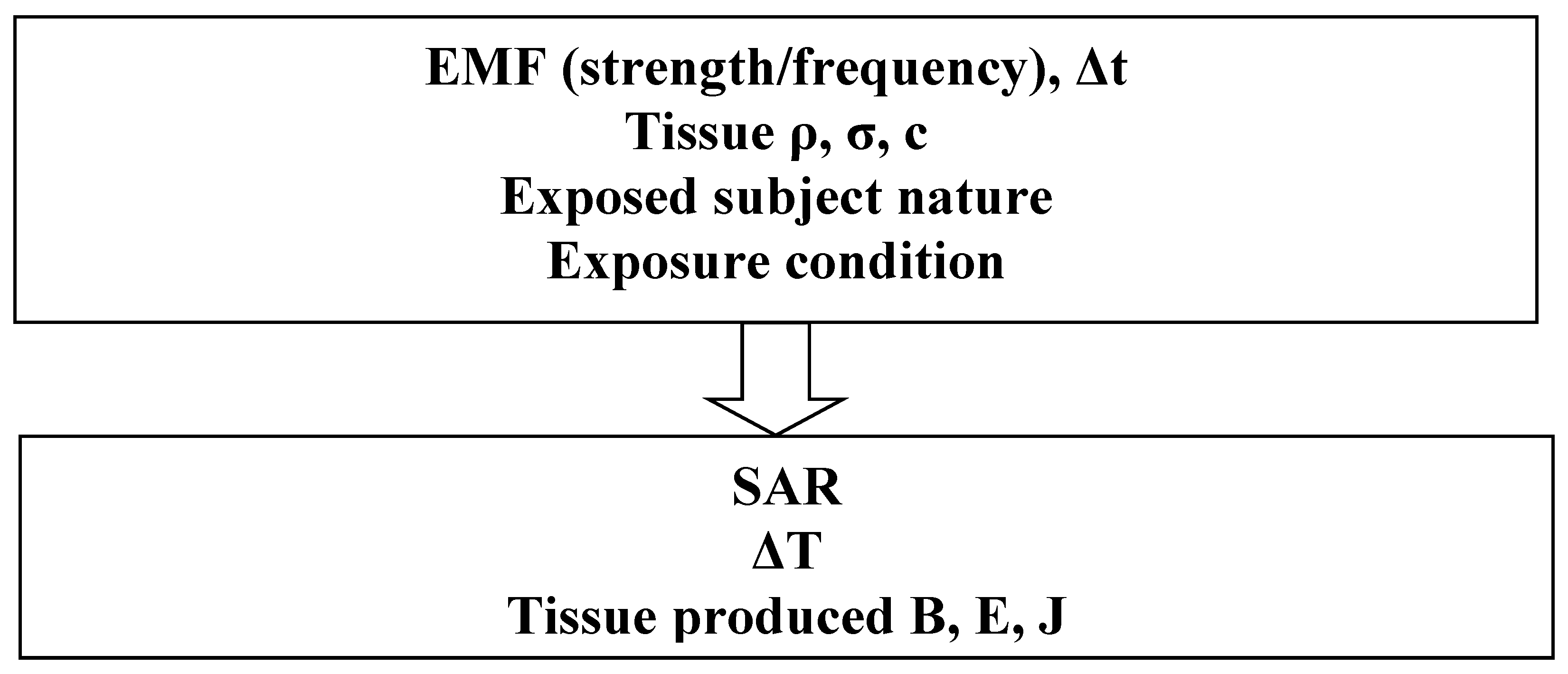
6. Governing Mathematical Equations

The governing equations are the Maxwell EMF equations and the Penne’s bio-heat tissue equation [30]. The external source in the bio-heat equation corresponds to the SAR, which is determined from the EMF equations. These governing equations can be given by:
× H = J
J = σ E + j ω D + Je
E = −∇ V − j ω A
B = × A
c ρ ∂T/∂t = (k T) + ρ (SAR) + qmet − 𝑐b 𝜌b 𝜔b (T − Tb)
In the EM Equations (3)–(6), H and E are the magnetic and electric fields, B and D are the magnetic and electric inductions, A and V are the magnetic vector and electric scalar potentials. J and Je are the total and source current densities, σ is the electric conductivity and ω is the frequency pulsation. The symbol is a vector of partial derivative operators, and its three possible implications are gradient (product with a scalar field), divergence and curl (dot and cross products, respectively, with a vector field). The magnetic and electric comportment laws between B/H and D/E are represented by the permeability μ and the permittivity ε, respectively.
As well in the Penne’s bio-heat Equation (7), k is tissue thermal conductivity, T is local temperature of tissue, qmet is the basal metabolic heat source in W/m3, cb is blood specific heat in J/(kg·°C), 𝜌b is blood density in kg/ m3, 𝜔b is blood perfusion rate (1/s), Tb blood temperature. · (k T) represents simple heat equation in differential form and ρ (SAR) represents the influence of electromagnetic energy absorbed in the human tissues.
The solution of Equations (3)–(6) allows us to determine the induced EMF, for a given frequency pulsation, in the body tissues. As well, the SAR (1) can be computed using the fields resulting from such solution. Additionally, the EMC analysis checking the perturbations due to EMF exposers of instruments (embedded or not) can be verified from such solution. Penne’s bio-heat equation [30] given by (7) is usually used to determine heat transfer in living tissues. Thermal behavior in tissues due to EMF exposure through the SAR is governed by Equations (1), (3)–(7). The EMF equations and the heat transfer equation must be solved in a coupled fashion. Due to slow thermal time behavior compared to fast EM time behavior, weak coupling with successive solution can be used.

This entry is adapted from the peer-reviewed paper 10.3390/app13031265

References

  1. Razek, A. Biological and Medical Disturbances Due to Exposure to Fields Emitted by Electromagnetic Energy Devices—A Review. Energies 2022, 15, 4455.
  2. Eriksson, N.; Höög, J.; Mild, K.H.; Sandström, M.; Stenberg, B. The psychosocial work environment and skin symptoms among visual display terminal workers: A case referent study. Int. J. Epidemiol. 1997, 26, 1250–1257.
  3. Lidén, S. “Sensitivity to electricity” a new environmental epidemic. Allergy 1996, 51, 519–524.
  4. Bergqvist, U.; Vogel, E. Possible Health Implications of Subjective Symptoms and Electromagnetic Fields; A Report Prepared by a European Group of Experts for the European Commission, DG V; National Institute for Working Life: Stockholm, Sweden, 1997; p. 135. Available online: https://gupea.ub.gu.se/bitstream/handle/2077/4156/ah1997_19.pdf?sequence=1 (accessed on 10 January 2023).
  5. Lindén, V.; Rolfsen, S. Video computer terminals and occupational dermatitis. Scand. J. Work Environ. Health 1981, 7, 62–64.
  6. Carpenter, D.O. The microwave syndrome or electro-hypersensitivity: Historical background. Rev. Environ. Health 2015, 30, 217–222.
  7. Lin, J.C. Early contributions to electromagnetic fields in living systems. In Advances in Electromagnetic Fields and Living Systems; Lin, J.C., Ed.; Plenum Press: New York, NY, USA, 1994; pp. 1–26.
  8. Bren, S. Historical introduction to EMF health effects. IEEE Eng. Med. Biol. 1996, 15, 24–30.
  9. Dieudonné, M. Controverses autour des champs électromagnétiques et de l’électrohypersensibilité. La construction de problèmes publics “à bas bruit”. St. Publique 2019, 31, 43.
  10. Schreier, N.; Huss, A.; Röösli, M. The prevalence of symptoms attributed to electromagnetic field exposure: A cross-sectional representative survey in Switzerland. Soz. Praventiv. Med. 2006, 51, 202–209.
  11. Schröttner, J.; Leitgeb, N. Sensitivity to electricity–temporal changes in Austria. BMC Public Health 2008, 8, 310.
  12. Blettner, M.; Schlehofer, B.; Breckenkamp, J.; Kowall, B.; Schmiedel, S.; Reis, U.; Potthoff, P.; Schuz, J.; Berg-Beckhoff, G. Mobile phone base stations and adverse health effects: Phase 1 of a population-based, cross-sectional study in Germany. Occup. Environ. Med. 2009, 66, 118–123.
  13. Karvala, K.; Sainio, M.; Palmquist, E.; Nyback, M.-H.; Nordin, S. Prevalence of various environmental intolerances in a Swedish and Finnish general population. Environ. Res. 2018, 161, 220–228.
  14. International Commission on Non-Ionizing Radiation Protection. Guidelines for limiting exposure to time-varying electric and magnetic fields for low frequencies (1 Hz–100 kHz). Health Phys. 2010, 99, 818–836.
  15. International Commission on Non-Ionizing Radiation Protection. Guidelines for limiting exposure to electromagnetic fields (100 kHz to 300 GHz). Health Phys. 2020, 118, 483–524.
  16. IEEE Standard C95.1; IEEE Standard for Safety Levels with Respect to Human Exposure to Electric, Magnetic, and Electromagnetic Fields, 0 Hz to 300 GHz. IEEE: Piscataway, NJ, USA, 2019.
  17. Sunstein, C.R. Beyond the precautionary principle. Univ. Pa Law Rev. 2003, 151, 1003–1058.
  18. Schmiedchen, K.; Driessen, S.; Oftedal, G. Methodological limitations in experimental studies on symptom development in individuals with idiopathic environmental intolerance attributed to electromagnetic fields (IEI-EMF)—A systematic review. Environ. Health 2019, 18, 88.
  19. Ishihara, T.; Yamazaki, K.; Araki, A.; Teraoka, Y.; Tamura, N.; Hikage, T.; Omiya, M.; Mizuta, M.; Kishi, R. Exposure to Radiofrequency Electromagnetic Field in the High-Frequency Band and Cognitive Function in Children and Adolescents: A Literature Review. Int. J. Environ. Res. Public Health 2020, 17, 9179.
  20. Dieudonné, M. Electromagnetic hypersensitivity: A critical review of explanatory hypotheses. Environ. Health 2020, 19, 48.
  21. Cirimele, V.; Freschi, F.; Giaccone, L.; Pichon, L.; Repetto, M. Human Exposure Assessment in Dynamic Inductive Power Transfer for Automotive Applications. IEEE Trans. Magn. 2017, 53, 5000304.
  22. Hariri, H.; Bernard, Y.; Razek, A. Dual piezoelectric beam robot: The effect of piezoelectric patches positions. J. Intell. Mater. Syst. Struct. 2015, 26, 2577–2590.
  23. Nunes, A.; Daniel, L.; Hage Hassan, M.; Domenjoud, M. Modeling of the magnetic behavior of permanent magnets including ageing effects. J. Magn. Magn. Mater. 2020, 512, 166930.
  24. Ren, Z.; Razek, A. New technique for solving three-dimensional multiply connected eddy-current problems. IEE Proc. 693 A Phys. Sci. Meas. Instr. 1990, 137, 135–140.
  25. Ren, Z.; Razek, A. Boundary edge elements and spanning tree technique in three-dimensional electromagnetic field computation. Int. J. Numer. Methods Eng. 1993, 36, 2877–2893.
  26. Jiao, D.; Jin, J.-M. An effective algorithm for implementing perfectly matched layers in time-domain finite-element simulation of open-region EM problems. IEEE Trans. Antennas Propag. 2002, 50, 1615–1623.
  27. Carpes, W.P.; Pichon, L.; Razek, A. A 3D finite element method for the modelling of bounded and unbounded electromagnetic problems in the time domain. Int. J. Numer. Model. Electron. Netw. Devices Fields 2000, 13, 527–540.
  28. Sun, Q.; Zhang, R.; Zhan, Q.; Liu, Q.H. 3-D Implicit–Explicit Hybrid Finite Difference/Spectral Element/Finite Element Time Domain Method without a Buffer Zone. IEEE Trans. Antennas Propag. 2019, 67, 5469–5476.
  29. Ding, P.; Bernard, L.; Pichon, L.; Razek, A. Evaluation of Electromagnetic Fields in Human Body Exposed to Wireless Inductive Charging System. IEEE Trans. Magn. 2014, 50, 1037–1040.
  30. Pennes, H.H. Analysis of tissue and arterial blood temperatures in the resting human forearm. J. Appl. Physiol. 1998, 85, 5–34.
More
This entry is offline, you can click here to edit this entry!