Pathophysiology of Exercise-Induced Pulmonary Hypertension: History
Please note this is an old version of this entry, which may differ significantly from the current revision.
Contributor: , , ,

Exercise-induced pulmonary hypertension (EIPH) has been defined as an increase in mean pulmonary arterial pressure (mPAP) during exercise in otherwise normal values at rest. EIPH reflects heart and/or lung dysfunction and may precede the development of manifest pulmonary hypertension (PH) in a proportion of patients. It is also associated with decreased life expectancy in patients with heart failure with reduced ejection fraction (HFrEF) or left ventricle (LV) valvular diseases. Diastolic dysfunction exacerbated during exercise relates to increased LV filling pressure and left atrial pressure (LAP). In this context backward, transmitted pressure alone or accompanied with backward blood flow promotes EIPH. 

  • exercise-induced pulmonary hypertension (EIPH)
  • pathophysiology
  • right heart catheterization (RHC)

1. Introduction

Exercise-induced pulmonary hypertension (EIPH) has been defined as an exaggerated increase in mean pulmonary artery pressure (mPAP) >30 mmHg and total pulmonary resistance (TPR) > 3 Wood units during exercise, while an mPAP <25 mmHg is present at rest [1]. Patients are free of symptoms at rest, and they complain only of fatigue or dyspnea during exercise.
It is increasingly recognized that EIPH may represent an early, mild stage of pulmonary arterial hypertension, and preliminary data suggest its association with an overall higher morbidity and mortality [2]. Past evidence has shown the relationship between exercise-induced elevation of pulmonary artery pressure (PAP) with exercise intolerance and exertional dyspnea in patients without established diagnosis of pulmonary hypertensions (PH), underlining its clinical importance [3]. In addition to this, EIPH has been associated with reduced exercise capacity and worse functional status in patients with heart failure with either reduced ejection fraction (HFrEF) or preserved ejection fraction (HFpEF) [4] or other cardiac disorders (cardiac restriction, chronotropic incompetence) [5]. Furthermore, the early appearance of EIPH in patients with left ventricle (LV) valvular diseases, such as mitral regurgitation (primary or secondary), and mitral and aortic stenosis, may be of clinical relevance, giving a valuable tool for the effective risk stratification of asymptomatic patients. EIPH may be undiagnosed in a significant proportion of patients with exertional dyspnea but normal resting PAP because of the considerable limitations of PAP measurements during exercise. Right heart catheterization (RHC) during exercise seems to be the most objective approach for conclusive diagnosis, but it is invasive and sometimes complicated, limiting its clinical applicability. Other non-invasive, objective, reproducible and easily performed diagnostic methods of EIPH assessment are required in clinical practice but they have made the exact definition of EIPH a subject of debate using variable cut-off values (e.g., pulmonary artery systolic pressure (PASP)).
The term “exercise induced pulmonary hypertension” was suspended at the 4th World Symposium of Pulmonary Hypertension in Dana Point, in 2008, largely due to failures at reaching a consensus. Since then, a plethora of studies have been published about EIPH, its pathophysiology and methods of assessment. A body of data increasingly supports EIPH as an important and promising entity for the research and clinical community. Despite this, its unambiguous association with exertional dyspnea and its clinical usage in the prevention, early diagnosis and management of cardiopulmonary pathologies remains controversial.

2. Pathophysiology of EIPH

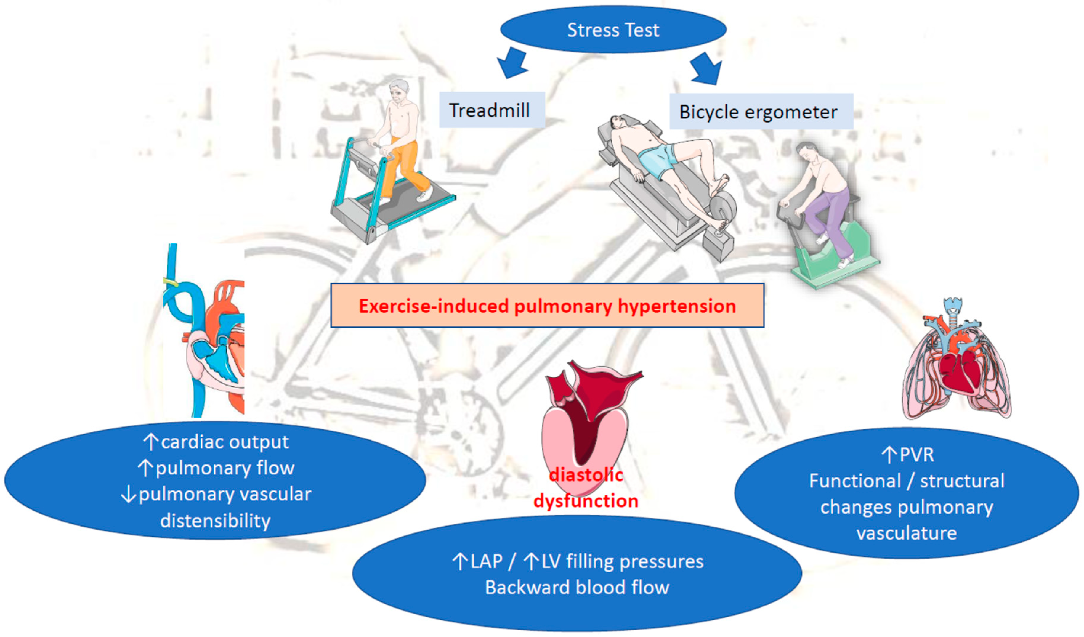
Significant progress has been made in understanding the pathophysiology of the hemodynamic abnormalities of cardiopulmonary circulation during exercise. Although EIPH pathogenesis is still not completely understood, it can be attributed to alterations in pulmonary artery flow affected by cardiac output (CO), pulmonary vascular resistance (PVR) and left atrial pressures (LAP) during exercise, as indicated by the Poiseuille–Hagen equation: mPAP = PVR × CO + LAP [6]. EIPH appears to be most similar to the WHO group 2 PH [5], and the following 3 pathophysiological mechanisms have been proposed:
(A) Increased pulmonary flow: Unambiguously, cardiac output (CO) and, consequently, pulmonary blood flow significantly increase during exercise in response to the increased oxygen uptake. In parallel, there is a significant pulmonary vascular dilatation as a compensatory mechanism. The magnitude of the inevitable elevation of PAP and, to a lesser extent of wedged PAP (wPAP), is a product of increased blood flow balanced by pulmonary vascular resistance (PVR). In healthy individuals, PVR as a result of pulmonary vascular distensibility is widely adapted to high pulmonary blood flow in order to maintain PAP within a normal range [7]. Previous invasive assessments have shown that wPAP and left ventricular filling pressure (named end-diastolic LV pressure) can rise >30 mmHg during exercise in athletes and exercising elderly [8]. A disproportional increase in CO over moderately declined PVR results in high wedged PAP (wPAP) and LAP during exercise. A possible explanation for this derives from the Frank–Starling mechanism matching of the LV flow output to the peripheral demand [8]. Moreover, the elite athletes can generate extraordinarily high cardiac flows and exceed mPAP of 30 mmHg at very high levels of exercise. It has been hypothesized that the additional effect of pericardial constrain, in particular, the increasing LV diastolic volume, competes for space with the right ventricle within the relatively non-distensible pericardium. In that case, the LV diastolic compliance is possibly impaired resulting in a remarkable elevation of LAP [7].
Another factor determining pulmonary blood flow and PAP elevation during exercise is the type of exercise. During aerobic exercise, pulmonary blood flow has shown a linear progressive increase and thus a linear adjustment in the mPAP–O correlation may occur, whereas resistance exercise is associated with exaggerated pulmonary vasoconstriction and higher PVR [3][6]. The fact that mPAP is a flow-dependent variable, which can reach “pathologic” values in subjects without cardiopulmonary diseases, remarkably confuses the definition of EIPH.
(B) Increased LAP: Evidence supports the relationship between EIPH and increased LAP as a result of left cardiac disorders, such as advanced diastolic dysfunction or severe mitral valvular diseases [9]. Notably, high LAP may induce an early rise in pulmonary artery systolic pressure (PASP) at early stage of the exercise test [9]. A retrograde transmission of high LAP to the pulmonary arteries explains most of the rise in mPAP [7]. Hence, EIPH appears to be associated with increased LV filling pressure upon exertion, in the context of significant diastolic dysfunction (grade II or III) [10][11][12]. In addition, increased LAP due to LV failure, aortic stenosis or mitral stenosis (MS) may result in elevated mPAP on exertion [3]. Beyond the transmission of augmented LV filling and LAP, the elevation of wPAP may also indicate exercise-induced volume overload due to mitral regurgitation (MR) [7]. Backward blood flow from the LA to pulmonary circulation leads to pulmonary congestion and further PAP increase during exercise, underlying the contribution of dynamic MR on the development of EIPH [13].
(C) Increased pulmonary vascular resistance (PVR): Another determinant of mPAP is PVR. Normally, an increase in CO and LAP, during exercise, distends the pulmonary circulation, leading to decreased vascular resistance [3][9]. When both disorders are present (increased LAP and PVR), this implicates a mixed cardiopulmonary disorder often observed in patients with chronic LV diseases (HFrEF, mitral disease, etc.). A prolong exposure of pulmonary circulation to repeated elevations in PAP in those patients may lead to initially functional/reversible and afterwards structural restriction of pulmonary vascular distensibility and thereby to progressive impairment of pulmonary vascular adaptation to exercise-induced pulmonary flow. This will end up in time causing permanent changes in the pulmonary vasculature and the development of PH at rest. The loss of pulmonary vascular distensibility alone results in poor exercise adaptation and reflects lung pathology (e.g., chronic obstructive pulmonary disease (COPD), interstitial lung disease) [3][10][14][15]. Therefore, EIPH without concomitant abnormal elevation in LAP more likely reflects early pulmonary vascular disease, rather than pathology of the left heart (e.g., heart failure, valvular diseases, etc.) (Figure 1).
Figure 1. Pathophysiology of EIPH. LAP: left atrial pressure, LV: left ventricular, PVR: pulmonary vascular resistance.
Overall, EIPH includes precapillary PH with abnormal pulmonary circulation and post-capillary PH with early rise in wPAP [16]. The occurrence of EIPH is present in a wide spectrum of left heart diseases, LV diastolic dysfunction, pulmonary vascular diseases, COPD, auto-immune diseases (e.g., scleroderma) and often a combination of those entities [17][18]. Furthermore, EIPH could be a potential marker for the risk of PH establishment in patients with connective tissue disorders [10]. Additional experimental and clinical studies are important for a better understanding of EIPH’s underlying pathology and interplay with nosology.

This entry is adapted from the peer-reviewed paper 10.3390/life13010128

References

  1. Kovacs, G.; Herve, P.; Barbera, J.A.; Chaouat, A.; Chemla, D.; Condliffe, R.; Garcia, G.; Grünig, E.; Howard, L.; Humbert, M.; et al. An official European Respiratory Society statement: Pulmonary haemodynamics during exercise. Eur. Respir. J. 2017, 50, 1700578.
  2. Kusunose, K.; Yamada, H.; Nishio, S.; Hirata, Y.; Saijo, Y.; Torii, Y.; Yamada, N.; Ise, T.; Yamaguchi, K.; Fukuda, D.; et al. Pulmonary Artery Hypertension-Specific Therapy Improves Exercise Tolerance and Outcomes in Exercise-Induced Pulmonary Hypertension. JACC Cardiovasc. Imaging 2019, 12, 2576–2579.
  3. Naeije, R.; Vanderpool, R.; Dhakal, B.P.; Saggar, R.; Saggar, R.; Vachiery, J.L.; Lewis, G.D. Exercise-induced pulmonary hypertension: Physiological basis and methodological concerns. Am. J. Respir. Crit. Care Med. 2013, 187, 576–583.
  4. Omote, K.; Sorimachi, H.; Obokata, M.; Reddy, Y.N.V.; Verbrugge, F.H.; Omar, M.; DuBrock, H.M.; Redfield, M.M.; A Borlaug, B. Pulmonary vascular disease in pulmonary hypertension due to left heart disease: Pathophysiologic implications. Eur. Heart J. 2022, 43, 3417–3431.
  5. Qutrio Baloch, Z.; Abbas, S.A.; Prasad, R.M.; Elamin, A.M.; Ali, A. Potential Role of Cardiopulmonary Exercise Testing as an Early Screening Tool for Patients with Suspected Pulmonary Hypertension Including Exercise-Induced Pulmonary Hypertension: Results from a Retrospective Analysis. Perm J. 2021, 25, 20.323.
  6. Wood, P. Pulmonary Hypertension with Special Reference to the Vasoconstrictive Factor. Heart 1958, 20, 557–570.
  7. Reeves, J.T.; Linehan, J.H.; Stenmark, K.R. Distensibility of the normal human lung circulation during exercise. Am. J. Physiol. Cell. Mol. Physiol. 2005, 288, L419–L425.
  8. Guseh, J.S. The Evolving Landscape of Exercise-Induced Pulmonary Hypertension. Curr. Treat. Options Cardiovasc. Med. 2016, 18, 41.
  9. Ha, J.-W.; Choi, D.; Park, S.; Shim, C.-Y.; Kim, J.-M.; Moon, S.-H.; Lee, H.-J.; Choi, E.-Y.; Chung, N. Determinants of exercise-induced pulmonary hypertension in patients with normal left ventricular ejection fraction. Heart 2008, 95, 490–494.
  10. Ojima, S.; Kubozono, T.; Saihara, K.; Miyauchi, T.; Kawasoe, S.; Kubota, K.; Shigemizu, S.; Ohtsubo, H.; Miyata, M.; Ohishi, M. Significant Clinical Indexes of Exercise-Induced Pulmonary Hypertension in Patients with Connective Tissue Disease. Circ. Rep. 2019, 1, 610–616.
  11. Voilliot, D.; Magne, J.; Dulgheru, R.; Kou, S.; Henri, C.; Laaraibi, S.; Sprynger, M.; Andre, B.; Pierard, L.A.; Lancellotti, P. Determinants of exercise-induced pulmonary arterial hypertension in systemic sclerosis. Int. J. Cardiol. 2014, 173, 373–379.
  12. Gargani, L.; Pignone, A.; Agoston, G.; Moreo, A.; Capati, E.; Badano, L.; Doveri, M.; Bazzichi, L.; Costantino, M.F.; Pavellini, A.; et al. Clinical and echocardiographic correlations of exercise-induced pulmonary hypertension in systemic sclerosis: A multicenter study. Am. Heart J. 2013, 165, 200–207.
  13. Suzuki, K.; Izumo, M.; Yoneyama, K.; Mizukoshi, K.; Kamijima, R.; Kou, S.; Takai, M.; Kida, K.; Watanabe, S.; Omiya, K.; et al. Influence of exercise-induced pulmonary hypertension on exercise capacity in asymptomatic degenerative mitral regurgitation. J. Cardiol. 2015, 66, 246–252.
  14. Schwaiblmair, M.; Faul, C.; von Scheidt, W.; Berghaus, T.M. Detection of Exercise-Induced Pulmonary Arterial Hypertension by Cardiopulmonary Exercise Testing. Clin. Cardiol. 2012, 35, 548–553.
  15. Miyanaga, S.; Kubota, K.; Iwatani, N.; Higo, K.; Miyata, M.; Horizoe, Y.; Ojima, S.; Kawasoe, S.; Kubozono, T.; Ohishi, M. Predictors of exercise-induced pulmonary hypertension in patients with connective tissue disease. Heart Vessel. 2019, 34, 1509–1518.
  16. Goda, A.; Takeuchi, K.; Kikuchi, H.; Finger, M.; Inami, T.; Sakata, K.; Soejima, K.; Satoh, T. Etiology of Exercise-Induced Pulmonary Hypertension Can Be Differentiated by Echocardiography—Insight from Patients with Chronic Pulmonary Thromboembolism With Normal Resting Hemodynamics by Balloon Pulmonary Angioplasty. Circ. J. 2019, 83, 2527–2536.
  17. Naeije, R.; Vonk Noordegraaf, A.; Kovacs, G. Exercise-induced pulmonary hypertension: At last! Eur. Respir. J. 2015, 46, 583–586.
  18. Fowler, R.M.; Maiorana, A.J.; Jenkins, S.C.; Gain, K.R.; O’Driscoll, G.; Gabbay, E. Implications of Exercise-Induced Pulmonary Arterial Hypertension. Med. Sci. Sports Exerc. 2011, 43, 983–989.
More
This entry is offline, you can click here to edit this entry!
ScholarVision Creations