Design and Preparation of Chitosan Hydrogels: History
Please note this is an old version of this entry, which may differ significantly from the current revision.
Contributor: , , , , , , ,

Flexible wearable sensors show great potential for applications in wearable devices, remote health monitoring, artificial intelligence, soft robotics, and artificial skin due to their stretchability, bendability, thinness and portability, and excellent electrical properties. Chitosan (CS) is the only alkaline polysaccharide present in nature, which is deacetylated from chitin, and has attracted great interest in the biomedical field due to biocompatibility, non-toxicity, biodegradability, antimicrobial ability and safety. Tremendous efforts have focused on the advancement of chitosan-based hydrogels (CS-Gels) to realize multifunctional wearable sensing by modifying hydrogel networks with additives/nanofillers/functional groups.

  • chitosan
  • hydrogels
  • flexible sensors
  • strain sensors

1. Introduction

Flexible wearable sensors are receiving increasing attention for their great potential in personalized health monitoring, human-machine interfaces, clinical diagnosis, soft robotics, and electronic skin [1][2][3]. Wearable sensors can convert external mechanical stimuli into easily detectable electrical signals with portability, high flexibility, fast response, and good compliance. Thin, lightweight, and portable flexible sensors facilitate the monitoring of health and exercise information, which is conducive to the timely detection, prevention, or rehabilitation of diseases. Currently, flexible wearable sensors have made remarkable progress and have been widely used for the detection of pulse, motion, temperature and biochemical parameters [4][5][6]. However, there are still many problems to be solved in the development of flexible wearable sensors. The conductive functional materials for flexible sensors mainly include carbon-based nanomaterials, liquid metals, and conductive polymers. Most of the conductive functional materials have complicated preparation processes, which increase the manufacturing cost of flexible sensors. In recent decades, many efforts have been made in designing high-performance electronic sensing materials [7][8][9][10]. In this regard, hydrogel-based flexible wearable sensing materials are ideal candidates for the fabrication of wearable sensors by easily tuning the mechanical properties, electrical conductivity, network structure, and biochemical properties of hydrogels.
Chitosan (CS) is the only alkaline polysaccharide present in nature, which is deacetylated from chitin, and has attracted great interest in the biomedical field due to biocompatibility, non-toxicity, biodegradability, antimicrobial ability and safety. Notably, the large number of hydroxyl and amino groups in the molecular structure of chitosan can lead to the development of physical/chemical crosslinked chitosan-based hydrogels (CS-Gels) in different fields. The main physical interactions that form CS-Gels mainly include molecular entanglement, van der Walls forces, electrostatic forces, hydrogen bonds, interpolymer complexes, ionic and hydrophobic linkages [11][12]. However, physically crosslinked CS-Gels also pose some problems, such as lack of elastic modulus, tensile strength, and toughness. Moreover, a variety of chemical interactions can occur between the -OH and -NH2 groups on CS and the function groups in the chemical crosslinkers. Among them, crosslinking agents are molecules with at least two reactive groups that allow for the formation of bridges between polymer chains. Currently, the most common chemical crosslinkers for the manufacture of CS-Gels are dialdehydes (e.g., glutaraldehyde, acetaldehyde) and epoxides (e.g., epichlorohydrin, ethylene glycol diglycidyl ether, genipin) [13][14][15]. The physicochemical structure and mechanical properties of physically crosslinked CS-Gels can be easily tuned by modifying parameters such as the type and concentration of CS crosslinking agents. In addition, CS-Gels also exhibit unique porous structures, high capacitance and flexibility and are considered as promising materials for developing flexible wearable sensors. Over the past few years, many reinforcing substances, such as metal nanomaterials, carbon-based materials, and polymers, have been incorporated into CS, and the resulting CS-Gels have significantly improved in flexible wearable sensors [16][17][18].

2. Structure and Properties of CS

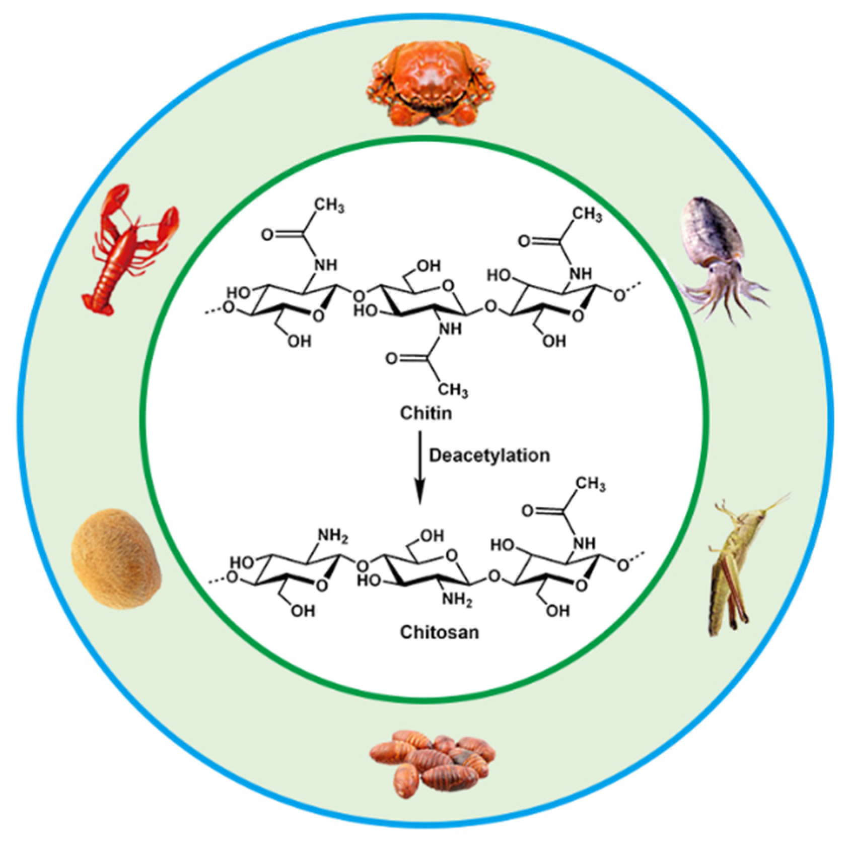
Chitosan is the product of N-deacetylation of chitin, which is the most basic and important derivative of chitin. Chitin, also called β-(1,4)-2-acetamido-2-deoxy-D-glucose, widely exists in the shells of crustaceans such as shrimps, carbs and insects, as well as in the cell walls of fungi and algae. Chitin and chitosan have been intensively used in many fields including biomedical materials, food additives, environmental protection, agriculture, cosmetics, medical treatment and drug development due to their biodegradability, biocompatibility and antibacterial abilities [19][20][21][22]. The annual biosynthesis of chitin on earth amounts to billions of tons, and it is a natural polymer compound whose output is second only to cellulose in production. Chitin, chitosan and cellulose have similar chemical structures, with fiber being hydroxyl at the C2 position, and chitin and chitosan are replaced by an acetylamino and amino group at the C2, respectively [23][24]. They are all polymers of six-carbon sugars with a molecular weight of more than one million. The sources and chemical structures of chitin and chitosan are shown in Figure 1.
Figure 1. Sources and chemical structures of chitin and chitosan.
The amino group in the molecular structure of CS is more reactive than the acetylamino group in the chitin molecule, which gives CS unique properties and enables chemical modification. CS exhibits a double helix structure with a helical pitch of 0.515 nm, and six sugar residues form a helical plane [23]. The amino groups, hydroxyl groups and N-acetylamino groups on the molecular chain of CS involved in the formation of intramolecular and intermolecular hydrogen bonds, forming the secondary structure of CS macromolecules. CS molecules are more likely to form crystalline regions because of the large number of hydrogen bonds, thus having high crystallinity and good physical and mechanical properties such as adsorption, film-forming, fiber-forming and moisturizing [25][26][27][28].

3. Design and Preparation of CS Hydrogels

Polymer hydrogels are highly water-swellable three-dimensional (3D) crosslinked networks that can rapidly swell in water and retain a large volume of water without dissolving in this swollen state [29][30]. The amount of water absorbed is closely related to the crosslinking density, the higher the degree of crosslinking, the lower the water absorption. This property of polymer hydrogels is similar to biological soft tissues, showing good physicochemical and biological properties, has wide application prospects in the controlled release of drugs, bioadhesion and biodegradable materials [31][32]. Chitosan acidic aqueous solution will immediately form a hydrogel when it encounters an alkaline environment, which is greatly affected by environmental pH. Therefore, the conventional preparation methods of CS hydrogels include physical crosslinking, chemical crosslinking and radiation crosslinking. In addition, the electrochemical deposition method developed in recent years for the preparation of CS gels has the characteristics of simple, rapid, non-polluting, reversible and controllable process, and is an economical and green new method for preparing chitosan gel [33][34][35][36].

3.1. Physically Crosslinked CS Hydrogels

Physically crosslinked hydrogels are mainly formed by crosslinking 3D network structures by physical interactions such as intermolecular entanglement, hydrogen bonds, hydrophobic interaction, van der Waals forces, coordination bonds, and ionic bonds [37][38][39][40]. Physically crosslinked hydrogels can be regarded as reversible hydrogels, and changes in physical states, such ionic strength, pH, temperature, stress, and solutes, could disrupt the structure of gels. Smart hydrogels with good biocompatibility and environmental responsiveness could be obtained under mild experimental conditions, which has become one of the current research hotspots [41][42][43].
Physically crosslinked hydrogels are generally prepared by the following methods: (1) Freeze-thaw cycles: hydrogels are prepared by microcrystals formed by repeated freeze-thaw processes [44][45]. (2) Ionic action: the principle of which is to use the electrostatic action of ions to form hydrogels. This method aggregates polyvalent ions with opposite charges in a polyelectrolyte solution to form a hydrogel [46][47]. The polycationic effect of CS and polyanionic polymers such as polyacrylic acid and alginate form hydrogels through electrostatic attraction between anions and cations. (3) Hydrogen bonding: the most common crosslinking method in physically crosslinked hydrogels [48][49][50]. The formed hydrogels are usually reversible, and hydrogen bonds are easily destroyed in salt solutions. (4) Hydrophobic interaction: when the polarity of the polymer solution changes, it will affect the hydrophobic interaction between molecular chains [51][52]. When this physical interaction increased, it can promote the self-assembly of molecules to form a gel. The solvent has a great dependence on hydrophobic interactions and affect the degree of crosslinking of polymer chains. Wearable strain sensors were prepared by dynamic physical crosslinking of polyacrylic acid, CS, and graphene oxide in a mixed solvent of water and glycerol [53]. The physical crosslinked CS gel had high stretchability (over 1000%), anti-freeze (use temperature range −20~70 °C), and excellent sensing performance (response time 40 ms).
Self-healing hydrogels can be obtained by using the physical interaction between two polysaccharide chains of soluble pectin and CS [54]. Cryo-SEM revealed the presence of nanogels in the crosslinked matrix. Due to the dynamic interaction between the pectin chains and the CS nanogels, the formed networks dissociated under the applied shear, allowing the hydrogel to flow. When the applied shear was removed, the storage modulus of the hydrogel can be quickly and fully recovered. The Young’s modulus of the hydrogels increased with increasing CS gel content indicating that higher crosslinking led to higher strength of the hydrogel.

3.2. Chemically Crosslinked CS Hydrogels

The chemical crosslinking method refers to the CS molecular chains involved in network through covalent bonds in the presence of crosslinking agents. Usually, glutaraldehyde (GA) [55][56][57], epichlorohydrin [58][59], formaldehyde [60][61], and genipin [14][62][63][64], are used as chemical crosslinking agents to form new covalent bonds at the crosslinking sites, so the chemical crosslinking process is generally irreversible and has good stability. Chemical crosslinking is an important method for preparing CS gels because it enhances the physical and mechanical properties of CS. The CS molecular chain contains a large number of hydrophilic groups, especially the amino group at the C2 position is often used as a crosslinking point, which can chemically interact with the functional groups on the crosslinking agent to form a crosslinking network [65][66]. To date, dialdehyde, especially glutaraldehyde (GA), are the most studied and prevalent crosslinkers for chemically modified chitosan. Dialdehyde formyl groups react with amino groups in CS to form covalent imine bonds. The semi-interpenetrating polymer network (IPN) hydrogel was prepared by crosslinking chitosan-polyvinylpyrrolidone with GA, and the gel showed good pH responsiveness [67]. In the presence of GA, crosslinked chitosan gels grafted with aminopropylsilane graphene oxide (GO) were prepared by sol-gel in acidic medium. The gel has high surface active sites and low swelling properties [68]. Khapre et al. modified chitosan with aniline in the presence of formaldehyde, and then further crosslinked chitosan with alginate using GA to obtain biocomposite gels [55]. Bilal et al. used 2.0% (v/v) GA to functionalize for 3 h in order to produce a fungal laccase-chitosan biocatalyst. This study demonstrates the efficient binding of laccase on the biopolymer network of glutaraldehyde-crosslinked chitosan, thereby enhancing the storage stability and substrate oxidation potential of the material [15].
The chemical reaction conditions using aldehyde groups and chitosan amino groups are mild and do not require the introduction of other auxiliary molecules such as reducing agents. However, the drawback of dialdehydes (GA, glyoxal, etc.) as crosslinkers is their high level of cytotoxicity and carcinogenic effects. The natural biological crosslinking agent genipin is an iridoid compound because its biological toxicity is much lower than other small organic molecules (formaldehyde, GA, etc.), and it is often used as a chemical crosslinking agent instead of aldehydes [69][70]. The biotoxicity of chitosan hydrogels can be greatly reduced. As a water-soluble bifunctional crosslinker, it reacts rapidly with chitosan to generate blue, fluorescent hydrogels. Under acidic and neutral conditions, genipin and chitosan are linked by amides and tertiary amines to form a crosslinked structure [71]. Delmar et al. studied genipin-crosslinked chitosan hydrogels and found that pH and crosslinking time significantly affected the properties of chitosan hydrogels [72]. Changing the pH in the range of 4.00–5.50 significantly affected the reaction, resulting in different appearance and properties of the hydrogel. Increasing the pH by 1.5 units resulted in a fourfold reduction in gel time and a more than tenfold equilibrium swelling. The swelling ability of the hydrogel was significantly pH dependent, which was attributed to the degree of protonation of CS and the inability of protonated CS to react with genipin. Tavares et al. investigated the effect of deacetylation degrees (DD, 83, 94 and 96%) on the properties of chitosan-genipin crosslinked gels. Using dynamic rheological tests, they confirmed that CS gel strength depends on frequency and temperature. The higher DD of CS, the lower the gelation temperature and the stronger the gel network structure. In addition, the high DD of CS is easier to crosslink with genipin, which can significantly improve the mechanical properties of the gel [62]. Nasrabadi et al. modeled two chitosan polymer sequences and six monomer units crosslinked by genipin [73]. The formation mechanism of genipin-crosslinked chitosan (GSC) was studied by calculating activation enthalpy and activation Gibbs free energy. The results suggested that H2O molecules were involved in the formation of the gel by substituting secondary amide bonds for the ester functional group of genipin through a tetrahedral intermediate (SN2 mechanism). Comparing the GCS model with a simple model of one polymer chain showed that the GCS model had more negative binding energies and stronger hydrogen bonds than the simple model. Muhammad Ubaid et al. prepared and optimized chitosan hydrogel membranes containing metformin using different concentrations of genipin as a crosslinker [14]. The gel membranes exhibited significant pH-sensitive behavior. The presence of hydrogen and ionic bonds between chitosan and genipin ensures that the drug is intact in the matrix system. The obtained hydrogels can be used for drug delivery. The ninhydrin assay allows for the measurement of the cross-linkage of the genipin crosslinked chitosan network and determination of the appropriate crosslinker concentration for gels used for swelling and thermomechanical analysis. Using a combined analysis of the modified Arrhenius and William Landel Ferry theories, Whitehead et al. determined the glass transition temperature range of −68 to −8 °C for genipin-crosslinked chitosan networks (40~60%, w/w solids), providing important guidance for the design and control of targeted delivery systems for biologically active compounds [74].

3.3. Irradiation-Crosslinked CS Hydrogels

The radiation crosslinking method refers to the gelation of CS by the interaction between molecular chains to form a crosslinked network structure under the action of high-energy light source such as ultraviolet rays, electron beams, γ-rays, etc. Irradiation can cause crosslinking and gel formation when the polysaccharide solution at the high concentration. Huh et al., introduced methacrylate groups on the CS molecular chain to obtain methacrylated hexanoyl glycol chitosan (M-HGC), and irradiated them with UV light at 220–260 nm [75]. It was found that there was a significant change at 15 min under UV irradiation and the signal of double-bonded peaks almost disappeared after 30 min and formed a gel, indicating that the crosslinking of C=C occurred under light irradiation. N-(2-hydroxyethyl)prop-2-enamide (HEPE) was grafted onto CS by reversible addition–fragmentation chain transfer (RAFT) radical polymerization under γ-ray irradiation, and the amino group need not be protected during the reaction. The drug was attached to the polymer by generating a Shiff base with the amino group of CS, and the product can self-assemble to form nanomicelles with pH and temperature sensitivity [76]. Nasef et al. crosslinked polyvinyl alcohol (PVA) and chitosan under γ-ray irradiation. The study showed that the dissolution rate of the composite hydrogel film decreased significantly with increasing radiation dose, which can be used as an elastic biomaterial for artificial skin [77]. Chan et al. prepared chitosan (CS)/corncob (CC) biocomposite gels by electron beam irradiation membrane [78]. When radiation was exposed to the biocomposite membrane, the free radicals generated by the radiolysis of water may attack CC to form CC radicals. These CC radicals may then attack long CS chains to form new CS-CC bonds. The induced crosslinking of CS/CC increased after electron beam irradiation. Compared with the unirradiated biocomposite films, the irradiated CS/CC biocomposite films showed better thermal stability and biodegradability.

3.4. Electrodeposited CS Hydrogels

CS is a weak electrolyte with specific pH responsiveness, and it is also the only natural cationic polymer that can be deposited to form hydrogels by electric field-induced deposition [79][80][81]. When the pH is less than its pKa (about 6.3), the protonation of the chitosan amino group leads to the dissolution of CS in cationic form; when the pH is close to or greater than its pKa, the amino group is deprotonated and precipitated. After applying an electric field, the H+ in the solution undergoes a reduction reaction at the cathode, causing the pH of the cathode surface to rise, inducing deprotonation of CS molecules and precipitation from the solution to form a CS gel. The CS gel film can be easily washed out by acid and could also be preserved after being crosslinked by a crosslinking agent. Compared with physical and chemical crosslinking methods, the preparation of CS hydrogels by electrodeposition shows unique advantages, such as simple operation, mild reaction, no need to add other chemical reagents, and can be carried out in the aqueous phase [82][83][84]. Recently, Yan et al. fabricated a vascular-like structured CS hydrogel with a diameter of about 0.4 mm by a templated electrodeposition process stimulated with an oscillating electrical signal [85]. The method spatially and temporally controls the internal multilayer structure of the hydrogel by using pulsed electrical signals (ON-OFF model), with short interruptions (OFF steps) forming tight boundaries between each individual layer. Their work provides a very promising self-assembly technique for constructing hydrogel coatings and artificial blood vessel regeneration. Yang et al. used electrodeposition-induced covalent crosslinking of CS and epichlorohydrin to obtain CS-based hydrogel contact lenses [86]. The electrodeposited hydrogel exhibits favorable optical properties, mechanical properties, and biocompatibility. The geometry of CS hydrogel could be simply tailored by electrode templates, the properties can be tuned by electrical signal and electrochemical crosslinking. In addition, the use of electrodeposition to print 3D CS hydrogels has received considerable attention for biomedical applications. Noriko Taira et al. performed 3D chitosan/gelatin hydrogel bioprinting by electrodeposition with a needle-shaped device [82]. This 3D design approach allows people to rapidly electrodeposit large hydrogels into several shapes, which holds promise for future tissue engineering, drug delivery, and on-chip applications.

This entry is adapted from the peer-reviewed paper 10.3390/chemosensors11010039

References

  1. Wang, L.; Xu, T.; Zhang, X. Multifunctional conductive hydrogel-based flexible wearable sensors. TrAC Trends Anal. Chem. 2021, 134, 116130.
  2. Chen, S.; Qi, J.; Fan, S.; Qiao, Z.; Yeo, J.C.; Lim, C.T. Flexible wearable sensors for cardiovascular health monitoring. Adv. Healthc. Mater. 2021, 10, 2100116.
  3. Cui, C.; Fu, Q.; Meng, L.; Hao, S.; Dai, R.; Yang, J. Recent progress in natural biopolymers conductive hydrogels for flexible wearable sensors and energy devices: Materials, structures, and performance. ACS Appl. Bio Mater. 2020, 4, 85–121.
  4. Jiang, L.; Liu, J.; He, S.; Liu, A.; Zhang, J.; Xu, H.; Shao, W. Flexible wearable sensors based on lignin doped organohydrogels with multi-functionalities. Chem. Eng. J. 2022, 430, 132653.
  5. Anwer, A.H.; Khan, N.; Ansari, M.Z.; Baek, S.-S.; Yi, H.; Kim, S.; Noh, S.M.; Jeong, C. Recent advances in touch sensors for flexible wearable devices. Sensors 2022, 22, 4460.
  6. del Bosque, A.; Sánchez-Romate, X.F.; Sánchez, M.; Ureña, A. Flexible wearable sensors based in carbon nanotubes reinforced poly (Ethylene Glycol) Diglycidyl ether (PEGDGE): Analysis of strain sensitivity and proof of concept. Chemosensors 2021, 9, 158.
  7. Fu, X.; Wang, L.; Zhao, L.; Yuan, Z.; Zhang, Y.; Wang, D.; Wang, D.; Li, J.; Li, D.; Shulga, V. Controlled Assembly of MXene Nanosheets as an Electrode and Active Layer for High-Performance Electronic Skin. Adv. Funct. Mater. 2021, 31, 2010533.
  8. Choi, S.; Han, S.I.; Kim, D.; Hyeon, T.; Kim, D.-H. High-performance stretchable conductive nanocomposites: Materials, processes, and device applications. Chem. Soc. Rev. 2019, 48, 1566–1595.
  9. Duan, Z.; Zhao, Q.; Wang, S.; Huang, Q.; Yuan, Z.; Zhang, Y.; Jiang, Y.; Tai, H. Halloysite nanotubes: Natural, environmental-friendly and low-cost nanomaterials for high-performance humidity sensor. Sens. Actuators B Chem. 2020, 317, 128204.
  10. Mohamed, M.E.B.; Attia, N.F.; Elashery, S.E. Greener and facile synthesis of hybrid nanocomposite for ultrasensitive iron (II) detection using carbon sensor. Microporous Mesoporous Mater. 2021, 313, 110832.
  11. Pita-López, M.L.; Fletes-Vargas, G.; Espinosa-Andrews, H.; Rodriguez-Rodriguez, R. Physically cross-linked chitosan-based hydrogels for tissue engineering applications: A state-of-the-art review. Eur. Polym. J. 2021, 145, 110176.
  12. Crini, G.; Torri, G.; Lichtfouse, E.; Kyzas, G.Z.; Wilson, L.D.; Morin-Crini, N. Dye removal by biosorption using cross-linked chitosan-based hydrogels. Environ. Chem. Lett. 2019, 17, 1645–1666.
  13. Iglesias, N.; Galbis, E.; Valencia, C.; Díaz-Blanco, M.J.; Lacroix, B.; de-Paz, M.-V. Biodegradable double cross-linked chitosan hydrogels for drug delivery: Impact of chemistry on rheological and pharmacological performance. Int. J. Biol. Macromol. 2020, 165, 2205–2218.
  14. Ubaid, M.; Murtaza, G. Fabrication and characterization of genipin cross-linked chitosan/gelatin hydrogel for pH-sensitive, oral delivery of metformin with an application of response surface methodology. Int. J. Biol. Macromol. 2018, 114, 1174–1185.
  15. Bilal, M.; Jing, Z.; Zhao, Y.; Iqbal, H.M. Immobilization of fungal laccase on glutaraldehyde cross-linked chitosan beads and its bio-catalytic potential to degrade bisphenol A. Biocatal. Agric. Biotechnol. 2019, 19, 101174.
  16. Cui, C.; Shao, C.; Meng, L.; Yang, J. High-strength, self-adhesive, and strain-sensitive chitosan/poly (acrylic acid) double-network nanocomposite hydrogels fabricated by salt-soaking strategy for flexible sensors. ACS Appl. Mater. Interfaces 2019, 11, 39228–39237.
  17. Jin, R.; Xu, J.; Duan, L.; Gao, G. Chitosan-driven skin-attachable hydrogel sensors toward human motion and physiological signal monitoring. Carbohydr. Polym. 2021, 268, 118240.
  18. Cong, J.; Fan, Z.; Pan, S.; Tian, J.; Lian, W.; Li, S.; Wang, S.; Zheng, D.; Miao, C.; Ding, W. Polyacrylamide/chitosan-based conductive double network hydrogels with outstanding electrical and mechanical performance at low temperatures. ACS Appl. Mater. Interfaces 2021, 13, 34942–34953.
  19. El Knidri, H.; Belaabed, R.; Addaou, A.; Laajeb, A.; Lahsini, A. Extraction, chemical modification and characterization of chitin and chitosan. Int. J. Biol. Macromol. 2018, 120, 1181–1189.
  20. Mohan, K.; Ganesan, A.R.; Muralisankar, T.; Jayakumar, R.; Sathishkumar, P.; Uthayakumar, V.; Chandirasekar, R.; Revathi, N. Recent insights into the extraction, characterization, and bioactivities of chitin and chitosan from insects. Trends Food Sci. Technol. 2020, 105, 17–42.
  21. Santos, V.P.; Marques, N.S.; Maia, P.C.; Lima, M.A.B.d.; Franco, L.d.O.; Campos-Takaki, G.M.d. Seafood waste as attractive source of chitin and chitosan production and their applications. Int. J. Mol. Sci. 2020, 21, 4290.
  22. Tao, F.; Cheng, Y.; Shi, X.; Zheng, H.; Du, Y.; Xiang, W.; Deng, H. Applications of chitin and chitosan nanofibers in bone regenerative engineering. Carbohydr. Polym. 2020, 230, 115658.
  23. Li, B.; Elango, J.; Wu, W. Recent advancement of molecular structure and biomaterial function of chitosan from marine organisms for pharmaceutical and nutraceutical application. Appl. Sci. 2020, 10, 4719.
  24. Jiménez-Gómez, C.P.; Cecilia, J.A. Chitosan: A natural biopolymer with a wide and varied range of applications. Molecules 2020, 25, 3981.
  25. Franca, E.F.; Freitas, L.C.; Lins, R.D. Chitosan molecular structure as a function of N-acetylation. Biopolymers 2011, 95, 448–460.
  26. Calixto, G.M.F.; Victorelli, F.D.; Dovigo, L.N.; Chorilli, M. Polyethyleneimine and chitosan polymer-based mucoadhesive liquid crystalline systems intended for buccal drug delivery. AAPS PharmSciTech 2018, 19, 820–836.
  27. Wang, T.; Chen, L.; Shen, T.; Wu, D. Preparation and properties of a novel thermo-sensitive hydrogel based on chitosan/hydroxypropyl methylcellulose/glycerol. Int. J. Biol. Macromol. 2016, 93, 775–782.
  28. Wu, S.; Li, K.; Shi, W.; Cai, J. Preparation and performance evaluation of chitosan/polyvinylpyrrolidone/polyvinyl alcohol electrospun nanofiber membrane for heavy metal ions and organic pollutants removal. Int. J. Biol. Macromol. 2022, 210, 76–84.
  29. Win, P.; Lin, C.-G.; Long, Y.; Chen, W.; Chen, G.; Song, Y.-F. Covalently cross-linked layered double hydroxide nanocomposite hydrogels with ultrahigh water content and excellent mechanical properties. Chem. Eng. J. 2018, 335, 409–415.
  30. Buenger, D.; Topuz, F.; Groll, J. Hydrogels in sensing applications. Prog. Polym. Sci. 2012, 37, 1678–1719.
  31. Feksa, L.R.; Troian, E.A.; Muller, C.D.; Viegas, F.; Machado, A.B.; Rech, V.C. Hydrogels for biomedical applications. In Nanostructures for the Engineering of Cells, Tissues and Organs; Elsevier: Amsterdam, The Netherlands, 2018; pp. 403–438.
  32. Li, Y.; Yang, J.; Yu, X.; Sun, X.; Chen, F.; Tang, Z.; Zhu, L.; Qin, G.; Chen, Q. Controlled shape deformation of bilayer films with tough adhesion between nanocomposite hydrogels and polymer substrates. J. Mater. Chem. B 2018, 6, 6629–6636.
  33. Wang, B.; Hua, J.; You, R.; Yan, K.; Ma, L. Electrochemically deposition of catechol-chitosan hydrogel coating on coronary stent with robust copper ions immobilization capability and improved interfacial biological activity. Int. J. Biol. Macromol. 2021, 181, 435–443.
  34. Zhao, P.; Liu, Y.; Xiao, L.; Deng, H.; Du, Y.; Shi, X. Electrochemical deposition to construct a nature inspired multilayer chitosan/layered double hydroxides hybrid gel for stimuli responsive release of protein. J. Mater. Chem. B 2015, 3, 7577–7584.
  35. Da Silva, A.C.; Wang, J.; Minev, I.R. Electro-assisted printing of soft hydrogels via controlled electrochemical reactions. Nat. Commun. 2022, 13, 1353.
  36. Helú, M.A.B.; Liu, L. Rational shaping of hydrogel by electrodeposition under fluid mechanics for electrochemical writing on complex shaped surfaces at microscale. Chem. Eng. J. 2021, 416, 129029.
  37. Hu, Y.; Du, Z.; Deng, X.; Wang, T.; Yang, Z.; Zhou, W.; Wang, C. Dual physically cross-linked hydrogels with high stretchability, toughness, and good self-recoverability. Macromolecules 2016, 49, 5660–5668.
  38. Yang, J.; Li, Y.; Yu, X.; Sun, X.; Zhu, L.; Qin, G.; Dai, Y.; Chen, Q. Tough and conductive dual physically cross-linked hydrogels for wearable sensors. Ind. Eng. Chem. Res. 2019, 58, 17001–17009.
  39. Gong, Z.; Zhang, G.; Zeng, X.; Li, J.; Li, G.; Huang, W.; Sun, R.; Wong, C. High-strength, tough, fatigue resistant, and self-healing hydrogel based on dual physically cross-linked network. ACS Appl. Mater. Interfaces 2016, 8, 24030–24037.
  40. Liu, X.; He, X.; Yang, B.; Lai, L.; Chen, N.; Hu, J.; Lu, Q. Dual physically cross-linked hydrogels incorporating hydrophobic interactions with promising repairability and ultrahigh elongation. Adv. Funct. Mater. 2021, 31, 2008187.
  41. Mahinroosta, M.; Farsangi, Z.J.; Allahverdi, A.; Shakoori, Z. Hydrogels as intelligent materials: A brief review of synthesis, properties and applications. Mater. Today Chem. 2018, 8, 42–55.
  42. Tan, Y.; Zhang, Y.; Zhang, Y.; Zheng, J.; Wu, H.; Chen, Y.; Xu, S.; Yang, J.; Liu, C.; Zhang, Y. Dual cross-linked ion-based temperature-responsive conductive hydrogels with multiple sensors and steady electrocardiogram monitoring. Chem. Mater. 2020, 32, 7670–7678.
  43. Yang, J.; Kang, Q.; Zhang, B.; Fang, X.; Liu, S.; Qin, G.; Chen, Q. Strong, tough, anti-freezing, non-drying and sensitive ionic sensor based on fully physical cross-linked double network hydrogel. Mater. Sci. Eng. C 2021, 130, 112452.
  44. Yi, Y.; Chiao, M.; Mahmoud, K.A.; Wu, L.; Wang, B. Preparation and characterization of PVA/PVP conductive hydrogels formed by freeze–thaw processes as a promising material for sensor applications. J. Mater. Sci. 2022, 57, 8029–8038.
  45. Kim, J.; Choi, J.; Hyun, J. Free-form three-dimensional nanocellulose structure reinforced with poly (vinyl alcohol) using freeze-thaw process. Carbohydr. Polym. 2022, 298, 120055.
  46. Wei, J.; Wang, R.; Pan, F.; Fu, Z. Polyvinyl Alcohol/Graphene Oxide Conductive Hydrogels via the Synergy of Freezing and Salting Out for Strain Sensors. Sensors 2022, 22, 3015.
  47. Yan, G.; He, S.; Ma, S.; Zeng, A.; Chen, G.; Tang, X.; Sun, Y.; Xu, F.; Zeng, X.; Lin, L. Catechol-based all-wood hydrogels with anisotropic, tough, and flexible properties for highly sensitive pressure sensing. Chem. Eng. J. 2022, 427, 131896.
  48. Du, C.; Zhang, X.N.; Sun, T.L.; Du, M.; Zheng, Q.; Wu, Z.L. Hydrogen-bond association-mediated dynamics and viscoelastic properties of tough supramolecular hydrogels. Macromolecules 2021, 54, 4313–4325.
  49. Liu, T.; Jiao, C.; Peng, X.; Chen, Y.-N.; Chen, Y.; He, C.; Liu, R.; Wang, H. Super-strong and tough poly (vinyl alcohol)/poly (acrylic acid) hydrogels reinforced by hydrogen bonding. J. Mater. Chem. B 2018, 6, 8105–8114.
  50. Chen, J.; Peng, Q.; Thundat, T.; Zeng, H. Stretchable, injectable, and self-healing conductive hydrogel enabled by multiple hydrogen bonding toward wearable electronics. Chem. Mater. 2019, 31, 4553–4563.
  51. Fang, X.; Li, Y.; Li, X.; Liu, W.; Yu, X.; Yan, F.; Sun, J. Dynamic hydrophobic domains enable the fabrication of mechanically robust and highly elastic poly (vinyl alcohol)-based hydrogels with excellent self-healing ability. ACS Mater. Lett. 2020, 2, 764–770.
  52. Qi, C.; Dong, Z.; Huang, Y.; Xu, J.; Lei, C. Tough, Anti-Swelling Supramolecular Hydrogels Mediated by Surfactant–Polymer Interactions for Underwater Sensors. ACS Appl. Mater. Interfaces 2022, 14, 30385–30397.
  53. Xia, S.; Song, S.; Li, Y.; Gao, G. Highly sensitive and wearable gel-based sensors with a dynamic physically cross-linked structure for strain-stimulus detection over a wide temperature range. J. Mater. Chem. C 2019, 7, 11303–11314.
  54. Shitrit, Y.; Davidovich-Pinhas, M.; Bianco-Peled, H. Shear thinning pectin hydrogels physically cross-linked with chitosan nanogels. Carbohydr. Polym. 2019, 225, 115249.
  55. Khapre, M.A.; Pandey, S.; Jugade, R.M. Glutaraldehyde-cross-linked chitosan–alginate composite for organic dyes removal from aqueous solutions. Int. J. Biol. Macromol. 2021, 190, 862–875.
  56. Bui, T.H.; Lee, W.; Jeon, S.-B.; Kim, K.-W.; Lee, Y. Enhanced Gold (III) adsorption using glutaraldehyde-crosslinked chitosan beads: Effect of crosslinking degree on adsorption selectivity, capacity, and mechanism. Sep. Purif. Technol. 2020, 248, 116989.
  57. Islam, N.; Wang, H.; Maqbool, F.; Ferro, V. In vitro enzymatic digestibility of glutaraldehyde-crosslinked chitosan nanoparticles in lysozyme solution and their applicability in pulmonary drug delivery. Molecules 2019, 24, 1271.
  58. Marrakchi, F.; Hameed, B.; Hummadi, E. Mesoporous biohybrid epichlorohydrin crosslinked chitosan/carbon–clay adsorbent for effective cationic and anionic dyes adsorption. Int. J. Biol. Macromol. 2020, 163, 1079–1086.
  59. Zhang, X.; Guo, H.; Xiao, N.; Ma, X.; Liu, C.; Zhong, L.; Xiao, G. Preparation and properties of epichlorohydrin-cross-linked chitosan/hydroxyethyl cellulose based CuO nanocomposite films. Cellulose 2022, 29, 4413–4426.
  60. Medellín-Castillo, N.A.; Isaacs-Páez, E.D.; Rodríguez-Méndez, I.; González-García, R.; Labrada-Delgado, G.J.; Aragón-Piña, A.; García-Arreola, M.E. Formaldehyde and tripolyphosphate crosslinked chitosan hydrogels: Synthesis, characterization and modeling. Int. J. Biol. Macromol. 2021, 183, 2293–2304.
  61. Atangana, E. Adsorption of Zn (II) and Pb (II) ions from aqueous solution using chitosan cross-linked formaldehyde adsorbent to protect the environment. J. Polym. Environ. 2019, 27, 2281–2291.
  62. Tavares, L.; Flores, E.E.E.; Rodrigues, R.C.; Hertz, P.F.; Noreña, C.P.Z. Effect of deacetylation degree of chitosan on rheological properties and physical chemical characteristics of genipin-crosslinked chitosan beads. Food Hydrocoll. 2020, 106, 105876.
  63. Vlasceanu, G.M.; Crica, L.E.; Pandele, A.M.; Ionita, M. Graphene oxide reinforcing genipin crosslinked chitosan-gelatin blend films. Coatings 2020, 10, 189.
  64. Liu, Y.; Liu, R.; Li, M.; Yu, F.; He, C. Removal of pharmaceuticals by novel magnetic genipin-crosslinked chitosan/graphene oxide-SO3H composite. Carbohydr. Polym. 2019, 220, 141–148.
  65. Kang, M.L.; Ko, J.-Y.; Kim, J.E.; Im, G.-I. Intra-articular delivery of kartogenin-conjugated chitosan nano/microparticles for cartilage regeneration. Biomaterials 2014, 35, 9984–9994.
  66. Xu, H.; Zhang, L.; Zhang, H.; Luo, J.; Gao, X. Green Fabrication of Chitin/Chitosan Composite Hydrogels and Their Potential Applications. Macromol. Biosci. 2021, 21, 2000389.
  67. Risbud, M.V.; Hardikar, A.A.; Bhat, S.V.; Bhonde, R.R. pH-sensitive freeze-dried chitosan–polyvinyl pyrrolidone hydrogels as controlled release system for antibiotic delivery. J. Control. Release 2000, 68, 23–30.
  68. Sharma, P.; Singh, A.K.; Shahi, V.K. Selective adsorption of Pb (II) from aqueous medium by cross-linked chitosan-functionalized graphene oxide adsorbent. ACS Sustain. Chem. Eng. 2018, 7, 1427–1436.
  69. Bryś, M.; Urbańska, K.; Olas, B. Novel Findings regarding the Bioactivity of the Natural Blue Pigment Genipin in Human Diseases. Int. J. Mol. Sci. 2022, 23, 902.
  70. Wang, C.; Gong, X.; Bo, A.; Zhang, L.; Zhang, M.; Zang, E.; Zhang, C.; Li, M. Iridoids: Research advances in their phytochemistry, biological activities, and pharmacokinetics. Molecules 2020, 25, 287.
  71. Muzzarelli, R.A. Genipin-crosslinked chitosan hydrogels as biomedical and pharmaceutical aids. Carbohydr. Polym. 2009, 77, 1–9.
  72. Delmar, K.; Bianco-Peled, H. The dramatic effect of small pH changes on the properties of chitosan hydrogels crosslinked with genipin. Carbohydr. Polym. 2015, 127, 28–37.
  73. Nasrabadi, M.; Morsali, A.; Beyramabadi, S.A. An applied quantum-chemical model for genipin-crosslinked chitosan (GCS) nanocarrier. Int. J. Biol. Macromol. 2020, 165, 1229–1240.
  74. Whitehead, F.A.; Young, S.A.; Kasapis, S. Swelling behaviour and glass transition in genipin-crosslinked chitosan systems. Int. J. Biol. Macromol. 2020, 164, 3075–3083.
  75. Cho, I.S.; Cho, M.O.; Li, Z.; Nurunnabi, M.; Park, S.Y.; Kang, S.-W.; Huh, K.M. Synthesis and characterization of a new photo-crosslinkable glycol chitosan thermogel for biomedical applications. Carbohydr. Polym. 2016, 144, 59–67.
  76. Huang, W.; Wang, Y.; Zhang, S.; Huang, L.; Hua, D.; Zhu, X. A facile approach for controlled modification of chitosan under γ-ray irradiation for drug delivery. Macromolecules 2013, 46, 814–818.
  77. Nasef, S.M.; Khozemy, E.E.; Kamoun, E.A.; El-Gendi, H. Gamma radiation-induced crosslinked composite membranes based on polyvinyl alcohol/chitosan/AgNO3/vitamin E for biomedical applications. Int. J. Biol. Macromol. 2019, 137, 878–885.
  78. Chan, M.Y.; Koay, S.C. Biodegradation and thermal properties of crosslinked chitosan/corn cob biocomposite films by electron beam irradiation. Polym. Eng. Sci. 2019, 59, E59–E68.
  79. Geng, Z.; Wang, X.; Guo, X.; Zhang, Z.; Chen, Y.; Wang, Y. Electrodeposition of chitosan based on coordination with metal ions in situ-generated by electrochemical oxidation. J. Mater. Chem. B 2016, 4, 3331–3338.
  80. Kim, E.; Xiong, Y.; Cheng, Y.; Wu, H.-C.; Liu, Y.; Morrow, B.H.; Ben-Yoav, H.; Ghodssi, R.; Rubloff, G.W.; Shen, J. Chitosan to connect biology to electronics: Fabricating the bio-device interface and communicating across this interface. Polymers 2014, 7, 1–46.
  81. Nawrotek, K.; Tylman, M.; Adamus-Włodarczyk, A.; Rudnicka, K.; Gatkowska, J.; Wieczorek, M.; Wach, R. Influence of chitosan average molecular weight on degradation and stability of electrodeposited conduits. Carbohydr. Polym. 2020, 244, 116484.
  82. Taira, N.; Ino, K.; Ida, H.; Nashimoto, Y.; Shiku, H. Electrodeposition-based rapid bioprinting of 3D-designed hydrogels with a pin art device. Biofabrication 2019, 11, 035018.
  83. Rajabi, M.; McConnell, M.; Cabral, J.; Ali, M.A. Chitosan hydrogels in 3D printing for biomedical applications. Carbohydr. Polym. 2021, 260, 117768.
  84. Nawrotek, K.; Grams, J. Understanding electrodeposition of chitosan–hydroxyapatite structures for regeneration of tubular-shaped tissues and organs. Materials 2021, 14, 1288.
  85. Yan, K.; Yang, C.; Zhong, W.; Lu, Z.; Li, X.; Shi, X.; Wang, D. Wire templated electrodeposition of vessel-like structured chitosan hydrogel by using a pulsed electrical signal. Soft Matter 2020, 16, 9471–9478.
  86. Yang, C.; Wang, M.; Wang, W.; Liu, H.; Deng, H.; Du, Y.; Shi, X. Electrodeposition induced covalent cross-linking of chitosan for electrofabrication of hydrogel contact lenses. Carbohydr. Polym. 2022, 292, 119678.
More
This entry is offline, you can click here to edit this entry!
ScholarVision Creations