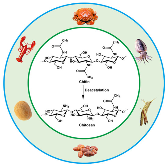
Chitosan is the product of N-deacetylation of chitin, which is the most basic and important derivative of chitin. Chitin, also called β-(1,4)-2-acetamido-2-deoxy-D-glucose, widely exists in the shells of crustaceans such as shrimps, carbs and insects, as well as in the cell walls of fungi and algae. Chitin and chitosan have been intensively used in many fields including biomedical materials, food additives, environmental protection, agriculture, cosmetics, medical treatment and drug development due to their biodegradability, biocompatibility and antibacterial abilities [
23,
24,
25,
26]. The annual biosynthesis of chitin on earth amounts to billions of tons, and it is a natural polymer compound whose output is second only to cellulose in production. Chitin, chitosan and cellulose have similar chemical structures, with fiber being hydroxyl at the C2 position, and chitin and chitosan are replaced by an acetylamino and amino group at the C2, respectively [
27,
28]. They are all polymers of six-carbon sugars with a molecular weight of more than one million. The sources and chemical structures of chitin and chitosan are shown in
Figure 1.
3. Design and Preparation of CS Hydrogels
Polymer hydrogels are highly water-swellable three-dimensional (3D) crosslinked networks that can rapidly swell in water and retain a large volume of water without dissolving in this swollen state [
33,
34]. The amount of water absorbed is closely related to the crosslinking density, the higher the degree of crosslinking, the lower the water absorption. This property of polymer hydrogels is similar to biological soft tissues, showing good physicochemical and biological properties, has wide application prospects in the controlled release of drugs, bioadhesion and biodegradable materials [
35,
36]. Chitosan acidic aqueous solution will immediately form a hydrogel when it encounters an alkaline environment, which is greatly affected by environmental pH. Therefore, the conventional preparation methods of CS hydrogels include physical crosslinking, chemical crosslinking and radiation crosslinking. In addition, the electrochemical deposition method developed in recent years for the preparation of CS gels has the characteristics of simple, rapid, non-polluting, reversible and controllable process, and is an economical and green new method for preparing chitosan gel [
37,
38,
39,
40].
3.1. Physically Crosslinked CS Hydrogels
Physically crosslinked hydrogels are mainly formed by crosslinking 3D network structures by physical interactions such as intermolecular entanglement, hydrogen bonds, hydrophobic interaction, van der Waals forces, coordination bonds, and ionic bonds [
41,
42,
43,
44]. Physically crosslinked hydrogels can be regarded as reversible hydrogels, and changes in physical states, such ionic strength, pH, temperature, stress, and solutes, could disrupt the structure of gels. Smart hydrogels with good biocompatibility and environmental responsiveness could be obtained under mild experimental conditions, which has become one of the current research hotspots [
45,
46,
47].
Physically crosslinked hydrogels are generally prepared by the following methods: (1) Freeze-thaw cycles: hydrogels are prepared by microcrystals formed by repeated freeze-thaw processes [
48,
49]. (2) Ionic action: the principle of which is to use the electrostatic action of ions to form hydrogels. This method aggregates polyvalent ions with opposite charges in a polyelectrolyte solution to form a hydrogel [
50,
51]. The polycationic effect of CS and polyanionic polymers such as polyacrylic acid and alginate form hydrogels through electrostatic attraction between anions and cations. (3) Hydrogen bonding: the most common crosslinking method in physically crosslinked hydrogels [
52,
53,
54]. The formed hydrogels are usually reversible, and hydrogen bonds are easily destroyed in salt solutions. (4) Hydrophobic interaction: when the polarity of the polymer solution changes, it will affect the hydrophobic interaction between molecular chains [
55,
56]. When this physical interaction increased, it can promote the self-assembly of molecules to form a gel. The solvent has a great dependence on hydrophobic interactions and affect the degree of crosslinking of polymer chains. Wearable strain sensors were prepared by dynamic physical crosslinking of polyacrylic acid, CS, and graphene oxide in a mixed solvent of water and glycerol [
57]. The physical crosslinked CS gel had high stretchability (over 1000%), anti-freeze (use temperature range −20~70 °C), and excellent sensing performance (response time 40 ms).
Self-healing hydrogels can be obtained by using the physical interaction between two polysaccharide chains of soluble pectin and CS [
58]. Cryo-SEM revealed the presence of nanogels in the crosslinked matrix. Due to the dynamic interaction between the pectin chains and the CS nanogels, the formed networks dissociated under the applied shear, allowing the hydrogel to flow. When the applied shear was removed, the storage modulus of the hydrogel can be quickly and fully recovered. The Young’s modulus of the hydrogels increased with increasing CS gel content indicating that higher crosslinking led to higher strength of the hydrogel.
3.2. Chemically Crosslinked CS Hydrogels
The chemical crosslinking method refers to the CS molecular chains involved in network through covalent bonds in the presence of crosslinking agents. Usually, glutaraldehyde (GA) [
59,
60,
61], epichlorohydrin [
62,
63], formaldehyde [
64,
65], and genipin [
14,
66,
67,
68], are used as chemical crosslinking agents to form new covalent bonds at the crosslinking sites, so the chemical crosslinking process is generally irreversible and has good stability. Chemical crosslinking is an important method for preparing CS gels because it enhances the physical and mechanical properties of CS. The CS molecular chain contains a large number of hydrophilic groups, especially the amino group at the C2 position is often used as a crosslinking point, which can chemically interact with the functional groups on the crosslinking agent to form a crosslinking network [
69,
70]. To date, dialdehyde, especially glutaraldehyde (GA), are the most studied and prevalent crosslinkers for chemically modified chitosan. Dialdehyde formyl groups react with amino groups in CS to form covalent imine bonds. The semi-interpenetrating polymer network (IPN) hydrogel was prepared by crosslinking chitosan-polyvinylpyrrolidone with GA, and the gel showed good pH responsiveness [
71]. In the presence of GA, crosslinked chitosan gels grafted with aminopropylsilane graphene oxide (GO) were prepared by sol-gel in acidic medium. The gel has high surface active sites and low swelling properties [
72]. Khapre et al. modified chitosan with aniline in the presence of formaldehyde, and then further crosslinked chitosan with alginate using GA to obtain biocomposite gels [
59]. Bilal et al. used 2.0% (
v/v) GA to functionalize for 3 h in order to produce a fungal laccase-chitosan biocatalyst. This study demonstrates the efficient binding of laccase on the biopolymer network of glutaraldehyde-crosslinked chitosan, thereby enhancing the storage stability and substrate oxidation potential of the material [
15].
The chemical reaction conditions using aldehyde groups and chitosan amino groups are mild and do not require the introduction of other auxiliary molecules such as reducing agents. However, the drawback of dialdehydes (GA, glyoxal, etc.) as crosslinkers is their high level of cytotoxicity and carcinogenic effects. The natural biological crosslinking agent genipin is an iridoid compound because its biological toxicity is much lower than other small organic molecules (formaldehyde, GA, etc.), and it is often used as a chemical crosslinking agent instead of aldehydes [
73,
74]. The biotoxicity of chitosan hydrogels can be greatly reduced. As a water-soluble bifunctional crosslinker, it reacts rapidly with chitosan to generate blue, fluorescent hydrogels. Under acidic and neutral conditions, genipin and chitosan are linked by amides and tertiary amines to form a crosslinked structure [
75]. Delmar et al. studied genipin-crosslinked chitosan hydrogels and found that pH and crosslinking time significantly affected the properties of chitosan hydrogels [
76]. Changing the pH in the range of 4.00–5.50 significantly affected the reaction, resulting in different appearance and properties of the hydrogel. Increasing the pH by 1.5 units resulted in a fourfold reduction in gel time and a more than tenfold equilibrium swelling. The swelling ability of the hydrogel was significantly pH dependent, which was attributed to the degree of protonation of CS and the inability of protonated CS to react with genipin. Tavares et al. investigated the effect of deacetylation degrees (DD, 83, 94 and 96%) on the properties of chitosan-genipin crosslinked gels. Using dynamic rheological tests, they confirmed that CS gel strength depends on frequency and temperature. The higher DD of CS, the lower the gelation temperature and the stronger the gel network structure. In addition, the high DD of CS is easier to crosslink with genipin, which can significantly improve the mechanical properties of the gel [
66]. Nasrabadi et al. modeled two chitosan polymer sequences and six monomer units crosslinked by genipin [
77]. The formation mechanism of genipin-crosslinked chitosan (GSC) was studied by calculating activation enthalpy and activation Gibbs free energy. The results suggested that H
2O molecules were involved in the formation of the gel by substituting secondary amide bonds for the ester functional group of genipin through a tetrahedral intermediate (SN2 mechanism). Comparing the GCS model with a simple model of one polymer chain showed that the GCS model had more negative binding energies and stronger hydrogen bonds than the simple model. Muhammad Ubaid et al. prepared and optimized chitosan hydrogel membranes containing metformin using different concentrations of genipin as a crosslinker [
14]. The gel membranes exhibited significant pH-sensitive behavior. The presence of hydrogen and ionic bonds between chitosan and genipin ensures that the drug is intact in the matrix system. The obtained hydrogels can be used for drug delivery. The ninhydrin assay allows for the measurement of the cross-linkage of the genipin crosslinked chitosan network and determination of the appropriate crosslinker concentration for gels used for swelling and thermomechanical analysis. Using a combined analysis of the modified Arrhenius and William Landel Ferry theories, Whitehead et al. determined the glass transition temperature range of −68 to −8 °C for genipin-crosslinked chitosan networks (40~60%,
w/
w solids), providing important guidance for the design and control of targeted delivery systems for biologically active compounds [
78].
3.3. Irradiation-Crosslinked CS Hydrogels
The radiation crosslinking method refers to the gelation of CS by the interaction between molecular chains to form a crosslinked network structure under the action of high-energy light source such as ultraviolet rays, electron beams, γ-rays, etc. Irradiation can cause crosslinking and gel formation when the polysaccharide solution at the high concentration. Huh et al., introduced methacrylate groups on the CS molecular chain to obtain methacrylated hexanoyl glycol chitosan (M-HGC), and irradiated them with UV light at 220–260 nm [
79]. It was found that there was a significant change at 15 min under UV irradiation and the signal of double-bonded peaks almost disappeared after 30 min and formed a gel, indicating that the crosslinking of C=C occurred under light irradiation. N-(2-hydroxyethyl)prop-2-enamide (HEPE) was grafted onto CS by reversible addition–fragmentation chain transfer (RAFT) radical polymerization under γ-ray irradiation, and the amino group need not be protected during the reaction. The drug was attached to the polymer by generating a Shiff base with the amino group of CS, and the product can self-assemble to form nanomicelles with pH and temperature sensitivity [
80]. Nasef et al. crosslinked polyvinyl alcohol (PVA) and chitosan under γ-ray irradiation. The study showed that the dissolution rate of the composite hydrogel film decreased significantly with increasing radiation dose, which can be used as an elastic biomaterial for artificial skin [
81]. Chan et al. prepared chitosan (CS)/corncob (CC) biocomposite gels by electron beam irradiation membrane [
82]. When radiation was exposed to the biocomposite membrane, the free radicals generated by the radiolysis of water may attack CC to form CC radicals. These CC radicals may then attack long CS chains to form new CS-CC bonds. The induced crosslinking of CS/CC increased after electron beam irradiation. Compared with the unirradiated biocomposite films, the irradiated CS/CC biocomposite films showed better thermal stability and biodegradability.
3.4. Electrodeposited CS Hydrogels
CS is a weak electrolyte with specific pH responsiveness, and it is also the only natural cationic polymer that can be deposited to form hydrogels by electric field-induced deposition [
83,
84,
85]. When the pH is less than its pKa (about 6.3), the protonation of the chitosan amino group leads to the dissolution of CS in cationic form; when the pH is close to or greater than its pKa, the amino group is deprotonated and precipitated. After applying an electric field, the H
+ in the solution undergoes a reduction reaction at the cathode, causing the pH of the cathode surface to rise, inducing deprotonation of CS molecules and precipitation from the solution to form a CS gel. The CS gel film can be easily washed out by acid and could also be preserved after being crosslinked by a crosslinking agent. Compared with physical and chemical crosslinking methods, the preparation of CS hydrogels by electrodeposition shows unique advantages, such as simple operation, mild reaction, no need to add other chemical reagents, and can be carried out in the aqueous phase [
86,
87,
88]. Recently, Yan et al. fabricated a vascular-like structured CS hydrogel with a diameter of about 0.4 mm by a templated electrodeposition process stimulated with an oscillating electrical signal [
89]. The method spatially and temporally controls the internal multilayer structure of the hydrogel by using pulsed electrical signals (ON-OFF model), with short interruptions (OFF steps) forming tight boundaries between each individual layer. The work provides a very promising self-assembly technique for constructing hydrogel coatings and artificial blood vessel regeneration. Yang et al. used electrodeposition-induced covalent crosslinking of CS and epichlorohydrin to obtain CS-based hydrogel contact lenses [
90]. The electrodeposited hydrogel exhibits favorable optical properties, mechanical properties, and biocompatibility. The geometry of CS hydrogel could be simply tailored by electrode templates, the properties can be tuned by electrical signal and electrochemical crosslinking. In addition, the use of electrodeposition to print 3D CS hydrogels has received considerable attention for biomedical applications. Noriko Taira et al. performed 3D chitosan/gelatin hydrogel bioprinting by electrodeposition with a needle-shaped device [
86]. This 3D design approach allows people to rapidly electrodeposit large hydrogels into several shapes, which holds promise for future tissue engineering, drug delivery, and on-chip applications.