Your browser does not fully support modern features. Please upgrade for a smoother experience.
Please note this is an old version of this entry, which may differ significantly from the current revision.
Subjects:
Biotechnology & Applied Microbiology
The diverse biological properties of platinum nanoparticles (PtNPs) make them ideal for use in the development of new tools in therapy, diagnostics, and other biomedical purposes. “Green” PtNPs synthesis is of great interest as it is eco-friendly, less energy-consuming and minimizes the amount of toxic by-products.
- platinum nanoparticles
- green synthesis
- capping agents
1. Introduction
Since the end of the 20th century, the popularity of metal nanoparticles has grown from year to year. This is not surprising because they are widely used in various spheres of human activity owing to their many valuable properties. Green technologies—eco-friendly technologies—are of particular importance as they are simple, cheap, practically waste-free and possess the ability to control the resulting nanoparticles characteristics (size, shape, stability), as well as being a popular topic to study in recent years. This is confirmed by the impressive dynamics of the number of publications on this topic that have emerged in the last twenty years. [1]. Different nanoparticles, nanocomposites and nanostructures possessing biocompatibility are of interest for a wide variety of human activity fields: in the food industry, food packaging and the development of functional food products to increase food safety, the detection of food pathogens and extend the shelf life of food products [2], as well as catalysts for biofuels (for example, Cs2O−MgO/MPC nanocomposite was used as the main nanocatalyst for the production of biodiesel from olive oil) [3], application in makeup and skin care [4], and, of course, use in diagnosis, medical treatment, theranostics, and tissue engineering (these are not only metal nanoparticles, but also, for instance, nanostructured CaPs with surface-rich–OH groups and Ca2+ cations, which can effectively adsorb therapeutic agents [5]). Biosynthesis strategies involve using living objects—microorganisms, fungi or plants—as bio-factories for metal nanoparticles production [6,7,8,9]. The obtained medicinal products reveal wide application prospects for the biomedical field for combating pathogens of various diseases, the prevention and treatment of oncological diseases, drugs delivery, diagnostic systems, etc.
Although metal nanoparticles such as silver and gold are more well known, the synthesis and analysis of other metal nanoparticles are also gaining momentum [10]. One of these metals is platinum. The Incas were the pioneers of its mining and application, but in the Old World, platinum was unknown until the 16th century. It was first introduced to the conquistadors from South America, and received its name from the Spanish word platina, literally meaning “little silver” according to its external similarity to real silver [11]. Possessing high refractory, platinum was not found worthy of use for a long time, being valued much lower than silver and gold. Between 1889 and 1960, 90% of platinum alloy was used as the international standard for one meter determining. Currently, South Africa and Russia are the leaders in platinum extraction. Platinum is an inert metal, and apparently does not play an important role in the vital activity of living organisms. In addition, this metal is non-toxic in metallic form.
Platinum finds its application in electroplating. It is used as a catalyst in various industries including coating microwave technology elements as well as jewelry. It is used for medical purposes in dentistry, and platinum compounds, cytostatics such as cisplatin, are applied for oncological disease therapy [12,13,14,15]. However, drugs such as cisplatin and carboplatin have nephrotoxic, neurotoxic and cytotoxic effects [16]. These effects can be neutralized with the help of green synthesis of platinum nanoparticles and become the key to solving many medical problems. Bio-factories (microorganisms, fungi, plants) are full of various cellular compounds such as proteins, enzymes, acids, etc., important for the characteristic features formation of platinum nanoparticles synthesized in different organisms. The bio-compound importance consists of not only their participation in the nanoparticle’s synthesis, but also the final assembly processes. Having their own remarkable properties, they will be able to multiply the positive effect of platinum nanoparticles use. Moreover, the agenda of “green” technologies, the absence of toxic side effects, and the targeted effect on the human body are still relevant. The present available information about PtNPs antibacterial properties [17], antitumor effects [18], and other potentially beneficial features make them an interesting topic for comprehensive research.
2. Application of Green PtNPs
2.1. Antibacterial Activity
The large-scale application of antibiotics to treat infectious disease has led to the emergence of a large number of pathogen strains resistant to antibiotics causing huge problems in medicine. Biofilms formed by pathogenic microorganisms are a separate issue. Metal nanoparticles are of great value in the solution to this problem, being an effective basis for the development of antimicrobial drugs. The bactericidal properties of metals such as gold and silver have been known to mankind for a long time, and modern methods of nanotechnology discover new possibilities for their use [106,107]. Metal nanoparticles, including PtNPs, exhibit remarkable biocidal properties against both gram-positive and gram-negative bacteria [108]. Such antimicrobial activity depends on the surface area in contact with microorganisms; their small size and high surface-to-volume ratio gives them the opportunity to interact closely with microbial membranes [107,109]. The nanoparticle shape also plays an important role in antibacterial activity, and capping agents, due to their own antimicrobial properties, are able to enhance it [110].
The mechanisms of nanoparticle action on bacterial cells include: destruction of the microbial cell wall and cell membrane, pump mechanisms damage; destruction of cellular components: ribosome breakdown, inhibition of deoxyribonucleic acid (DNA) replication and enzyme dysfunction; formation of reactive oxygen species (ROS) and induction of oxidative stress; and triggering of both congenital and adaptive host immune responses and inhibition of biofilm formation (Figure 1) [20,111]. Metallic nanoparticles interact with the bacterial cell wall by attraction between the negative charge of the cell wall and the PtNPs positive charge [20,112]. The main functions of the bacterial cell wall and cell membrane are protection from external influences and the transport of nutrients into and out of the cell. Both Gram-negative and Gram-positive bacteria surface are negatively charged due to the presence of lipoteichoic acid (in Gram-positive) and lipopolysaccharide (in Gram-negatives) [113]. The outer membrane of Gram-negative bacteria is a lipid bilayer, whereas the inner membrane is composed of phospholipids. One of the major differences between Gram-positive and Gram-negative is a thicker peptidoglycan layer in the cell wall of Gram-positive, which makes them less vulnerable to metal nanoparticles [114]. In addition, the greater nanoparticle efficiency to Gram-negative may be the result of the presence of lipopolysaccharides (LPS) carrying a negative charge, which ensures the PtNPs adhesion to the bacterial cell wall. Due to the presence of the rigid cell wall of Gram-positive bacteria, the antimicrobial activity of Gram-negative bacteria was higher compared to Gram-positive bacteria. Therefore, the biogenic nanoparticles carrying amino groups on their surfaces can attach more effectively to both Gram-negative and Gram-positive bacterial cell walls and destroy them [50]. As a result of interaction between PtNPS and the bacterial cell wall, morphological and permeability function changes in the membrane is observed, the integrity of the bacterial cell is disrupted and death occurs [20]. In case of PtNPs, their strong negative zeta potential enhances antibacterial activity [110].
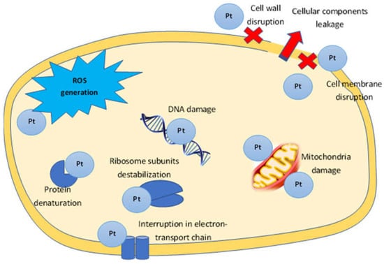
Figure 1. The proposed mechanism of PtNPs antibacterial activity.
Penetrating into the cell, platinum nanoparticles complete the process: binding to DNA, they transfer it from the normal state to the condensed one, leading to the loss of replication ability; binding to thiol, phosphorous or sulfhydryl (-SH), amine and carboxylic groups of enzymes results in their inactivation [101]. The loss of hydrogen ions in proteins leads to the cell membrane destruction, increasing its permeability to PtNPs, ultimately inhibiting bacterial metabolism and initiating cell death [77]. The antibacterial activity of PtNPs is associated with ROS (reactive oxygen species) generation: they increase ROS number in bacterial cells. ROS include highly reactive radicals −OH, H2O2 and less toxic radicals O2−, able to affect DNA, RNA and proteins, causing the death of bacteria [77]. These ROS interact directly with PtNPs, causing protein degradation and lipid breakdown, resulting in the down-regulation of DNA, oxidative stress, and finally apoptosis of bacterial cells [77,115]. An important role in maintaining the intracellular redox environment is played by the reduced glutathione (GSH), a non-protein tripeptide that protects cells from oxidative damage by absorbing ROS. However, excessive ROS formation can oxidize GSH to glutathione disulfide. The decrease in the concentration of GSH in cells treated with platinum nanoparticles was determined [116]. Interaction PtNPs with the 30S ribosome subunit induces protein synthesis to stop [117]. Some studies suggested that bacterial growth inhibition is interconnected with ATP production and mitochondrial membrane potential [118]. The effect of platinum nanoparticles on DNA synthesis was shown by PtNPs ability to inhibit the Taq DNA polymerase and affect the secondary structure of DNA in higher concentrations [119]; a one-hour treatment of Salmonella enteritidis with PtNPs removed part of the DNA from the bacterial cell [120]. The influence on the proposed mechanism of platinum nanoparticles on the bacterial cell is presented in Figure 1. The size of platinum nanoparticles is of great importance in antibacterial activity: small particle sizes often have a higher surface area and so are more effective than those with larger particle sizes; for example, a smaller size PtNPs could pass more effectively through the thick cell wall of Gram-positive bacteria [50].
All of the above is confirmed by the influence of platinum nanoparticles on different bacterium types. Thus, an inhibitory effect is shown for gram-negative bacteria Pseudomonas aeruginosa [50], S. typhimurium [100], Klebsiella oxytoca and Klebsiella aerogenes [101], S. typhi [116], Ps. aeruginosa [118], Enterobacter aerogenes, S. enteritidis [120], Klebsiella pneumoniae [84,121], B. licheniformis [122], E. coli [77,123], Aeromonas hydrophila [63,124], Proteus vulgaris [121], Enterobacter cloacae [121]; Gram-positive bacterium—Staphylococcus aureus [50], Listeria innocua [50], Streptococcus pneumonia [68], Lactococcus lactis, Bacillus subtilis [89], St. epidermis [101], St. haemolyticus [125], Enterococcus faecium [3,125], St. pyogens and Vibrio cholerae [126].
3. Anti-Fungal Activity
There are numerous antifungal drugs, but most of them have many side effects. Green PtNPs represent an alternative in solving this problem. The received data allow us to characterize them as an effective antifungal agent. Anti-fungal activity was found for Fusarium oxysporum [66], and also plant pathogenic fungi such as Colletotrichum acutatum, and Cladosporium fulvum [98]. PtNPs prepared using X. strumarium leaves extracts showed significant anti-fungal activity against Candida albicans, C. tropicalis, C. parapsilosis, Aspergillus flaves and A. niger [91]. The fungicidal effect of platinum nanoparticles obtained with gum kondagogu was found for A. parasiticus and A. flavus [127]. The inhibitory activity of Pt nanocomposite on A. parasiticus and A. flavusfungi is mediated through the induction of oxidative stress, resulting in the formation of ROS and subsequent damage to fungal mycelial morphology and membrane, ultimately leading to cellular damage and fungal growth [127].
4. Anti-Cancer Activity
Cancer is one of the biggest threats to humanity, occupying second place in terms of mortality. Due to the lack of high-quality diagnosis and treatment in the early disease stages, cancer mortality rates are significantly higher in low- and middle-income than in developed countries. Moreover, most modern drugs targeted at treating cancer have a huge number of adverse effects that can seriously worsen the quality of a patient’s life. Therefore, searching for new, inexpensive drugs possessing a low toxic effect would allow this problem to be solved on a qualitatively new level.
Platinum–based anticancer drugs have been known for a long time. One of these is cisplatin, with strong cytotoxic, bactericidal and mutagenic properties. The anticancer activity of cisplatin was discovered back in 1965 by Barnett Rosenberg [128]. Its action is based on the ability to form strong specific bonds with DNA that induce chemical damage to DNA bases. However, there are many negative effects of such drugs, including nephrotoxicity, fatigue, emesis, alopecia, peripheral neuropathy, and myelosupression. Platinum nanoparticles can open a new page for cancer treatment. The most promising are PtNPs mediated by plant extracts containing essential oils, acids, alkaloids, phytoncides, which show efficacy in various types of cancers [129]. For example, an anti-cancer effect was found for Aloe vera, Catharanthus roseus, hot pepper (Capsicum annuum), tulasi (Ocimum sanctum) and many other medicinal plants [130]. The combined effect of platinum nanoparticles, capping by different herbs compounds, could enhance the anti-cancer effect and eliminate the toxic effect of the metal.
To create new therapeutic agents against cancer, it is very significant to develop inducers of apoptosis: programmed cell death coordinated by a cascade of interdependent cellular reactions. Moreover, it is the most important process for maintaining homeostasis between cell proliferation and mortality [131].
Although the action mechanism of platinum nanoparticles on cancer cells is not fully studied, the basic principles, based on in vitro experiments, can be selected: (a) cell cycle arrest; (b) penetration into the nuclear, nucleus and DNA fragmentation; (c) the level of glutathione; (d) inducing mitochondrial dysfunction; (e) expression of caspases; (f) expression of different proinflammatory cytokines; (g) increasing the level of different enzymes (superoxide dismutase, lactate dehydrogenase etc.); and (h) ROS generation (Figure 2).
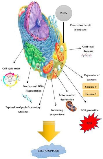
Figure 2. The proposed mechanism of PtNPs anti-cancer activity.
Apoptosis induction by platinum nanoparticles was determined through G0/G1 cell cycle arrest [32]. The influence of PtNPs on cell cycle progression was shown [16]. A significant increase in the percentage of cells in the sub-G1 phase was found after treatment with nanoparticles, and the G0/G1 phase was shown to decrease along with the increase in the sub-G1 phase [16]. Apoptosis was noted in the part of the G0/G1 region, indicating that the G1 phase cells were lost by programmed cell death [32]. PtNPs-treated cells showed higher cell growth at the G2/M phase, which revealed that in the G2/M phase, induced cell cycle was arrested and cell numbers were increased in the sub G0 cell death phase, which revealed the application of PtNPs for the treatment of cervical cancer treatment [31]. Platinum nanoparticles also induced changes in cell morphology: for instance, the appearance of long cytoplasmic protrusions was determined in SH-SY5Y cells, a cloned subline of a neuroblastoma cell line [132], and density; membrane disintegration increased cell population in apoptosis and caused chromatin condensation and nucleus fragmentation [16,103,133]. The mechanism of anticancer activity was found to be an externalization of phosphatidyl serine and an increase in membrane permeability, which are considered to be the hallmarks of apoptosis [74]. Additionally, in A549 cells, the proliferative activity decreased gradually over time in proportion to the increasing concentration of the platinum nanoparticles [102].
The literature data reveal that the antitumor activity of PtNPs may be associated with penetration into the nucleus and mitochondria of the cell, inhibition of DNA replication and mitosis by binding to a DNA molecule, leading to apoptosis [134]. The PtNPs inhibited the DNA replication and affected the secondary structure of DNA at higher concentrations [114]. Mitochondria are responsible for energy production and are also necessary for the primary pathway regulation of the apoptosis and non-apoptotic cell death [135]. The mitochondrial membrane potential (MMP) loss evokes disturbances in the ATP synthesis, leading to ATP accumulation in the mitochondria. Exposed to platinum nanoparticles, low levels of MMP and ATP indicated mitochondrial dysfunction. Higher expression of pro-apoptotic genes and lower expression of anti-apoptotic genes were also observed by the influence of PtNPs [136]. Inhibition of mitochondrial respiration was found in HeLa cells, and the polarization of mitochondria under the action of PtNPs induced a large loss in ATP and mitochondrial dysfunction and led to apoptosis in human cervical cancer cells [95]. PtNPs decreased the level of MMP in various types of cancer cells, including human neuroblastoma cancer cells SH-SY5Y, human monocytic THP-1 cells, and human bone osteosarcoma epithelial cells [132].
PtNPs increased the expression of caspases, playing a substantial role in cell apoptosis [137]. The downregulation of p53 (protein p53) may also be a key element of anticancer activity, because it is a transcription factor regulating the cell cycle and acting as a suppressor of malignant tumor formation [138]. p53 also induces the expression of pro-apoptotic Bcl-2 family members such as Bax, Puma, and Noxa in response to the mitochondrial apoptosis pathway activation. On the surface of mitochondria, these pro-apoptotic proteins meet with anti-apoptotic members of the Bcl-2 family. DNA damage is determined by the ratio of pro-apoptotic and anti-apoptotic proteins. Furthermore, pro-apoptotic signals result in the release of cytochrome C from mitochondria, followed by the activation of cysteine caspases [139]. Bcl-2 protein plays an essential role in the apoptosis process, which activates caspase-9 and caspase-3, triggering the apoptosis cascade (with the participation of another caspases-7,8) [140]. Almeer et al. suggested that PtNPs synthesized using leaf extract of Azadirachta indica initiate an internal apoptosis pathway, mediated by an increase and decrease in the expression of Bax and bcl-2 in mitochondria [137]. PtNPs exposure in vitro leads to p53activation in cells caused by genotoxic stress, followed by activation of p21, leading to a stop in the growth of proliferating cells in the S-phase and subsequent apoptosis [119].
Cellular redox homeostasis change in a significant part of the signaling pathway due to the overproduction of intracellular ROS puts the cell on the apoptosis pathway [141]. Moreover, ROS-mediated transcription factors control the expression of various genes involved in inflammation, cell transformation, tumor cells death or survival, proliferation, invasion, angiogenesis and metastasis. Platinum nanoparticles from T. involucrata are able to enhance cellular apoptosis due to ROS over-production [95]. Similar data were obtained for HEK293 (human embryonic kidney), MCF-7 (human breast cancer) and HepG2 (hepatocellular carcinoma) cells, where platinum nanoparticles induced cytotoxicity and apoptosis via the generation of ROS [137,142].
Possessing antioxidant properties, GSH not only protects the cell from toxic free radicals, but also generally determines the redox characteristics of the intracellular environment. ROS generation was discovered to convert GSH to GSSG (Glutathione disulfide) through the oxidation process [143]. Oxidized glutathione is reduced by the enzyme glutathione reductase induced by oxidative stress. The most important parameters showing the oxidative stress level are the ratio of reduced and oxidized glutathione forms in the cell. ROS generation in a concentration and time-dependent manner, and a decrease in GSH levels in HEK293 (human embryonic kidney) cells treated with platinum nanoparticles led to damage to the cellular component [137]. PtNPs can decrease the various antioxidant levels in different types of cancer cells, initiating their apoptosis [132].
Additionally, nanoparticles also promoted the expression of different proinflammatory proteins: cytokines such as tumor necrosis factor TNF-α, TGF-β, and NF-κB, and interleukin-1β (IL-1β), IL-6, IL-8 [32,133,144]. Platinum nanoparticles enhance the lactate dehydrogenase level and increase apoptosis and oxidative DNA damage [145], as well as superoxide dismutase activity (SOD), lipid peroxide (LPO) and malondialdehyde (MDA) levels [137,146]. Lactate dehydrogenase is a characteristic cell death marker, released into the surrounding extracellular space when cell membranes are disrupted [132]. LPO arises from the oxidation of fatty acids induced by oxidants; therefore, it is also a characteristic sign of negative cellular effects. The production of LPO-derived aldehydes in cancer cells depends on the presence of ROS. The increased ROS level can increase the formation of LPO products and eventually increase oxidative damage to DNA [147].
Abnormally increased production of Nitric Oxide (NO) triggers cell damage [148]. An increased NO level results in mitochondria damage through a change in membrane potential and inhibition of respiratory chain, and also induces a sequence of events in the cell, leading to the ROS generation, loss of mitochondrial membrane potential, the release of cytochrome C into the cytosol, activation of caspases, DNA fragmentation and, ultimately, apoptosis [148,149,150]. PtNPs in combination with retinoic acid have the ability to enhance NO production in SH-SY5Y cells and cause cancer cell death [132].
Apoptosis stimulation is possible by the induction of ERS (Endoplasmic reticulum stress). As a result, an unfolded protein response (UPR) is initiated to restore cellular homeostasis or induce apoptosis [151]. UPR is regulated by various transmembrane proteins, such as protein kinase-like ER kinase (PARK), inositol-requiring enzyme 1 (IRE1), activating transcription factor (ATF6), andATF4, involved in maintaining homeostasis. The inducing apoptosis through the induction of ERS was shown to be possible via platinum nanoparticles [132].
Various research display anticancer activity in vitro against HeLa cells [85,91,152], HEK293 cells [137,153], MCF-7 (breast cancer) and HepG-2 (hepatocellular carcinoma) [32,49,103,104,142], MDA-MB-231 breast cancer cell line [154], lung carcinoma cells A549 [102,131], SH-SY5Y cells [132], ovarian teratocarcinoma (PA-1), pancreatic cancer(Mia-Pa-Ca-2) cells [16], colon carcinoma cells (HCT-116) [90], sarcoma-180 (S-180) cells [155], 4T1 breast cancer cells [156], A431 cell lines (epidermoid carcinoma) [55] and myoblast C2C12 carcinoma cells [144].
The anticancer activity data are not limited by in vitro studies. Thus, in vivo experiments showed PtNPs at the medium and high doses effectively inhibited and delayed the growth of lung cancer in severe combined immune deficient mice [157]. The effect of platinum nanoparticles on breast cancer cells was found for PtNPs that contribute to breast cancer metastasis by damaging the vascular endothelial barrier. PtNPs disrupt the proliferation and migration of endothelial cells and the formation of tubular structures, destroy endothelial junction adhesions and induce the impermeability of the endothelial barrier in vitro. It is assumed that such a stimulation occurs through ROS generation, changing in the expression and conformation of endothelial connective proteins, thereby contributing to intravasation and extravasation of implanted breast cancer cells 4T1 and leading to cancer metastasis in female mice BALB/c nude in vivo [156].
5. Antioxidant Activity
Reactive oxygen species such as hydroxyl, epoxyl, superoxide, peroxylnitrile, and singlet oxygen generate oxidative stress, leading to the growth of various diseases such as inflammation, atherosclerosis, aging, cancer, and neurodegenerative disorders [158]. Free radical activity suppression by antioxidants helps to support the immune system and allows it to fight against viruses and other foreign invaders more effectively. An evaluation of the antioxidant properties of PtNPs in vitro, as a rule, is performed by removing DPPH (2,2-diphenyl-1-picrylhydrazyl) radicals, as one of the most important and widespread free radicals that can harm human cells [159]. DPPH is an uncharged free radical that can accept hydrogen or free electrons to produce a stable diamagnetic molecule, that is why it has long been used to test the free radical scavenging capacity of antioxidants [159,160]. The antioxidant activity is reflected as a percentage in the DPF absorption or removal [159,160]. It was shown that antioxidant activity was found to be dose-dependent for PtNPs and may also correlate with the nanoparticle size and the zeta potential, depending on the used “bio-factory” [50,66,68,96,123]. For example, the smallest nanoparticles produced by gram-negative bacteria showed better antioxidant activity than gram-positive ones [50]. In addition, capping agents also seemed to play quite an important role in the PtNPs antioxidant activity [77]. It should be noted that platinum nanoparticles synthesized by plants with antioxidant potential are of great importance, because plant compounds can prevent ROS-triggered oxidative damage when the endogenous antioxidant system does not cope independently. Ascorbic acid can be an example, possessing an important antioxidant value in physiological conditions and pro-oxidant in pathological conditions (bacterial infections or cancer) [161]. Antioxidant activity data were found for plant-mediated platinum nanoparticles from T. involucrata [95], A. halimus [89], D. bulbifera [86], Salix tetraspeama [87] and Cordyceps militaris [100]. Antioxidant and neuroprotective activities studies of platinum nanoparticles synthesized by Bacopa monnieri leaf extract showed a decrease in ROS generation and the free radical’s removal, thereby increasing the levels of dopamine, its metabolites, GSH, catalase, SOD, and complex I, and decreasing MDA levels along with enhanced motor activity with MPTP-induced (1-methyl 4-phenyl 1,2,3,6 tetrahydropyridine) neurotoxicity in a model of Parkinson’s disease in Danio fish [85]. The obtained data may become a potential option for the fight against Parkinson’s disease. Suppression of reactive oxygen species by means of PtNPs interacting with antioxidant enzymes such as superoxide dismutase, catalase and glutathione peroxidase was shown using Drosophila melanogaster as an in vivo model system. Moreover, platinum nanoparticles interacted with hemocytes without any toxic cell effect and significantly accelerated the wound healing process in a short time [162].
6. Anti-Diabetic Activity
Diabetes mellitus (diabetes) is one of the most urgent problems faced by mankind, despite the significant amount of well-established diagnosis and treatment methods for this disease. Alpha-amylase and α-glucosidase are known to be key enzymes in carbohydrate metabolism, so their inhibition is one of the most significant strategies for diabetes therapy [163,164]. In addition, α-glucosidase is also considered as the main enzyme involved in carbohydrate metabolism, catalyzing the cleavage of oligosaccharides and disaccharides into monosaccharides [163,164]. An amylase inhibitor, jointly with starchy foods, reduces the usual upturn in blood sugar. Amylase inhibitors or starch blockers, including silver and other metal nanoparticles, were proved to prevent the absorption of food starches by organism [163]. The anti-diabetic effect was discovered in vitro for PtNPs from P. salicifolium: a mild inhibitory effect against α-amylase and a higher effect against α-glucosidase was exhibited [96]. The authors suggested that the antidiabetic effect could be attributed to the direct correlation between the phytoconstituents surrounding PtNPs and α-amylase and/or α- glucosidase inhibitory action, excessive ROS production or imbalanced antioxidant protection mechanisms [96]. Furthermore, a significant decrease in glucose levels after PtNPs injection was observed in streptozotocin-induced diabetic rats [165]. Type 2 Diabetes is characterized by an increase in ROS production level, induced by chronic extracellular hyperglycemia as a result of a violation of the cell redox state, causing the abnormal expression of insulin sensitivity genes [166,167]. In this regard, the enzyme-like antioxidant properties of platinum nanoparticles to absorb free radicals and decline ROS concentration can promote the diabetes struggle. For instance, under the influence of PtNPs, the induction of the gene expression of the antioxidant enzyme catalase (CAT), glutathione peroxidase (GPx) and hemoxoigenase, suppression of fasting blood glucose levels and an improvement in the impaired ability to sugar tolerance in obese insulin-resistant type 2 diabetic KK-Ay mice was shown [168].
7. Anti-Inflammation Activity
ROS overproduction is associated with the pathogenesis of inflammatory diseases. Antioxidant therapy to solve this problem is possible in the face of platinum nanoparticles. Rehman et al. demonstrated that in vitro anti-inflammatory activity of PtNPs may be attributed to their down regulation of the NFjB signaling pathway in macrophages in lipopolysaccharide-stimulated RAW 264.7 cells [169]. PtNPs showed direct anti-inflammatory activity in RAW264.7 macrophages through a mechanism involving the intracellular ROS uptake by suppressing lipopolysaccharide-triggered production of proinflammatory mediators, including nitric oxide, tumor necrosis factor-α and interleukin-6 [170]. The high antioxidant activity of platinum nanoparticles was found in a cavernous malformation cellular model of the human brain [171]. PtNPs had a significant neuroprotective effect on the ischemic mouse brain [172] and effectively protected keratinocytes from UV-induced inflammation [173] and suppressed chronic obstructive lung inflammation provoked by acute cigarette smoking [174]. Platinum nanoparticles, alone and in combination with palladium nanoparticles, showed antioxidant activity and weakened aging-related skin pathologies in vivo in mice, without causing morphological abnormalities such as cellular infiltration, fat deposition, or cell damage in mouse skin [175]. It was supposed that the catalase activity shown by the nanoparticle combination may be useful in the treatment of vertigo: an acquired pigmentation disorder characterized by H2O2/peroxynitrite-mediated oxidative and nitrative stress in the skin.
8. Other Application
Photothermal therapy and radiotherapy. Antitumor chemotherapy has many negative diverse consequences for patients, therefore developing less harmful and cancer-specific strategies is an extremely important task. Photothermal therapy may be the one of the decisions. This non-invasive treatment assumes that PtNPs increase the cellular temperature upon irradiation, causing DNA/RNA damage, membrane rupture, protein denaturation and finally apoptosis [176,177]. Such photothermal therapy using PtNPs, 5–6 nm in size, induce damage of a selective cellular component and cell death [38,176]. Polypyrrole-coated iron-platinum nanoparticles were used for photothermal therapy and photoacoustic imaging. In vitro investigation experimentally demonstrated the effectiveness of these NPs in killing cancer cells with NIR laser irradiation. Moreover, the phantom test of PAI used in conjunction with FePt@PPyNPs showed a strong photoacoustic signal [177]. Cysteine surface modified FePtNPs can be potential sensitizers for chemoradiotherapy: in vitro NPS FePt-Cys induced ROS, suppressed the antioxidant protein expression and induced cell apoptosis, and also facilitated the chemoradiotherapy effects by activating the caspase system and disrupting DNA damage repair. The drug safety and the synergistic effect with cisplatin and irradiation was confirmed by in vivo studies [178]. FePtNPs could potentially be a new strategy for increasing radiation therapy efficiency in cancer cells overexpressing hCtr1 due to enhanced uptake and targeting of mitochondria [179]. The synergistic antitumor effect with radiation to eliminate tumors for MFP-FePt-GONCs nanoparticles was determined [180]. These nanoparticles improved the radiation effects by activating internal mitochondrial-mediated apoptosis and worsening DNA damage repair. Additionally, they induced ROS release, which suppressed antioxidant protein expression and induced cell apoptosis [180]. A combination of PtNPs with irradiation by fast ions effectively enhances the strong, lethal damage to DNA [181]. In spite of the data presented above, which were obtained for platinum nanoparticles/nanocomposites synthesized by physicochemical methods, “green” PtNPs may have serious potential in this method. Platinum nanoparticles data obtained by Prosopis farcta fruit extract indicate their stability and biocompatibility for application as a contrast agent in computed tomography as an alternative to low molecular weight agents with toxic effects [153].
Catalytic activity. The excellent catalytic activity of “green” PtNPs was shown for removing pharmaceutical products (PhP). The platinum nanoparticles produced via D. vulgaris worked as an effective biocatalyst in the removal of four PHPs classes: ibuprofen, ciprofloxacin, sulfamethoxazole and 17β-estradiol, which are most relevant in the environment [182]. It is important to note that only 13% of catalytic activity was lost during recycling, indicating the possibility of bacteriogenic PtNPs reuse for technological development in the pharmaceutical wastewater treatment [182].
Detection. Platinum nanoparticles obtained by physicochemical approaches can be used for the detection of DNA, cancer cells, antibiotics, glucose, proteins, bacteria, viruses and antibodies [38,183,184,185,186,187,188,189]. The peroxidase activity of plant-mediated PtNPs makes it possible to quickly detect Hg ions [190], as well as hydrogen peroxide [82]. Additionally, PtNPs were successfully used for hydrazine detection in spiked water samples [82].
This entry is adapted from the peer-reviewed paper 10.3390/jfb13040260
This entry is offline, you can click here to edit this entry!