Beneficial Effects of Ursolic Acid against Lung Cancer: History
Please note this is an old version of this entry, which may differ significantly from the current revision.

Lung cancer is the leading cause of cancer-related deaths globally. Despite current treatment approaches that include surgery, chemotherapy, radiation and immunotherapies, lung cancer accounted for 1.79 million deaths worldwide in 2020, emphasizing the urgent need to find novel agents and approaches for more effective treatment. Traditionally, chemicals derived from plants, such as paclitaxel and docetaxel, have been used in cancer treatment, and in recent years, research has focused on finding other plant-derived chemicals that can be used in the fight against lung cancer. Ursolic acid is a polyphenol found in high concentrations in cranberries and other fruits and has been demonstrated to have anti-inflammatory, antioxidant and anticancer properties. Here, researchers summarize studies examining the effects of ursolic acid and its derivatives on lung cancer. Data from in vitro cell culture and in vivo animal studies show potent anticancer effects of ursolic acid and indicate the need for clinical studies.

  • lung cancer
  • ursolic acid
  • proliferation
  • survival
  • invasion
  • metastasis
  • signaling cascades

1. Effects of Ursolic Acid against Lung Cancer: In Vitro Studies

Studies that have examined the effects of UA in cultured human lung cancer cells are summarized below and in Table 1.
Human A549 adenocarcinoma lung cancer cells treated with ursolic acid (2–40 µM) had significantly reduced proliferation, G1-phase cell cycle arrest and induction of apoptosis (determined via DNA fragmentation), (Table 1). Western blot analysis revealed that UA-treated A549 cells had increased p53 protein levels and decreased levels of cyclin D1, D2, E and NF-kB/p65 proteins. The levels of the antiapoptotic proteins B-cell lymphoma 2 (Bcl-2) and Bcl-XL were significantly reduced, whereas Bax protein levels were increased. Treatment with UA induced p21/WAF1 and Fas/APO-1 signaling [1], (Table 1). Loss or reduced levels of the tumor suppressor p53 contributes to cancer initiation and progression [2][3], and the evidence from this study showing an increase in p53 levels with UA treatment is of major significance, indicating its tumor suppression potential. In addition, the study provided strong evidence of induction of lung cancer cell apoptosis with UA treatment, indicating its potential to be used in the treatment of lung cancer.
Table 1. Evidence of the effects of ursolic acid in lung cancer cells: a summary of in vitro studies.
Human NSCLC H460 cells treated with 10 µM UA for 24 h exhibited increased apoptosis, characterized by activation of caspase-3 and DNA fragmentation, which was associated with a significant increase in gene expression of matrix metalloproteinases (MMPs) 1, 2, 3, 9 and 10, as well as an increase in glucocorticoid receptor (GR) cytosolic localization. These data suggest that members of the MMP family may be involved not only in invasion but also apoptosis of cancer cells and that cytosolic localization of GR may be an important factor in the upregulation of MMP during ursolic-acid-induced apoptosis of H460 cells [4].
Treatment of A549, H3255 and Calu-6 human lung cancer cells with UA decreased cell viability, migration and invasion and increased DNA fragmentation and apoptosis. Treatment with UA led to decreased protein expression of angiogenic factors vascular endothelial growth factor (VEGF) and transforming growth factor beta1 (TGF-beta1), both of which are biomarkers associated with cancer cell metastasis. In addition, mRNA levels of intercellular adhesion molecule-1 (ICAM-1) fibronectin and matrix metalloproteinases (MMP) 2 and 9, all important for cell adhesion, migration and invasion, were reduced. The levels of lactate dehydrogenase (LDH) in the medium, an indicator of plasma membrane damage, were increased, whereas Na+-K+-ATPase and PKC activities were reduced. Taken together, these data suggest that UA treatment increases apoptosis and decreases viability and migration of human lung cancer cells [5]. Although the reduction in the angiogenic factors VEGF and TGF-beta1 is a strong indicator of reduced migration, the study did not include any assays assessing cell migration/metastasis. The use of the scratch-wound and/or transwell cell invasion assays would have benefited the study. In addition, protein levels (not only mRNA levels) of ICAM-1, MMPs and fibronectin should have been examined.
Apart from examining the effects of UA on lung cancer cells, researchers have examined the potential of UA to counteract the effects of cigarette smoke in lung cells. Normal human bronchial epithelial (NHBE) cells were exposed to cigarette smoke extract (CSE) to induce cytotoxicity and cell death in order to mimic the in vivo conditions of cigarette smoking. Treatment with UA resulted in reversal of many of the negative effects of CSE, including reduced CSE-induced cytotoxicity, reduced CSE-induced lactate dehydrogenase efflux and recovery of CSE-induced glutathione loss. CSE activated the Nrf2 pathway, as shown by the increased levels of NQO1, GST and Nrf2 proteins. Treatment with UA normalized these results to near-control levels and reduced the CSE-induced DNA damage. In this cell model, UA alleviated the cytotoxic effects of CSE and recovered the intracellular redox balance, suggesting that UA has a protective effect against CSE-induced cell injury. The authors concluded that UA has a potential role in both the prevention and treatment of lung tumors caused by cigarette smoking [19]. The model created by this research group provides preliminary evidence of UA acting against cigarette-smoke-induced changes in lung cells. However, this is an in vitro model, and further work utilizing animal models is required to determine whether UA protects against cigarette-smoke-induced damage. Strong evidence of a protective role of UA in animal models would provide a justification for clinical trials.
A549 cells treated with UA showed reduced proliferation, as well as reduced adhesion, wound healing and transwell migration, indicating inhibition of metastasis. Western blot analysis revealed an upregulation of E-cadherin and a downregulation of N-cadherin and vimentin, indicating an inhibition of epithelial–mesenchymal transition (EMT). Furthermore, decreased expression of astrocyte-elevated gene-1 (AEG-1) correlated with repression of NF-kB signaling was observed in cells treated with UA [6].
Treatment of A549 and H460 human lung cancer cells with UA significantly decreased cell viability and proliferation. UA increased chromatin condensation and induced apoptosis. Flow cytometry revealed an increase in cell cycle arrest in the G0/G1 phase. Western blot analysis revealed an increase in cleaved caspases 3 and 9, increased phosphorylation of the proapoptotic protein Bax and a decrease in phosphorylated Bcl-2, an anti-apoptotic protein, suggesting that UA induces apoptosis in these cells via a caspase-dependent pathway. Treatment with UA increased phosphorylation/activation of AMP-activated protein kinase (AMPK) and inhibited the phosphorylation/activation of mTOR and p70S6K proteins, which are involved in protein synthesis and cell growth. In addition, the levels and activity of the lipogenic enzymes acetyl-CoA carboxylase (ACC) and fatty acid synthase (FASN) were decreased due to increased AMPK activation [7]. These data clearly show that UA is a strong activator of the AMPK energy sensor. UA-induced AMPK activation was abolished by an siRNA approach, which downregulated LKB1. Activated AMPK leads to downstream inhibition of mTOR and p70S6K, resulting in inhibition of protein synthesis and, ultimately, inhibition of cell proliferation. The inhibition of lipogenesis is also the result of activation of AMPK, resulting in limited availability of lipids, which are important components for cell proliferation. Although the study did not provide any evidence, it is possible that the activation of AMPK is also responsible for the induction of apoptosis. An approach to knockout/downregulate AMPK would have provided stronger evidence of the role of AMPK in the UA-induced effects.
Treatment of A549 human lung cancer cells with ursolic acid (0–50 µM) resulted in reduced cell viability and increased DNA damage associated with inhibition of vaccinia-related kinase 1 (VRK1) autophosphorylation and reduced phosphorylation of its downstream substrates, CREB and histone H3. This UA treatment also decreased cyclin D1 mRNA levels, a downstream effector of CREB and histone H3. Nuclear magnetic resonance (NMR) titration experiments and in silico modeling showed that UA is able to bind directly to VRK1, causing inhibition of kinase activity. A combination of UA with doxorubicin resulted in greater reduction in cell viability compared to either compound alone, indicating a synergic effect. Furthermore, when UA was combined with the PARP-1 inhibitor veliparib, a significant reduction in cell viability was observed compared to UA alone, indicating that UA exerts synergistic effects when combined with drugs that target DNA damage repair genes [8]. The finding reported in this study, that UA binds directly to the catalytic domain of VRK1 inhibiting its kinase activity, is based on in silico modeling. Further work is required to confirm that UA binds to VRK1 in cells, and the use of VRK1 overexpression/knockdown (siRNA) approaches can provide evidence of the role of VRK1 in UA-induced apoptosis. The finding that UA enhanced the effect of doxorubicin and veliparib is significant, suggesting that UA has the potential to be used as an adjuvant chemotherapy agent against lung cancer.
Wu et al. reported significantly reduced cell viability and induction of apoptosis in NSCLC (H1299, H1650, A549, PC9 and H1975) cells treated with UA (5–80 µM). UA treatment increased phosphorylation/activation of stress-activated protein kinases/Jun amino-terminal kinases (SAPK/JNK) while simultaneously decreasing the protein levels of SP1, DNMT1 and EZH2. Detection of caspase 3/7 activity was increased by UA treatment, as observed with a Caspase 3/7 assay kit. Use of the SAPK/JNK inhibitor SP600125 abolished the effects of UA on SP1, DNMT1 and EZH2, confirming the involvement of this signaling pathway. Exogenous expression of SP1 or DNMT1 also abolished the UA-induced effects, providing strong evidence of their involvement. Based on these data, the authors concluded that UA inhibits NSCLC cell growth via SAPK/JNK-mediated inhibition of SP1, DNMT1 and EZH2 [9].
Treatment of mesothelioma cells (H28, H2452 and MSTO-211H) with 10–40 µM UA inhibited proliferation and colony formation in a dose-dependent manner. Cell-cycle analysis showed an accumulation in the sub-G1 phase compared to untreated controls, indicating induction of apoptosis. Western blot analysis showed reduced total PARP and reduced caspase 3, both of which are apoptosis-related proteins. UA treatment attenuated the levels of EMT-related proteins; E-cadherin expression was increased, whereas levels of vimentin, N-cadherin, twist, B-catenin and pGSK3a/b were decreased. The expression of the survival genes cyclin D1, p-AKT and NF-kB were also reduced with UA treatment. MicroRNA array and qRT-PCR results indicated that let7b was upregulated in H2452 and H28 cells treated with UA. Overall, the data reported in this study suggest that UA treatment can induce apoptosis in lung cancer cells through inhibition of EMT and increased expression of let7b [10]. Downregulation of let7b would have provided stronger evidence of its role in the UA-induced effects.
Song et al. (2017) examined the ability of UA to increase the radiosensitivity of H1299 NSCLC cells [20]. They established radioresistant H1299 NSCLC cells by expressing a mutant hypoxia inducible factor-1α (HIF-1α) and found that treatment with UA increased their radiosensitivity (as evidenced by reduced cell survival), and this effect was associated with reduced endogenous GSH and HIF-1α levels [20]. Although these findings suggest that UA has the potential to radiosensitize lung cancer cells in vitro, further studies are required to examine whether UA causes radiosensitization in animals xenografted with NSCLC cells. Once strong evidence is reported from in vivo animal studies, clinical studies will be required to examine whether UA causes radiosensitization in NSCL patients.
Treatment of A549 cells with UA (10–100 µM) for 24 h inhibited cell proliferation and induced apoptosis. UA decreased the number of cells in the G0/G1 phase in a dose-dependent manner and increased the number of cells in the S and G2/M phases, suggesting UA-induced cell cycle arrest in the S and G2/M phase. This UA treatment decreased protein expression of Bcl-2 and total PARP, increased levels of cleaved-PARP and increased markers for autophagy, including the LC3-II/LC3-I ratio and p62 protein expression [11].
Human lung cancer A549 cells treated with UA (10, 20 and 40 µg/mL) for 24 or 48 h showed reduced cell viability, as well as increased autophagy and mitophagy. Treatment with UA increased the levels of autophagy-associated protein LC3-II and increased the LC3-II/LC-I ratio. In addition, the mitochondria of cells treated with UA appeared to be fragmented, and the levels of p62 protein, which is required for mitophagy, were increased and colocalized with fragmented mitochondria. Furthermore, treatment with UA increased ROS production, increased Nrf2 and PTEN-induced kinase 1 (PINK1) protein levels and reduced phosphorylated Akt and mTOR levels [12]. The data clearly show inhibition of Akt and mTOR as a result of UA treatment and not activation, as stated in the abstract (possible typographical error). The data from the two above-mentioned studies [11][12] suggest induction of autophagy and mitophagy by UA treatment; however, whether autophagy and mitophagy are induced in lung tumors in vivo is not known.
Ursolic acid treatment of NSCLC H1975 cells with an EGFR T790M mutation, a major cause of epidermal growth factor receptor tyrosine kinase inhibitor (EGFR-TKI/erlotinib) resistance, resulted in inhibition of proliferation and motility, as well as induction of apoptosis. UA treatment resulted in inhibition of CT45A2 expression via β-catenin/TCF4 inhibition. These data show that UA may be a potential treatment for NSCLC with the EGFRl858R/T790M mutation [14]. This study included examination of the effects of UA in animals xenografted with lung cancer cells expressing the EGFR T790M mutation; the findings are reported below (see in vivo section).
Chen et al. constructed a paclitaxel resistance cell line, A549-PR, with increased expression of stemness markers CD133, Oct-4, Notch3, Nanog and Sox2. Treating these chemotherapy-resistant cells with UA resulted in decreased expression of these marker proteins, reduced aldehyde dehydrogenase 1 (ALDH1) activity and, importantly, increased sensitivity to paclitaxel. Treatment of A549-PR cells with UA resulted in reduced miR-149-5/MyD88 signaling. The UA-mediated inhibition of chemoresistance and stemness was partially reversed by overexpression of miR-149 and knockdown of MyD88 [13]. Paclitaxel is a chemotherapy agent commonly used in the treatment of lung cancer. Unfortunately, many patients treated with paclitaxel develop resistance; therefore, the identification of methods to overcome such resistance is highly desirable, as it will improve patient survival. The findings of this study are significant; however, they are derived from in vitro studies only. Further in vivo animal studies and human clinical trials are required to confirm whether UA has the ability to overcome paclitaxel resistance.
Human NSCLC cells (H1975) treated with UA exhibited inhibited mesenchymal-like responses, such as migration, invasion and matrix metallopeptidase (MMP) 2 and 9 activity [15]. Furthermore, treatment with UA attenuated the transforming growth factor-β1 (TGF-β1)-induced decrease in E-cadherin and increased N-cadherin levels. TGF-β1-induced αVβ5 integrin levels and cell migration and invasion (using scratch and transwell invasion assays) were attenuated by UA treatment. The use of a pharmacological inhibitor of integrin (SB273005), similarly to UA treatment, reduced TGF-β1-stimulated cell migration and invasion, whereas combined treatment with UA and the inhibitor did not have any synergic effect. Although these data indicate that UA may target/reduce αVβ5 levels, leading to reduced epithelial-to-mesenchymal transition in NSCLC [15], more robust evidence is required to confirm this phenomenon. The use of inhibitors in general may lead to inhibition of other off-target molecules, and the specificity of SB273005 against αVβ5 versus other integrins is not clear. In addition, it is not known whether UA can bind to αVβ5. Further studies are required to answer these questions.
Human lung cancer cells (NCI-H292) treated with UA showed significantly reduced viability and increased apoptosis, as evidenced by increased levels of cleaved PARP and increased cytochrome C levels. Western blotting analysis revealed increased levels of endonuclease G (Endo G) and apoptosis inducing factor (AIF), whereas levels of the antiapoptotic proteins B-cell lymphoma 2 (BCL2) and BH3 interacting domain death agonist (BID) were reduced with UA treatment. In these cells, UA increased intracellular calcium levels by causing a time-dependent release and reduced mitochondrial membrane potential without affecting ROS levels. Taken together, these results suggest that UA treatment can induce apoptosis through AIF and Endo G release via a mitochondria-dependent pathway [16].
A variety of lung cancer cells (NSCLC: A549, H1975, H1299, H460 and H520; SCLC: H82 and H446; murine Lewis lung carcinoma cell line, LLC), when treated with UA, exhibited significantly reduced cell proliferation, whereas apoptosis was induced, as evidenced by an annexin V-FITC/PI double staining assay. UA treatment increased levels of cleaved PARP and decreased levels of Bcl-2, both indicators of apoptosis. In addition, treatment of H460, H1975 and A549 lung cancer cells with UA induced autophagy, as revealed by the elevation in the protein levels of the autophagy marker LC-3-II. Furthermore, UA treatment decreased phosphorylation/activation of Akt and its downstream targets, p70S6K and 4EBP1. Inhibition of autophagy, using siRNA for autophagy-related gene 5 (ATG5) or chloroquine, enhanced the effects of UA (inhibition of proliferation and induction of apoptosis), suggesting that the autophagy observed following UA treatment acts as a pro-survival mechanism in these cells [17].
Human A549 and H460 lung cancer cells treated with UA exhibited reduced proliferation, as well as reduced CDK4, cyclin D1 and cyclin E protein and mRNA levels, whereas the levels of p21 and p27 tumor suppressor proteins were increased. In addition, treatment with UA resulted in decreased VEGFR and pSTAT3 levels, suggesting inhibition of angiogenesis. Decreases in cell invasion and wound healing/migration were observed, suggesting antimetastatic activity of UA in NSCLC cells. Cancer stem cell (CSC) proliferation was inhibited with UA treatment, as evidenced by a significant reduction in the levels of CSC markers (NANOG, POU5F1 and SOX2). Molecular binding studies showed a high-affinity interaction between UA and EGFR, suggesting that UA interferes with EGFR signaling. This was further tested by treating cells with human recombinant EGF combined with 20 µM of UA. In the presence of UA, the EGF-mediated EGFR phosphorylation was reduced, and the downstream targets of the EGFR pathway, JAK2 and STAT3, showed reduced phosphorylation, whereas the total protein levels remained unchanged. Chromatin DNA extracted from NSCLC cells that had been treated with UA exhibited a significant reduction in the binding of STAT3 to promoters MMP2 and PD-L1. Based on these results, the authors concluded that UA has anticancer activity dependent on STAT3 signaling, that STAT3 acts as a bridge between EGFR and PD-L1 and that UA may be a drug candidate for PD-L1-based targeted cancer therapies [18]. Although the authors suggested that UA acts as an inhibitor of EGFR, further studies are required to confirm this hypothesis. Future in vitro studies utilizing recombinant EGFR and UA can clarify this issue.
According to the results of the studies presented above, it is evident that the effects of UA were examined in a variety of lung cancer cells representing distinct subtypes of the disease. Small cell lung cancer (SCLC) and non-small cell lung cancer (NSCLC) adenocarcinoma, squamous cell carcinoma and large cell carcinoma cell lines were utilized. The effective concentration of UA appears to be in the range of 20–50 µM in the majority of the studies reviewed herein. Unfortunately, no studies have been conducted that examine and directly compare the UA concentration required for half-maximum inhibition (IC50 values) of proliferation of different lung cancer cell lines.
Overall, the in vitro studies presented above and in Table 1 show that treatment of lung cancer cells with UA results in inhibition of proliferation, induction of apoptosis and autophagy through inhibition of Akt, NF-kB and mTOR; increased LC3-II/LC-3-I ratio; and increased cleavage of caspase 3/7 and PARP (Figure 1).
Figure 1. Ursolic acid reduced proliferation and induced apoptosis and autophagy of lung cancer cells in vitro. UA inhibited the phosphorylation/activation of Akt [10][17], mTOR [7][12] and NF-kB [1][10] and increased p53 protein levels [1], leading to inhibition of proliferation and survival. Furthermore, UA increased the proapoptotic protein Bax and decreased the antiapoptotic protein Bcl-2 [1][7][11]. An increase in cleaved caspases 3 and 7 was observed, leading to an increase in cleaved PARP and apoptosis [4][10][16]. An increase in LC3-II protein levels, a marker of autophagy, was also observed as a result of UA treatment [11][12][17]. The figure was created using BioRender.com (accessed on 9 September 2022) based on data from [1][4][6][7][10][11][12][16][17].

2. Effects of Ursolic Acid against Lung Cancer: In Vivo Studies

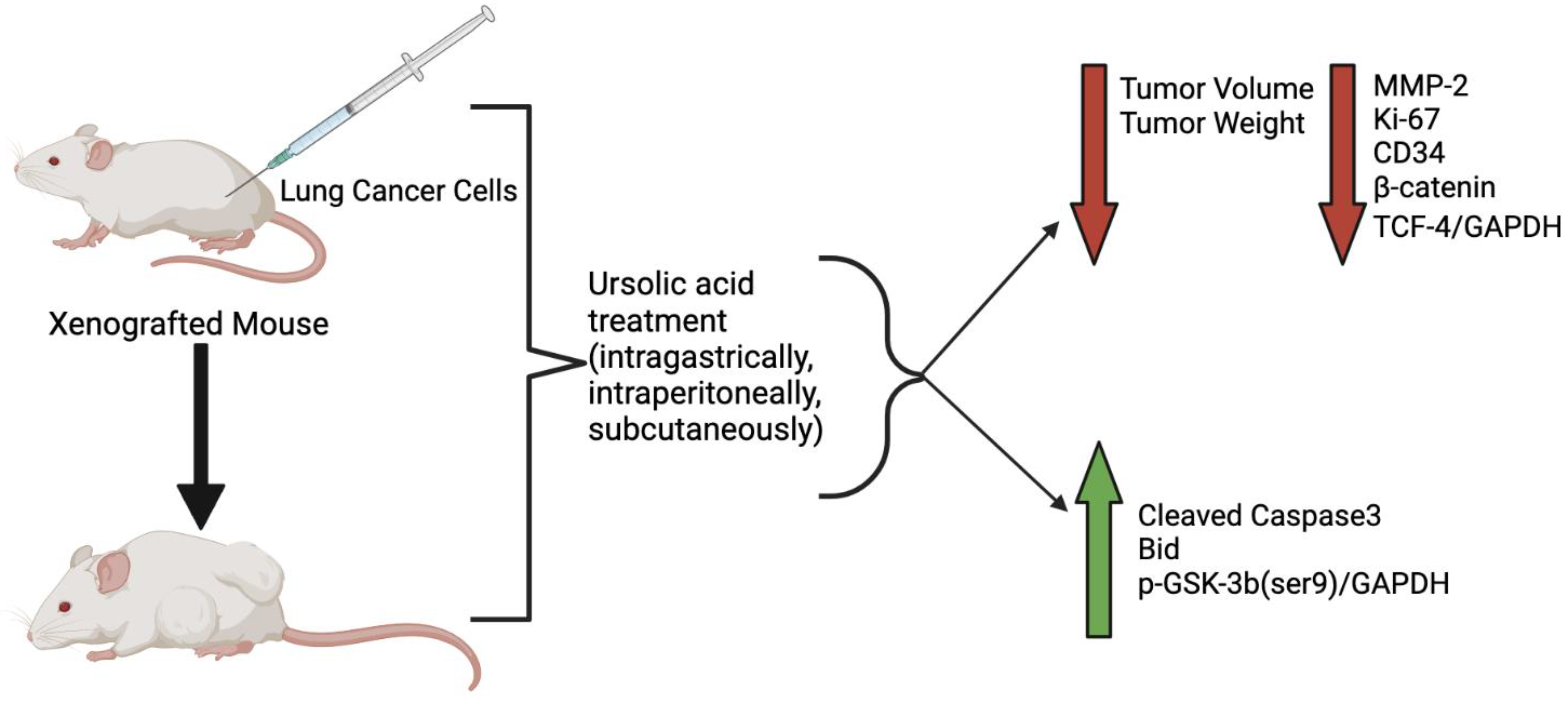
A limited number of studies have examined the effects of UA administration in mice xenografted with lung cancer cells. All the available studies are presented below and in Table 2.
Table 2. Effects of ursolic acid against lung cancer: in vivo evidence.

3. Ursolic Acid Derivatives and Their Effect against Lung Cancer

Owing to it anticancer properties but limited bioavailability in its natural state, researchers are looking for ways to manipulate UA and increase its effectiveness. A number of analogs of UA have been produced, and their effects have been examined using lung cancer cells. All the available studies are presented below and in Table 3.
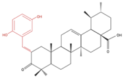
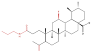
Table 3. Ursolic acid derivatives and their effect against lung cancer in vitro. Chemical structures created with BioRender.com (accessed on 9 September 2022). Black portions indicate parent UA structure, and red segments indicate modifications.
Cell Line Derivative Name Derivative Structure Findings Mechanism Reference
A549
SF-295 (CNS)
UA-9
10 nM
Molecules 27 07466 i001 ↓ Cell Density Not investigated [22]
A549 UA-triazolyl derivative Molecules 27 07466 i002 ↓ Cell Density Not investigated [23]
A549 Compound 3B

50 µM, 48h
Molecules 27 07466 i003 ↓ Cell Growth Not investigated [24]
H460
H322
Compound 17 Molecules 27 07466 i004 ↑ Apoptosis
↑ Autophagy
↑ Cleaved caspases 8 and 7
↑ Cleaved PARP
↑ LC3A/B-II ratio
↓ Bcl-2 protein
↓ mTOR protein
[25]
A549
H460
UA232
24, 48 and 72 h
Molecules 27 07466 i005 ↑ Cell cycle arrest
↑ Apoptosis
↓ Proliferation
↓ Cyclin D1 protein
↓ CDK4 protein
↑ CHOP protein
↑ Cleaved PARP
[26]
NCI-H460 5Y8
5 and 10 µM
Molecules 27 07466 i006 ↑ Apoptosis
G1 phase cell cycle arrest
↓ p-NF-kB
↓ p-IKKα/β
↓ TAK1
↓ TAB1
↑ ROS
[27]
A549 8c
5, 10 and 20 µM
24 h
Molecules 27 07466 i007 ↑ Apoptosis
G1 phase cell cycle arrest
↓ Cell migration
↑ Caspase-3 cleavage
↓ p-NF-kB
↓ p-IKBα
↓ p-IKKα/β
[28]
Kanali et al. examined the effects of 32 UA derivatives utilizing human A549 lung cancer cells and found that the analog 4-bromoanilamideursolic acid (UA-9) had the greatest anticancer effect. Accordingly, UA derivatives with electron-donating groups (UA-9) were suggested to have the most potent anticancer activity [22]. However, all of the examined analogs had higher molecular weights and more limited polarity than UA, indicating potentially low solubility and therefore difficulty in passing through cell membranes.
Ursolic acid-triazolyl derivatives with o-bromo, o-chloro or o-methoxy substitutions on the aromatic ring showed inhibition of cell growth of cancer cells, including A549 lung cancer cells [23]. These UA-derivative compounds displayed higher anti-cancer effects than the parent ursolic acid, suggesting these derivatives should be further examined as potential cancer treatment options [23].
A group of ursolic acid benzylidene derivatives were created through an oxidation/condensation procedure and tested against cultured cancer cells, including A549 lung cancer cells [24]. Each of the compounds exhibited greater cancer cell cytotoxicity than UA and less cytotoxicity to normal FR-2 lung epithelial cells. The most promising derivative created was a UA derivative with 2,5-dihydroxy substitution on the aromatic ring, named 3b. Although this compound was further tested against colon cancer cells (HCT-116) and shown to induce apoptosis [24], studies utilizing lung cancer cells are lacking.
A series of A-ring cleaved UA derivatives were prepared and evaluated in NSCLC cells (H460, H322 and H460LKB1+/+). UA with a cleaved A-ring and a secondary amide at C3 (compound 17) was found to be the most active in inducing apoptosis. Treatment of lung cancer cells with this UA derivative induced apoptosis, as evidenced by the increased cleaved caspase-8 and caspase-7 levels and the decrease in Bcl-2 protein levels. Increased levels of Beclin-1 and LC3A/B-II suggested an induction of autophagy with this UA-derivative treatment. Decreases in mTOR and p62 protein levels were also observed. Based on these findings, UA derivative compound 17 may be a potential candidate for lung cancer treatment and should be further researched [25].
Human lung cancer cell (A549 and H460) treatment with the UA derivative UA232 resulted in reduced cell proliferation and induction of apoptosis. These effects were more prominent with UA232 than treatment with the parent UA compound, whereas the cytotoxic effects on normal cells (HEK293T) were the same. UA232 treatment of lung cancer cells caused the cells to arrest in the G0/G1 phase of the cell cycle in association with downregulation of cyclin D1 and CDK4. This novel UA derivative significantly increased apoptosis and increased cleaved-PARP1 protein levels compared to standard UA treatment. There was no significant change in the levels of Bax, Bcl-2 or caspase-8, indicating that apoptosis was not induced through either the mitochondrial apoptosis pathway or the death receptor pathway. UA232 treatment increased expression of CHOP, indicating a mechanism involving the ER stress pathway. Pretreatment with 4-PBA, an ER stress inhibitor, attenuated the UA232-induced apoptosis, a further indication of the role of ER stress [26].
Another UA derivative with functionalized aniline or amide side chains was synthesized and used to treat lung adenocarcinoma NCI-H460 cells. Compound 5Y8 had the most potent antiproliferative activity, which was significantly higher than that associated with treatment with the UA parent compound. Molecular docking studies revealed that compound 5Y8 had a key interaction with the active site of NF-kB, blocking the activity and signaling pathway, which led to apoptosis. These findings indicate that the 5Y8 UA derivative has potential as a new class of NF-kB inhibitor for the treatment of lung cancer and may contribute to overcoming chemotherapy resistance [27].
Treatment of A549 lung cancer cells with a number of UA derivatives containing long-chain diamine moieties resulted in significant inhibition of viability, with IC50 values in a micromolar range (5.22–8.95 μM) [28]. The UA derivative compound 8C was the most potent (IC50: 5.22 μM) and caused G1 phase cell cycle arrest and increased caspase 3 cleavage, an indicator of apoptosis. In addition, cancer cell migration was inhibited, suggesting that this compound may help to prevent metastasis. Treatment with UA compound 8C inhibited the NF-kB signaling pathway, as evidenced by the significantly reduced levels of phosphorylated IKKα/β and IKBα and reduced NF-kB levels. Furthermore, molecular docking studies showed a key interaction between compound 8C and the active site of NF-kB, blocking its activity [28]. Activated NF-kB is associated with increased cancer cell proliferation and inhibition of apoptosis; therefore, NF-kB inhibitors have potential as anticancer agents. This study [28] shows clear evidence of UA compound 8C targeting/inhibiting NF-kB activity in lung cancer cells; therefore, its anticancer potential should be further explored.
Although the parent ursolic acid compound has been shown to be an effective anticancer agent, the above-described studies have shown that derivatives of the parent UA compound have enhanced potency, indicating that modification may be an effective approach to drug development.
UA has limited bioavailability [29][30] due, in part, to its low water solubility and high molecular weight of 456.7 g/mol. A limited number of studies have examined UA bioavailability. Mice fed a diet containing 0.05% UA for 8 weeks were euthanized, and plasma UA levels and tissue distribution were measured utilizing high-performance liquid chromatography–mass spectrometry (HPLC-MS) [29]. Plasma UA levels of 580 ng/mL (1.26 µM) were observed, with the highest distribution in the liver (9.7 µg/g), followed by the colon (6.4 µg/g), kidney (5.9 µg/g), heart (3.9 µg/g), bladder (2.9 µg/g) and brain (1.6 µg/g). These data indicate that after oral administration of UA, significant plasma levels (micromolar) and tissue distribution can be achieved. In another study, Yang et al. prepared UA nanoparticles and measured plasma levels in rats after oral administration. One hour after rats were orally administered the parent UA or the nanoparticles at a dosage of 100 mg/kg of body weight, the resulting plasma levels were approximately 300 ng/mL (656.89 nM) and 1200 ng/mL, respectively. Plasma concentration dropped within 4 h in both groups of animals and remained at approximately 100 ng/mL (218.96 nM) for the next 12 h [30]. This study clearly indicates that although UA nanoparticles have increased gastrointestinal absorption, the parent UA compound is sufficiently absorbed to reach significant plasma levels (nanomolar). In another study UA or UA-phospholipid complex was administered at a dose of 10 mg/kg body weight to male rats, and the same trend was observed: an increase in plasma concentration to approximately 60 ng/mL (131.38 nM) and 125 ng/mL, respectively, after one hour, which rapidly reduced to approximately 10 ng/mL (21.90 nM) by hour five and remained at that level for the remining 24 h of the experiment [31]. However, no studies have been conducted examining UA plasma levels in humans after oral administration. In one study, subjects administered intravenous infusion of UA nanoliposomes (98 mg/m2) had increased plasma concentration with the peak UA concentration of 3404.6 ng/mL (7454.78 nM) four hours post infusion, followed by a rapid decline between hours 4 and 6. The concentration slowly declined from post-infusion hour six (approximately 300 ng/mL (656.89 nM)) to hour sixteen (approx. 30 ng/mL (65.69 nM)) [32].
Collectively, the limited animal studies indicate that oral administration of UA can result in plasma UA levels in the nano-to-micromolar range, at concentrations close to those used in the majority of the in vitro studies showing potent anticancer effects.
Another point that deserves consideration is the role of the gut microbiome in UA-induced effects. The gut microbiota may influence UA metabolism and result in the generation of UA metabolites with potent anticancer properties. Unfortunately, the anticancer properties of UA metabolites have not been studied to date.

4. Patent Applications and Clinical Trials Related to Ursolic Acid Use

A search of patent applications (using GooglePatents.com, accessed on 9 September 2022) revealed that in the past 10 years, 25,638 patent applications have been filed globally pertaining to UA, of which 5793 were related to UA use in cancer. In Canada, 354 applications were filed for UA patents, with 140 related to cancer. In the US, the numbers were higher, with a total of 1341 applications related to UA and 542 specifically related to UA and cancer. A search of European Union (EU) Clinical Trials Registry and the Government of Canada clinical trials registry yielded no registered trials related to ursolic acid use specifically for cancer treatment or any other use.
A search of ClinicalTrials.gov (accessed on 9 September 2022) related to ursolic acid revealed four trials: three completed and one withdrawn. Among the completed clinical trials, one (NCT02401113) examined the effect of ursolic acid (derived from loquat extract) in preventing sarcopenia in 54 adults, although no data/results have been published (trial completed Oct 2015). Another trial examined the bioavailability of ursolic acid in 18 healthy male adult participants (NCT04421716); although the study was completed in April 2021, no results have been published. The third trial (NCT02337933, trial completed Sept 2015) examined the effects of 12-week ursolic acid administration (150 mg administered orally once a day) in 24 adult participants with metabolic syndrome [33], with findings of reduced body weight, BMI, waist circumference and fasting blood glucose levels and improved insulin sensitivity. Approximately 50% of patients had transient remission of their metabolic syndrome. Patients with insulin resistance and metabolic syndrome are at an increased risk of developing cancer in general; although this clinical trial was not focused on cancer patients, the data are indirectly relevant to cancer. The reduced metabolic syndrome symptoms with UA use suggest improved metabolic control and potentially reduced cancer risk. One clinical trial (NCT04403568) involving the use of UA (150 mg, twice a day) for the treatment of prostate cancer was posted in May 2020. Unfortunately, this trial was withdrawn due to lack of funding.
These data clearly indicate that although there is evidence from in vitro studies and limited in vivo animal studies of the anticancer potential of ursolic acid, interest from the scientific community to perform clinical trials is limited, possibly due to a lack of strong in vivo animal studies, leading to a lack of funding.
One potential limitation of clinical translation of UA is its low bioavailability due to its low water solubility. An innovation that can combat this limitation would be to successfully encapsulate UA into micelles, nanoparticles or liposomes. These encapsulation strategies can increase the water solubility of UA, hopefully resolving the low bioavailability issue. In one preliminary clinical trial, the maximum tolerated dose (MTD), as well as the dose-limiting toxicity (DLT) of ursolic acid liposomes (11–130 mg/m2, administered by a 4 h intravenous infusion), was investigated in a group of 63 volunteer subjects. The DLT was found to be between 74–130 mg/m2 and mainly consisted of diarrhea and hepatotoxicity, and the MTD was determined to be 98 mg/m2 [34]. The above study [34] and the study by Xia et al. [32] described in Section 3, (intravenous infusion of UA nanoliposomes in humans) and the study by Yang et al. [30] described in Section 3 (oral administration of UA nanoparticles in rats) are the only studies published to date that attempted to examine dose, bioavailability and toxicity of UA nanoparticles. Furthermore, no studies have been conducted examining the effects of such UA nanoparticles against lung cancer (or any other cancer).
Although few UA derivatives have been found to be more effective than the parent compound in cell culture studies (studies presented in Section 3), none of them have been tested in animal or human studies. The few studies showing chemosensitizing [8][13][14] and radiosensitizing [20] properties of UA, have all been conducted in cell cultures. One potential future application of UA and UA derivatives is to be used as chemo- and/or radiosensitizing agents; therefore, researchers hope that in vivo studies utilizing lung cancer animal models will be performed in the future to evaluate such a potential.

5. Conclusions

In vitro studies reported here suggest that treatment of lung cancer cells with UA reduces proliferation and viability while increasing apoptosis and autophagy. Some studies showed cell cycle arrest in the G0/G1 phase, as well as reduced cell migration. Several studies showed inhibition of NF-kB and/or mTOR pathways, which are involved in cell proliferation and survival. In most of the studies examined herein, apoptosis was confirmed through increased levels of cleaved PARP, as well as caspases 3, 7 and 9. Some studies showed an increase in the expression of the proapoptotic protein Bax and a significant decrease in the expression of the antiapoptotic protein Bcl-2, highlighting the apoptotic effects of ursolic acid on lung cancer cells.
In vivo studies revealed that treatment of lung cancer xenografted animals with UA resulted in a decrease in tumor volume and weight; however, it is not yet clear whether the mechanisms involved are the same as those reported with cell cultures.
A number of UA derivatives and analogs have been developed and tested for their effect against lung cancer, with some showing higher anticancer effects than the parent UA compound. Further studies with derivatives are needed to examine the signaling pathways involved.
Drugs currently used for the treatment of lung cancer include cisplatin, gemcitabine, docetaxel, etoposide, paclitaxel and vinorelbine. Only a few studies have compared the effects of UA to the effects of currently used lung cancer drugs. Kim et al. [8] found that UA had similar effects as doxorubicin and veliparib in A549 lung cancer cells. Furthermore, when used in combination, UA enhanced the effect of doxorubicin and veliparib. In another study [13], UA was reported to overcome paclitaxel resistance in A549 cells.
In vivo animal studies have shown that UA treatment of animals xenografted with lung cancer cells had a similar effect in reducing tumor volume as cyclophosphamide [19], etoposide [8] and doxorubicin [8] and enhanced the effects of etoposide and doxorubicin when used in combination [8]. Although very limited, these studies provide strong evidence of the anticancer potential of UA.
Overall, the studies summarized in the present discussion collectively show that treatment of lung cancer cells with ursolic acid considerably reduces key cancer features, including cell viability, proliferation, colony formation and migration, in addition to inducing cancer cell death. Treatment of animal models of lung cancer with UA resulted in reduced tumor volume. Future research is required to further determine the effects of UA on both cancerous and normal tissues, as well as to elucidate the cellular signaling pathways involved.
It should be noted that UA has low water solubility and limited bioavailability. Future studies should be conducted to better understand the best route of administration, pharmacokinetics, bioavailability and tumor-reducing potential of UA, its derivatives and metabolites.
Importantly, to further investigate the anticancer potential of UA in cancer patients, clinical trials are necessary.

This entry is adapted from the peer-reviewed paper 10.3390/molecules27217466

References

  1. Hsu, Y.-L.; Kuo, P.-L.; Lin, C.-C. Proliferative Inhibition, Cell-Cycle Dysregulation, and Induction of Apoptosis by Ursolic Acid in Human Non-Small Cell Lung Cancer A549 Cells. Life Sci. 2004, 75, 2303–2316.
  2. Bykov, V.J.N.; Eriksson, S.E.; Bianchi, J.; Wiman, K.G. Targeting Mutant P53 for Efficient Cancer Therapy. Nat. Rev. Cancer 2018, 18, 89–102.
  3. Kontomanolis, E.N.; Koutras, A.; Syllaios, A.; Schizas, D.; Mastoraki, A.; Garmpis, N.; Diakosavvas, M.; Angelou, K.; Tsatsaris, G.; Pagkalos, A.; et al. Role of Oncogenes and Tumor-Suppressor Genes in Carcinogenesis: A Review. Anticancer Res. 2020, 40, 6009–6015.
  4. Lai, M.Y.; Leung, H.W.C.; Yang, W.H.; Chen, W.H.; Lee, H.Z. Up-Regulation of Matrix Metalloproteinase Family Gene Involvement in Ursolic Acid-Induced Human Lung Non-Small Carcinoma Cell Apoptosis. Anticancer Res. 2007, 27, 145–153.
  5. Huang, C.-Y.; Lin, C.-Y.; Tsai, C.-W.; Yin, M.-C. Inhibition of Cell Proliferation, Invasion and Migration by Ursolic Acid in Human Lung Cancer Cell Lines. Toxicol. Vitr. 2011, 25, 1274–1280.
  6. Liu, K.; Guo, L.; Miao, L.; Bao, W.; Yang, J.; Li, X.; Xi, T.; Zhao, W. Ursolic Acid Inhibits Epithelial–Mesenchymal Transition by Suppressing the Expression of Astrocyte-Elevated Gene-1 in Human Nonsmall Cell Lung Cancer A549 Cells. Anti-Cancer Drugs 2013, 24, 494–503.
  7. Way, T.-D.; Tsai, S.-J.; Wang, C.-M.; Ho, C.-T.; Chou, C.-H. Chemical Constituents of Rhododendron Formosanum Show Pronounced Growth Inhibitory Effect on Non-Small-Cell Lung Carcinoma Cells. J. Agric. Food Chem. 2014, 62, 875–884.
  8. Kim, S.-H.; Ryu, H.G.; Lee, J.; Shin, J.; Harikishore, A.; Jung, H.-Y.; Kim, Y.S.; Lyu, H.-N.; Oh, E.; Baek, N.-I.; et al. Ursolic Acid Exerts Anti-Cancer Activity by Suppressing Vaccinia-Related Kinase 1-Mediated Damage Repair in Lung Cancer Cells. Sci. Rep. 2015, 5, 14570.
  9. Wu, J.; Zhao, S.; Tang, Q.; Zheng, F.; Chen, Y.; Yang, L.; Yang, X.; Li, L.; Wu, W.; Hann, S.S. Activation of SAPK/JNK Mediated the Inhibition and Reciprocal Interaction of DNA Methyltransferase 1 and EZH2 by Ursolic Acid in Human Lung Cancer Cells. J. Exp. Clin. Cancer Res. 2015, 34, 99.
  10. Sohn, E.J.; Won, G.; Lee, J.; Yoon, S.W.; Lee, I.; Kim, H.J.; Kim, S.-H. Blockage of Epithelial to Mesenchymal Transition and Upregulation of Let 7b Are Critically Involved in Ursolic Acid Induced Apoptosis in Malignant Mesothelioma Cell. Int. J. Biol. Sci. 2016, 12, 1279–1288.
  11. Lin, Y.-J.; Liang, W.-M.; Chen, C.-J.; Tsang, H.; Chiou, J.-S.; Liu, X.; Cheng, C.-F.; Lin, T.-H.; Liao, C.-C.; Huang, S.-M.; et al. Network Analysis and Mechanisms of Action of Chinese Herb-Related Natural Compounds in Lung Cancer Cells. Phytomedicine 2019, 58, 152893.
  12. Castrejón-Jiménez, N.S.; Leyva-Paredes, K.; Baltierra-Uribe, S.L.; Castillo-Cruz, J.; Campillo-Navarro, M.; Hernández-Pérez, A.D.; Luna-Angulo, A.B.; Chacón-Salinas, R.; Coral-Vázquez, R.M.; Estrada-García, I.; et al. Ursolic and Oleanolic Acids Induce Mitophagy in A549 Human Lung Cancer Cells. Molecules 2019, 24, 3444.
  13. Chen, Q.; Luo, J.; Wu, C.; Lu, H.; Cai, S.; Bao, C.; Liu, D.; Kong, J. The MiRNA-149-5p/MyD88 Axis Is Responsible for Ursolic Acid-mediated Attenuation of the Stemness and Chemoresistance of Non-small Cell Lung Cancer Cells. Environ. Toxicol. 2020, 35, 561–569.
  14. Yang, K.; Chen, Y.; Zhou, J.; Ma, L.; Shan, Y.; Cheng, X.; Wang, Y.; Zhang, Z.; Ji, X.; Chen, L.; et al. Ursolic Acid Promotes Apoptosis and Mediates Transcriptional Suppression of CT45A2 Gene Expression in Non-small-cell Lung Carcinoma Harbouring EGFR T790M Mutations. Br. J. Pharm. 2019, 176, 4609–4624.
  15. Ruan, J.S.; Zhou, H.; Yang, L.; Wang, L.; Jiang, Z.S.; Sun, H.; Wang, S.M. Ursolic Acid Attenuates TGF-Β1-Induced Epithelial-Mesenchymal Transition in NSCLC by Targeting Integrin AVβ5/MMPs Signaling. Oncol. Res. 2019, 27, 593–600.
  16. Chen, C.-J.; Shih, Y.-L.; Yeh, M.-Y.; Liao, N.-C.; Chung, H.-Y.; Liu, K.-L.; Lee, M.-H.; Chou, P.-Y.; Hou, H.-Y.; Chou, J.-S.; et al. Ursolic Acid Induces Apoptotic Cell Death Through AIF and Endo G Release Through a Mitochondria-Dependent Pathway in NCI-H292 Human Lung Cancer Cells In Vitro. Vivo 2019, 33, 383–391.
  17. Wang, M.; Yu, H.; Wu, R.; Chen, Z.; Hu, Q.; Zhang, Y.; Gao, S.; Zhou, G. Autophagy Inhibition Enhances the Inhibitory Effects of Ursolic Acid on Lung Cancer Cells. Int. J. Mol. Med. 2020, 46, 1816–1826.
  18. Kang, D.Y.; Sp, N.; Lee, J.-M.; Jang, K.-J. Antitumor Effects of Ursolic Acid through Mediating the Inhibition of STAT3/PD-L1 Signaling in Non-Small Cell Lung Cancer Cells. Biomedicines 2021, 9, 297.
  19. Liu, W.; Tan, X.; Shu, L.; Sun, H.; Song, J.; Jin, P.; Yu, S.; Sun, M.; Jia, X. Ursolic Acid Inhibits Cigarette Smoke Extract-Induced Human Bronchial Epithelial Cell Injury and Prevents Development of Lung Cancer. Molecules 2012, 17, 9104–9115.
  20. Song, B.; Zhang, Q.; Yu, M.; Qi, X.; Wang, G.; Xiao, L.; Yi, Q.; Jin, W. Ursolic Acid Sensitizes Radioresistant NSCLC Cells Expressing HIF-1α through Reducing Endogenous GSH and Inhibiting HIF-1α. Oncol. Lett. 2017, 13, 754–762.
  21. Gao, Y.S.; Yuan, Y.; Song, G.; Lin, S.Q. Inhibitory Effect of Ursolic Acid and Oleanolic Acid from Eriobotrya Fragrans on A549 Cell Viability in Vivo. Genet. Mol. Res. 2016, 15, 1–8.
  22. Kalani, K.; Yadav, D.K.; Khan, F.; Srivastava, S.K.; Suri, N. Pharmacophore, QSAR, and ADME Based Semisynthesis and in Vitro Evaluation of Ursolic Acid Analogs for Anticancer Activity. J. Mol. Model 2012, 18, 3389–3413.
  23. Rashid, S.; Dar, B.A.; Majeed, R.; Hamid, A.; Bhat, B.A. Synthesis and Biological Evaluation of Ursolic Acid-Triazolyl Derivatives as Potential Anti-Cancer Agents. Eur. J. Med. Chem. 2013, 66, 238–245.
  24. Dar, B.A.; Lone, A.M.; Shah, W.A.; Qurishi, M.A. Synthesis and Screening of Ursolic Acid-Benzylidine Derivatives as Potential Anti-Cancer Agents. Eur. J. Med. Chem. 2016, 111, 26–32.
  25. Mendes, V.I.S.; Bartholomeusz, G.A.; Ayres, M.; Gandhi, V.; Salvador, J.A.R. Synthesis and Cytotoxic Activity of Novel A-Ring Cleaved Ursolic Acid Derivatives in Human Non-Small Cell Lung Cancer Cells. Eur. J. Med. Chem. 2016, 123, 317–331.
  26. Gou, W.; Luo, N.; Wei, H.; Wu, H.; Yu, X.; Duan, Y.; Bi, C.; Ning, H.; Hou, W.; Li, Y. Ursolic Acid Derivative UA232 Evokes Apoptosis of Lung Cancer Cells Induced by Endoplasmic Reticulum Stress. Pharm. Biol. 2020, 58, 707–715.
  27. Huang, R.-Z.; Hua, S.-X.; Liao, Z.-X.; Huang, X.-C.; Wang, H.-S. Side Chain-Functionalized Aniline-Derived Ursolic Acid Derivatives as Multidrug Resistance Reversers That Block the Nuclear Factor-Kappa B (NF-ΚB) Pathway and Cell Proliferation. Med. Chem. Commun. 2017, 8, 1421–1434.
  28. Jiang, W.; Huang, R.-Z.; Zhang, J.; Guo, T.; Zhang, M.-T.; Huang, X.-C.; Zhang, B.; Liao, Z.-X.; Sun, J.; Wang, H.-S. Discovery of Antitumor Ursolic Acid Long-Chain Diamine Derivatives as Potent Inhibitors of NF-ΚB. Bioorg. Chem. 2018, 79, 265–276.
  29. Yin, M.-C.; Lin, M.-C.; Mong, M.-C.; Lin, C.-Y. Bioavailability, Distribution, and Antioxidative Effects of Selected Triterpenes in Mice. J. Agric. Food Chem. 2012, 60, 7697–7701.
  30. Yang, L.; Sun, Z.; Zu, Y.; Zhao, C.; Sun, X.; Zhang, Z.; Zhang, L. Physicochemical Properties and Oral Bioavailability of Ursolic Acid Nanoparticles Using Supercritical Anti-Solvent (SAS) Process. Food Chem. 2012, 132, 319–325.
  31. Wang, W.; Zhang, W.; Jiang, Y.; Wang, X.; Zhang, X.; Liu, H.; Zhang, T. Preparation of Ursolic Acid–Phospholipid Complex by Solvent-Assisted Grinding Method to Improve Dissolution and Oral Bioavailability. Pharm. Dev. Technol. 2020, 25, 68–75.
  32. Xia, Y.; Wei, G.; Si, D.; Liu, C. Quantitation of Ursolic Acid in Human Plasma by Ultra Performance Liquid Chromatography Tandem Mass Spectrometry and Its Pharmacokinetic Study. J. Chromatogr. B 2011, 879, 219–224.
  33. Ramírez-Rodríguez, A.M.; González-Ortiz, M.; Martínez-Abundis, E.; Acuña Ortega, N. Effect of Ursolic Acid on Metabolic Syndrome, Insulin Sensitivity, and Inflammation. J. Med. Food 2017, 20, 882–886.
  34. Wang, X.-H.; Zhou, S.-Y.; Qian, Z.-Z.; Zhang, H.-L.; Qiu, L.-H.; Song, Z.; Zhao, J.; Wang, P.; Hao, X.-S.; Wang, H.-Q. Evaluation of Toxicity and Single-Dose Pharmacokinetics of Intravenous Ursolic Acid Liposomes in Healthy Adult Volunteers and Patients with Advanced Solid Tumors. Expert Opin. Drug Metab. Toxicol. 2013, 9, 117–125.
More
This entry is offline, you can click here to edit this entry!
Video Production Service