Extracellular Hemoglobin: History
Please note this is an old version of this entry, which may differ significantly from the current revision.

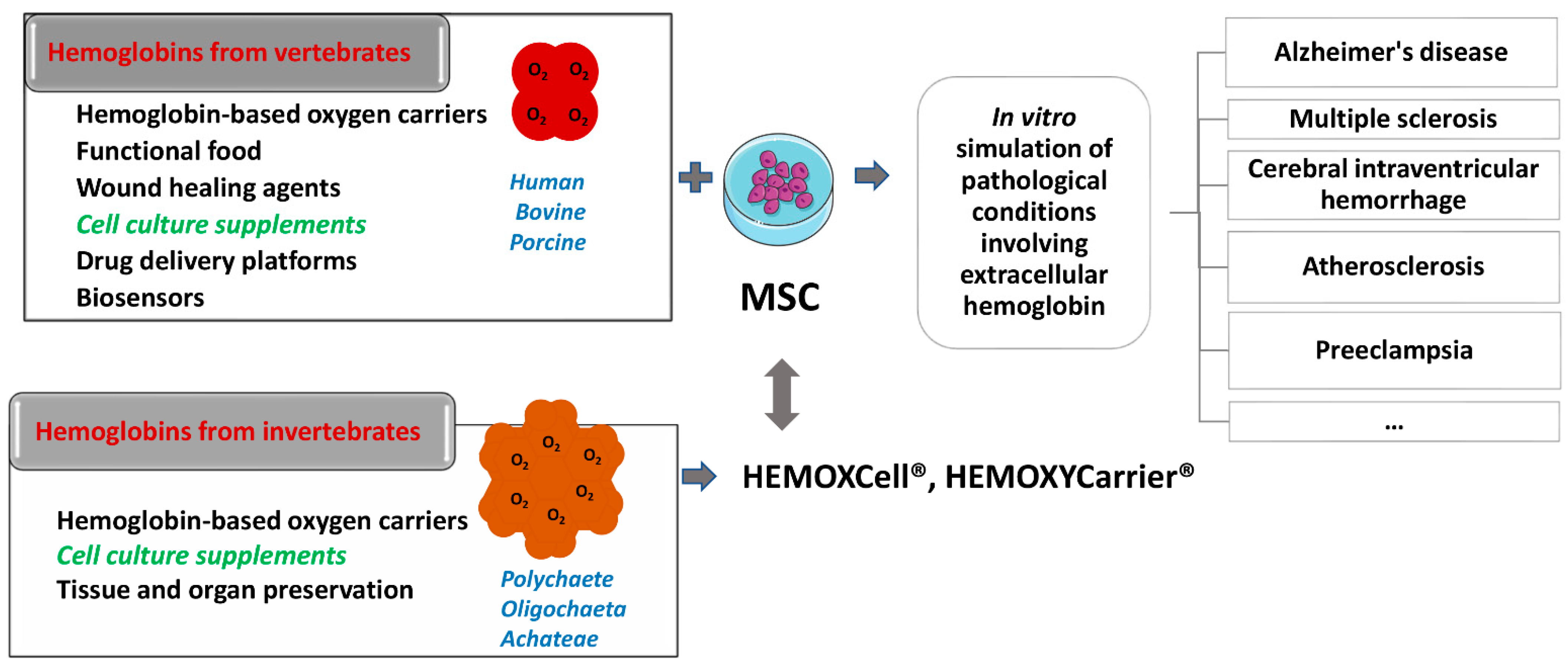
Hemoglobin is essential for maintaining cellular bioenergetic homeostasis through its ability to bind and transport oxygen to the tissues. Besides its ability to transport oxygen, hemoglobin within erythrocytes plays an important role in cellular signaling and modulation of the inflammatory response either directly by binding gas molecules (NO, CO, and CO2) or indirectly by acting as their source. Once hemoglobin reaches the extracellular environment, it acquires several secondary functions affecting surrounding cells and tissues. By modulating the cell functions, this macromolecule becomes involved in the etiology and pathophysiology of various diseases. The up-to-date results disclose the impact of extracellular hemoglobin on (i) redox status, (ii) inflammatory state of cells, (iii) proliferation and chemotaxis, (iv) mitochondrial dynamic, (v) chemoresistance and (vi) differentiation.


  • extracellular hemoglobin
  • oxygen carriers
  • cell culture additives

1. Introduction

Hemoglobin, a highly conserved protein, due to its ability to reversibly bind oxygen, is involved in the processes that underlie the aerobic life on planet Earth. The primary role of this protein is reflected in the maintenance of cellular homeostasis, by supporting its energy requirements. However, due to nearly 200 years of research into hemoglobin, it is now known that this protein also plays important roles in cellular signaling and modulation of the inflammatory response. Hemoglobin performs these functions directly, by binding gas molecules (NO, CO and CO2), or indirectly, by acting as their source. In vertebrates, hemoglobin performs most of its functions while inside the erythrocytes [1][2][3]. However, once hemoglobin reaches the extracellular environment, in conditions such as trauma, inflammation or infection, hemoglobin exerts other potentially harmful effects on cells and tissues and may be involved in the etiology and pathophysiology of various diseases [4][5][6]. This dichotomy in function makes hemoglobin the so-called “two-edged sword”, a friend in physiological conditions, and an enemy or potential danger in stress conditions [1][6][7][8]. Furthermore, through applied biomedical research which implies application of exogenous hemoglobin originating from different sources, novel insights into the modulatory capacities of this macromolecule are obtained.

2. Extracellular Hemoglobin in Mammals

2.1. Endogeous Extracellular Hemoglobin Removal and Degradation

Under physiological conditions, about 80–90% of erythrocytes are destroyed without releasing hemoglobin into plasma, in a process termed extravascular hemolysis. The remaining erythrocytes are removed under physiological conditions by a process designated as intravascular hemolysis—hemolysis within blood vessels [8][9]. In intravascular hemolysis, hemoglobin is released directly into the circulation, where this molecule and its degradation products can cause cell and tissue damage [10][11] if they exceed the capacity of the mechanisms involved in their removal. Free hemoglobin in plasma is rapidly oxidized to methemoglobin, which readily and non-enzymatically dissociates into heme and αβ dimers. At low plasma hemoglobin release, haptoglobin (Hp) irreversibly binds all αβ-globin dimers present [12][13]. Hp binding sites are located on α-globin chains [14]. Hp is synthesized in parenchymal cells of the liver, and the half-life of this glycoprotein in the circulation is 3.5–5 days. However, if it binds αβ-globin dimers, the half-life of such a complex becomes only 9 to 30 min. These complexes are rapidly removed from the circulation by phagocytosis by monocytes and tissue macrophages after binding to their CD163 receptor [15]. Since there is no Hp recycling, once the reserves from circulation are depleted it takes 5–7 days for the pool to be recovered because a reduced Hp concentration does not lead to an increase in the synthesis of this molecule.
In addition to Hp, both hemopexin and albumin act as mechanisms that limit the effects of extracellular hemoglobin [3][13]. These two molecules can bind free heme, maintain it in soluble form and thus prevent it from exhibiting its oxidative and proinflammatory effects [16]. Hemopexin is a plasma heme-binding glycoprotein with an affinity for heme higher than all known heme-binding proteins. Moreover, hemopexin mediates the intake of heme in hepatocytes in which this prosthetic group is removed. Hemopexin is synthesized in the liver and in healthy individuals has a half-life of an average of seven days, while in the complex with heme, its half-life is reduced to 7–8 h [17]. The heme-hemopexin complex enters hepatocytes through receptor-mediated endocytosis via a lipoprotein receptor-related protein-1 receptor (LRP), also known as CD91. After the endocytosis, the heme-hemopexin complex dissociates, and the hemopexin is released and returned to the plasma as an intact protein. Transport of heme in the cytoplasm occurs by an internal heme-binding membrane protein, and iron is rapidly removed by the action of heme oxygenase [18][19].
Heme can also bind to albumin in the circulation and form methemalbumin. When added to human serum, heme is initially primarily bound to albumin, probably due to its high concentration relative to hemopexin. Removing methemalbumin from circulation is a kinetically complex process [16]. Previous research shows that plasma heme can also bind α1-microglobulin (A1M), a 26 kDa glycoprotein synthesized in the liver and secreted into the blood. This protein has reductase activity, prevents intracellular oxidation, and reduces the expression of heme-induced heme oxygenase-1 (HO-1) and reactive oxygen species (ROS) production due to the presence of hemoglobin in the extracellular environment [20][21][22]. The newest data suggest the use of apohemoglobin or apohemoglobin-Hp as a novel therapeutic strategy in scavenging and clearing excess heme through the monocyte/macrophage CD163 surface receptor [23]. Subramanian et al. [24] identified a “shortcut” for detoxification of extracellular hemoglobin within plasma, which functions Hp-independently via the capture and quench mechanism and involves the CD163 monocyte/macrophage receptor. Following hemoglobin recruitment, membrane CD163 (mCD163) directly suppresses the pseudoperoxidase activity of hemoglobin in situ on the monocyte membrane. Hemoglobin induces the release of mCD163 into plasma, and the resulting soluble CD163 (sCD163) further captures and quenches residual redox-reactive hemoglobin. These authors showed that sCD163 and immunoglobulin G (IgG) interact with extracellular hemoglobin in plasma. The resulting sCD163-Hb-IgG complex then triggers an autocrine loop of endocytosis via Fcγ receptors on monocytes and consequent recycling of internalized sCD163 via endosomes to establish mCD163 homeostasis, while internalized hemoglobin is catabolized by HO-1. Additionally, this complex induces paracrine transactivation of vascular endothelial cells, stimulates HO-1 expression in them, and secretes cytokines that will trigger a systemic defense response directed toward extracellular hemoglobin [24].

2.2. Endogenous Extracellular Hemoglobin Mode of Action

If extracellular hemoglobin exceeds the homeostatic mechanisms for its removal during intravascular hemolysis, it can affect surrounding cells and tissues. Various clinical aspects associated with circulating extracellular hemoglobin excess have been attributed to hemoglobin molecule-specific structural and biochemical characteristics through four proposed interacting mechanisms [1][10]. The first of these is the extravascular translocation of hemoglobin (Figure 1A) [10]. After hemolysis, hemoglobin exists in the dynamic equilibrium of tetramers and αβ heterodimers, with a predominant dimer state at low plasma hemoglobin concentrations. Heterodimers are relatively small and capable of translocation and access to vulnerable anatomical sites (for example, glomeruli in the kidneys or vascular wall). Tissue exposure to hemoglobin most often accompanies cases of excessive hemoglobinuria after massive intravascular hemolysis, but hemoglobin can also translocate through endothelial barriers and thus enter the subendothelial and perivascular spaces and lymph [25][26]. The hemoglobin dimer/oligomer is considered a damage/danger-associated molecular pattern (DAMP), while heme is independently recognized as an “alarmin” [27][28][29].
Figure 1. Scheme illustrating the mechanisms of action of endogenous extracellular hemoglobin: (A) extravascular translocation of hemoglobin, (B) prooxidative reactivity of hemoglobin in plasma or within tissues after extravasation, (C) release of hemin from Hb-Fe3+ as the main product of oxidative reactions and (D) hemin induced changes in cell activation, gene expression, and metabolism.
Another mechanism by which extracellular hemoglobin exerts its effects is the prooxidative reactivity of hemoglobin in plasma or within tissues after extravasation (Figure 1B) [10]. The reactions of hemoglobin with NO and physiological oxidants (hydrogen peroxide and lipid peroxides) are best studied. NO consumption and consequent oxidation of hemoglobin occur through two reactions: (1) NO dioxygenation of oxyhemoglobin in which nitrates (NO3) and ferrihemoglobin (Hb-Fe3+) are generated (Equations (1) and (2)) through nitrosylation of iron deoxyhemoglobin which occurs in direct binding of NO to iron of ferrohemoglobin (Hb-Fe2+) [10][30][31]. The binding of NO by cell-free hemoglobin leads to depletion of this critical vasodilator produced in the vascular endothelium; it is reported that this reaction is the fundamental cause of hypertension [31], since even the recombinant hemoglobin, with diverse chemistries [32], exerts a similar rate of reaction (Equation (1)). The reaction of extracellular hemoglobin and NO also results in the generation of Hb-Fe3+ within the tissue parenchyma. The accumulation of Hb-Fe3+ can stimulate the release and/or transfer of hemin to other proteins and lipids, thus manifesting the secondary toxicity of free hemin. Although hemoglobin and peroxide reactions have been the subject of numerous studies for over 40 years, the pathophysiology underlying these reactions is still not completely clarified [2][10][18][33].
HbFe2+ + NO → NO3 + HbFe3+
It has been shown that in vitro, the reaction of hemoglobin and peroxide leads to the formation of Hb-Fe3+ and also to chemical species in which iron is in the ferryl form (Hb-Fe4+) and its radical forms (Equations (2) and (3)) [4][31][34][35].
HbFe2+ + O2 → HbFe3+ + O2
HbFe3+ + H2O2HbFe4+ =O + H2O
The outcome of such reactions is hemoglobin degradation, hemin loss, cross-linking, or precipitation of globin chains, which lead to tissue damage. It is still unknown how much Hb-Fe4+ and radicals are formed in vivo during hemolysis and to what extent they contribute to the development of the disease. The only oxidized type of hemoglobin that can be consistently quantified in vivo is Hb-Fe3+. In other words, it should be taken into account that the disparity observed in vitro and in vivo may be due to a shifted balance between oxidation and reduction reactions in vivo [33][36]. The third proposed mechanism by which extracellular hemoglobin exerts its effects is the release of hemin from Hb-Fe3+ as the main product of oxidative reactions (Figure 1C) [10]. The release of hemin allows the transfer of reactive porphyrin to the cell membrane or soluble plasma proteins and lipids and provides free hemin as a ligand for various signaling pathways. As hemin is a hydrophobic molecule, it is unlikely that more significant amounts of free hemin will be found in plasma; it binds rapidly to albumin or lipids and forms complexes. Depending on what it binds to, hemin can transform a molecule from a complex into a reactive product, such as oxidized low-density lipoprotein, further damaging the vasculature [8][37][38]. The fourth mechanism refers to the effects achieved by hemin (Figure 1D) [10]. Hemin can selectively bind to several receptors and transcription factors and lead to changes in the state of cell activation, gene expression, and metabolism. The best-characterized interaction is hemin binding to transcriptional repressor 1, which shows homology to BTB and CNC homology 1, Bach-1, which regulates the transcription of HO-1 and other antioxidant enzymes necessary for an adaptive response to increased intracellular hemin levels [39]. Hemin is also a ligand for the nuclear receptor of the hormone REV-ERB, which regulates circadian rhythm, glucose metabolism, and adipogenesis [40].

3. Exogenous Administration of Extracellular Hemoglobin

The very first beginnings of hemoglobin application in biomedicine are related to its use as a blood substitute, i.e., more precisely as oxygen carriers (hemoglobin-based oxygen carriers, HBOCs) [31][41][42][43] (Figure 2). The first HBOC was hemoglobin isolated from lysed erythrocytes given to patients intravenously. However, it has been observed that soon after applying hemoglobin solution in this way, besides its short intravascular persistence, kidney damage, high blood pressure, and cardiovascular complications occur [44][45][46] due to the effects of extracellular hemoglobin. These features and mechanisms of the act of extracellular hemoglobin are well known today and explained in detail in several comprehensive reviews and in the previous sections. Stroma-free hemoglobin rapidly dissociates into dimers and monomers, activates liver and spleen macrophages, and has the ability to bind the potent vasodilator, NO, resulting in vasoconstriction [31]. Due to hemoglobin dimer precipitation in proximal tubules, nephrotoxic action occurs. Additionally, outside of erythrocytes, hemoglobin quickly oxidizes to methemoglobin, which cannot bind oxygen; instead, it releases heme, which participates in the creation of free radicals that exert harmful effects on surrounding cells [42][43][47][48]. After such reports on outcomes of extracellular hemoglobin administration, researchers have focused on obtaining recombinant hemoglobin by mutagenesis with desired features and reduced side effects. With the use of recombinant technology, a low oxygen affinity human hemoglobin variant was produced in Escherichia coli by Somatogen Inc. by introducing the substitution of amino acid Lys for Asn at the β108 position [31]. To further stabilize this hemoglobin in tetrameric form, a site-specific mutation was introduced by inserting glycine into the region where two α subunits almost touch each other, fusing them (rHb1.0) [49]. The second generation of recombinant hemoglobin (rHb2.0) included a change of heme pocket chemistry by introducing site-directed mutagenesis followed by polymerization [50], such as providing HbA mutants with lower NO reactivity. A comprehensive summary of the impressive work done by biochemists in this field of recombinant hemoglobin is disclosed in the review of Varnado et al. [51]. Progress in the development of recombinant hemoglobin enabled (i) adjustment of oxygen (O2) affinity over a 100-fold range, (ii) reduction of nitric oxide (NO) scavenging activity over 30-fold without compromising dioxygen binding, (iii) decrease of antioxidant activity, (iv) reduction in hemin loss rate, (v) regulation of subunit dissociation rate, and (vi) decrease of irreversible subunit denaturation [51]. Since the relatively high cost of large-quantity production of these proteins for emergency management of trauma patients is the main obstacle for their further progress, more cost-effective chemical modifications of the hemoglobin molecules have been launched [42][52][53][54][55][56][57][58][59][60][61][62]. The procedure includes hemoglobin cross-linking/polymerization with succinyldisalicylic acid [53], glycine [54], glutaraldehyde [55], O-raffinose [56][57] or modification of the surface area of hemoglobin molecules with polyethylene glycol (PEG) [58][59] or human serum albumin with bound platinum nanoparticles [60][61]. Another successful approach towards overcoming the side effects of a “naked hemoglobin molecule” administration involves the use of encapsulation as a method. Hemoglobin encapsulated in different biomaterials revealed longer circulatory lifetime, decreased hypertensive response and phagocytic uptake [42]. From the pioneering steps in this approach by Chang et al. [62], encapsulating material for hemoglobin varied from cellulose nitrate or PEG-polylactide to poly(ε-caprolactone)/poly(l-lactic acid) and poly(l-lysine), poly(lactic-coglycolicacid)/PEG copolymers and various lipid vesicles [42]. Encapsulation of hemoglobin within emulsion with hydrophobic nanodrops (where hydrophobicity features of the nanoemulsion structures influence the binding of the hydrophobic niche of hemoglobin and thus partially act as a “stunt” -2, 3-DPG moderator) is patented demonstrating very promising results in the model of hemorrhagic rats [63]. Recent studies on hemoglobin encapsulation in lipid vesicles demonstrated remarkable progress by means of larger-scale production without affecting the biophysical properties of hemoglobin molecules [64] or introduction to clinical trials [65].
Figure 2. Schematic presentation of the potential use of vertebrates and invertebrates’ hemoglobin preparations in biomedicine and biotechnology (left side of the scheme), emphasizing already commercially available cell culture additives based on invertebrate hemoglobin (HEMOXCell®, HEMOXYCarriers®, HEMARINA SA, Morlaix, France). Up-to-date research on the impact of such hemoglobin preparation on mesenchymal stromal cell (MSC) cultures could significantly contribute to a better understanding of the onset and progression of pathological conditions involving extracellular hemoglobin as presented on the right side of the scheme.
Both allogeneic and xenogeneic hemoglobins were used as a source of extracellular hemoglobin. Among xenogeneic hemoglobins, the most studied are bovine and porcine hemoglobin, which show a high degree of homology with human hemoglobin (84.4% and 85%, respectively). Apart from the use as blood substitute, porcine hemoglobin was used as a wound healing agent in the form of spray if applied at regular intervals of 2–4 weeks. Namely, this product has been shown to reduce pain, wound size, and the necrotic tissue layer that impairs wound healing [66][67]. Moreover, there are reports on bovine hemoglobin used as a drug platform itself, i.e., as a pH-sensitive nano-vehicles for potential cancer detection and therapy [68] and as an effective glucose biosensor in vitro [69]. One part of the research relies on examining rodent hemoglobin as well, aiming to simulate extracellular hemoglobin presence in vivo [70]. Furthermore, hemoglobin from invertebrates has also become a significant subject of research. Thus, potential of extracellular hemoglobin as an additive for organ and tissue preservation [71][72] and as a growth stimulator of mesenchymal stromal cells (MSC), which maintain ‘‘stemness” in vitro [73], have been described. Therefore, one should be aware that under the same term “hemoglobin”, the studies on extracellular hemoglobin functions imply both vertebrate and evolutionary distant invertebrate hemoglobin.
Among invertebrate hemoglobins, the most investigated are extracellular hemoglobin of annelids (also named erythrocruorins and chlorocruorins after the nature of the porphyrin group) [74][75], present in three classes of annelids: Polychaete, Oligochaeta, and Achateae. Annelid hemoglobins are huge biopolymers with a high molecular weight of 3000–4000 kDa [75][76]. The beginning of the application of EHb in biotechnology is related to the group led by Andre Tulmond and Franck Zal. This research group started an examination on hemoglobin from the marine polychaete Arenicola marina (HbAm) in 1993 and described the structure of several extracellular hemoglobins in detail [77]. It has been discovered that the hemoglobin of Arenicola marina (HbAm) had all the sought traits of the universal oxygen carrier: it is naturally extracellular and polymerized, and its molecular mass is 50 times greater than the mass of human hemoglobin. It had properties of O2-binding and release like human hemoglobin within erythrocytes. Each HbAm molecule can bind 156 oxygen molecules (human Hb can bind four) and has natural antioxidant properties. Additionally, HbAm has functional properties independent of secondary molecules such as 2,3-DPG in humans. The HbAm molecule displayed activity without any additional chemical modifications. It appears to be stable at temperatures ranging from 4 to >30 °C and does not show any vasoconstriction effects upon application [75][76]. HbAm dissociates in human plasma from a hexagonal bilayer into globin dodecamers (although the dodecamer still appears to function as desired without extravasating) [78][79]. It is important to note erythrocruorin of Lumbricus terrestris (LtEc), which has been shown to be extremely stable, resistant to oxidation, and may interact with NO differently than mammalian hemoglobin molecules [79].

This entry is adapted from the peer-reviewed paper 10.3390/biom12111708

References

  1. Quaye, I.K. Extracellular Hemoglobin: The Case of a Friend Turned Foe. Front. Physiol. 2015, 6, 96.
  2. Coates, C.J.; Decker, H. Immunological Properties of Oxygen-Transport Proteins: Hemoglobin, Hemocyanin and Hemerythrin. Cell. Mol. Life Sci. 2016, 74, 293–317.
  3. Olson, J.S. Lessons Learned from 50 Years of Hemoglobin Research: Unstirred and Cell-Free Layers, Electrostatics, Baseball Gloves, and Molten Globules. Antioxid. Redox Signal. 2020, 32, 228–246.
  4. Rifkind, J.M.; Mohanty, J.G.; Nagababu, E. The Pathophysiology of Extracellular Hemoglobin Associated with Enhanced Oxidative Reactions. Front. Physiol. 2015, 5, 500.
  5. Lara, F.A.; Kahn, S.A.; Da Fonseca, A.C.C.; Bahia, C.P.; Pinho, J.P.C.; Graca-Souza, A.V.; Houzel, J.C.; De Oliveira, P.L.; Moura-Neto, V.; Oliveira, M.F. On the fate of extracellular hemoglobin and heme in brain. J. Cereb. Blood Flow Metab. 2009, 29, 1109–1120.
  6. Rother, R.P.; Bell, L.; Hillmen, P. The clinical sequelae of Intravascular Hemolysis A Novel Mechanism of Human Disease. JAMA 2015, 293, 1653–1662.
  7. Ogasawara, N.; Oguro, T.; Sakabe, T.; Matsushima, M.; Takikawa, O.; Isobe, K.I.; Nagase, F. Hemoglobin Induces the Expression of Indoleamine 2,3-Dioxygenase in Dendritic Cells through the Activation of PI3K, PKC, and NF-ΚB and the Generation of Reactive Oxygen Species. J. Cell. Biochem. 2009, 108, 716–725.
  8. Jeney, V.; Eaton, J.W.; Balla, G.; Balla, J. Natural History of the Bruise: Formation, Elimination, and Biological Effects of Oxidized Hemoglobin. Oxid. Med. Cell. Longev. 2013, 2013, 703571.
  9. Bahl, N.; Winarsih, I.; Tucker-Kellogg, L.; Ding, J.L. Extracellular Haemoglobin Upregulates and Binds to Tissue Factor on Macrophages: Implications for Coagulation and Oxidative Stress. Thromb. Haemost. 2014, 111, 67–78.
  10. Schaer, D.J.; Buehler, P.W.; Alayash, A.I.; Belcher, J.D.; Vercellotti, G.M. Hemolysis and Free Hemoglobin Revisited: Exploring Hemoglobin and Hemin Scavengers as a Novel Class of Therapeutic Proteins. Blood 2013, 121, 1276–1285.
  11. Jeney, V. Pro-Inflammatory Actions of Red Blood Cell-Derived DAMPs. In Inflammasomes: Clinical and Therapeutic Implications, Experientia Supplementum; Cordero, M., Alcocer-Gómez, E., Eds.; Springer: Cham, Switzerland, 2018; Volume 108, pp. 211–233.
  12. Garton, T.; Keep, R.F.; Hua, Y.; Xi, G. CD163, a Hemoglobin/Haptoglobin Scavenger Receptor, After Intracerebral Hemorrhage: Functions in Microglia/Macrophages Versus Neurons. Transl. Stroke Res. 2017, 8, 612–616.
  13. Smith, A.; McCulloh, R.J. Hemopexin and Haptoglobin: Allies against Heme Toxicity from Hemoglobin Not Contenders. Front. Physiol. 2015, 6, 187.
  14. McCormick, D.J.; Atassi, M.Z. Hemoglobin Binding with Haptoglobin: Delineation of the Haptoglobin Binding Site on the α-Chain of Human Hemoglobin. J. Protein Chem. 1990, 9, 735–742.
  15. Kaempfer, T.; Duerst, E.; Gehrig, P.; Roschitzki, B.; Rutishauser, D.; Grossmann, J.; Schoedon, G.; Vallelian, F.; Schaer, D.J. Extracellular Hemoglobin Polarizes the Macrophage Proteome toward Hb-Clearance, Enhanced Antioxidant Capacity and Suppressed HLA Class 2 Expression. J. Proteome Res. 2011, 10, 2397–2408.
  16. Smeds, E.; Romantsik, O.; Jungner, Å.; Erlandsson, L.; Gram, M. Pathophysiology of Extracellular Haemoglobin: Use of Animal Models to Translate Molecular Mechanisms into Clinical Significance. ISBT Sci. Ser. 2017, 12, 134–141.
  17. Belcher, J.D.; Chen, C.; Nguyen, J.; Abdulla, F.; Zhang, P.; Nguyen, H.; Nguyen, P.; Killeen, T.; Miescher, S.M.; Brinkman, N.; et al. Haptoglobin and Hemopexin Inhibit Vaso-Occlusion and Inflammation in Murine Sickle Cell Disease: Role of Heme Oxygenase-1 Induction. PLoS ONE 2018, 13, e0196455.
  18. Lee, S.K.; Ding, J.L. A Perspective on the Role of Extracellular Hemoglobin on the Innate Immune System. DNA Cell Biol. 2013, 32, 36–40.
  19. Ponka, P.; Sheftel, A.D.; English, A.M.; Scott Bohle, D.; Garcia-Santos, D. Do Mammalian Cells Really Need to Export and Import Heme? Trends Biochem. Sci. 2017, 42, 395–406.
  20. Sverrisson, K.; Axelsson, J.; Rippe, A.; Gram, M.; Åkerström, B.; Hansson, S.R.; Rippe, B. Extracellular Fetal Hemoglobin Induces Increases in Glomerular Permeability: Inhibition with A1 -Microglobulin and Tempol. Am. J. Physiol. Ren. Physiol. 2014, 306, 442–448.
  21. Penders, J.; Delanghe, J.R. Alpha 1-Microglobulin: Clinical Laboratory Aspects and Applications. Clin. Chim. Acta 2004, 346, 107–118.
  22. Olsson, M.; Olofsson, T.; Tapper, H.; Åkerström, B. The Lipocalin Aa1-Microglobulin Protects Erythroid K562 Cells against Oxidative Damage Induced by Heme and Reactive Oxygen Species. Free Radic. Res. 2008, 42, 725–736.
  23. Munoz, J.C.; Pires, S.I.; Baek, H.J.; Buehler, W.P.; Palmer, F.A.; Cabrales, P. Apohemoglobin-Haptoglobin Complex Attenuates the Pathobiology of Circulating Acellular 2 Hemoglobin and Heme. Am. J. Physiol. Circ. Physiol. 2020, 318, H1296–H1307.
  24. Subramanian, K.; Du, R.; Tan, N.S.; Ho, B.; Ding, J.L. CD163 and IgG Codefend against Cytotoxic Hemoglobin via Autocrine and Paracrine Mechanisms. J. Immunol. 2013, 190, 5267–5278.
  25. Nakai, K.; Sakuma, I.; Ohta, T.; Ando, J.; Kitabatake, A.; Nakazato, Y.; Takahashi, T.A. Permeability Characteristics of Hemoglobin Derivatives across Cultured Endothelial Cell Monolayers. J. Lab. Clin. Med. 1998, 132, 313–319.
  26. Matheson, B.; Razynska, A.; Kwansa, H.; Bucci, E. Appearance of Dissociable and Cross-Linked Hemoglobins in the Renal Hilar Lymph. J. Lab. Clin. Med. 2000, 135, 459–464.
  27. Schechter, A.N. Hemoglobin Research and the Origins of Molecular Medicine. Blood 2008, 112, 3927–3938.
  28. Akira, S.; Hemmi, H. Recognition of Pathogen-Associated Molecular Patterns by TLR Family. Immunol. Lett. 2003, 85, 85–95.
  29. Bozza, M.T.; Jeney, V. Pro-inflammatory Actions of Heme and Other Hemoglobin-Derived DAMPs. Front. Immunol. 2020, 30, 1323.
  30. Helms, C.; Kim-Shapiro, D.B. Hemoglobin-Mediated Nitric Oxide Signaling. Free Radic. Biol. Med. 2013, 61, 464–472.
  31. Alayash, A.I. Oxygen therapeutics: Can we tame haemoglobin? Nat. Rev. Drug Discov. 2004, 3, 152–159.
  32. Doherty, D.H.; Doyle, M.P.; Curry, S.R.; Vali, R.J.; Fattor, T.J.; Olson, J.S.; Lemon, D.D. Rate of reaction with nitric oxide determines the hypertensive effect of cell-free hemoglobin. Nat. Biotechnol. 1998, 16, 672–676.
  33. Reeder, B.J. The Redox Activity of Hemoglobins: From Physiologic Functions to Pathologic Mechanisms. Antioxidants Redox Signal. 2010, 13, 1087–1123.
  34. Bamm, V.V.; Henein, M.E.L.; Sproul, S.L.J.; Lanthier, D.K.; Harauz, G. Potential Role of Ferric Hemoglobin in MS Pathogenesis: Effects of Oxidative Stress and Extracellular Methemoglobin or Its Degradation Products on Myelin Components. Free Radic. Biol. Med. 2017, 112, 494–503.
  35. Chintagari, N.R.; Jana, S.; Alayash, A.I. Oxidized Ferric and Ferryl Forms of Hemoglobin Trigger Mitochondrial Dysfunction and Injury in Alveolar Type i Cells. Am. J. Respir. Cell Mol. Biol. 2016, 55, 288–298.
  36. Buehler, P.W.; D’Agnillo, F.; Hoffman, V.; Alayash, A.I. Effects of Endogenous Ascorbate on Oxidation, Oxygenation, and Toxicokinetics of Cell-Free Modified Hemoglobin after Exchange Transfusion in Rat and Guinea Pig. J. Pharmacol. Exp. Ther. 2007, 323, 49–60.
  37. Balla, G.; Jacob, H.S.; Eaton, J.W.; Belcher, J.D.; Vercellotti, G.M. Hemin: A Possible Physiological Mediator of Low Density Lipoprotein Oxidation and Endothelial Injury. Arterioscler. Thromb. Vasc. Biol. 1991, 11, 1700–1711.
  38. Nagy, E.; Eaton, J.W.; Jeney, V.; Soares, M.P.; Varga, Z.; Galajda, Z.; Szentmiklósi, J.; Méhes, G.; Csonka, T.; Smith, A.; et al. Red Cells, Hemoglobin, Heme, Iron, and Atherogenesis. Arterioscler. Thromb. Vasc. Biol. 2010, 30, 1347–1353.
  39. Ogawa, K.; Sun, J.; Taketani, S.; Nakajima, O.; Nishitani, C.; Sassa, S.; Hayashi, N.; Yamamoto, M.; Shibahara, S.; Fujita, H.; et al. Heme Mediates Derepression of Maf Recognition Element through Direct Binding to Transcription Repressor Bach1. EMBO J. 2001, 20, 2835–2843.
  40. Raghuram, S.; Stayrook, K.R.; Huang, P.; Rogers, P.M.; Nosie, A.K.; McClure, D.B.; Burris, L.L.; Khorasanizadeh, S.; Burris, T.P.; Rastinejad, F. Identification of Heme as the Ligand for the Orphan Nuclear Receptors REV-ERBα and REV-ERBβ. Nat. Struct. Mol. Biol. 2007, 14, 1207–1213.
  41. Alayash, A.I. Hemoglobin-Based Blood Substitutes: Oxygen Carriers, Pressor Agents, or Oxidants? Nat. Biotechnol. 1999, 17, 545–549.
  42. Bialas, C.; Moser, C.; Sims, C.A. Artificial Oxygen Carriers and Red Blood Cell Substitutes: A Historic Overview and Recent Developments toward Military and Clinical Relevance. J. Trauma Acute Care Surg. 2019, 87 (Suppl. S1), S48–S58.
  43. Baron, J.F. Blood substitutes. Haemoglobin therapeutics in clinical practice. Crit. Care 1999, 3, R99–R102.
  44. Amberson, W.R. Clinical experience with hemoglobin-saline solutions. Science 1947, 106, 117.
  45. Savitsky, J.P.; Doczi, J.; Black, J.; Arnold, J.D. A clinical safety trial of stroma-free hemoglobin. Clin. Pharmacol. Ther. 1978, 23, 73–80.
  46. Bunn, H.F.; Esham, W.T.; Bull, R.W. The renal handling of hemoglobin. I. Glomerular filtration. J. Exp. Med. 1969, 129, 909–923.
  47. Sen Gupta, A. Hemoglobin-based Oxygen Carriers: Current State-of-the-art and Novel Molecules. Shock 2019, 52 (Suppl. S1), 70–83.
  48. Sakai, H.; Sou, K.; Horinouchi, H.; Kobayashi, K.; Tsuchida, E. Review of hemoglobin-vesicles as artificial oxygen carriers. Artif. Organs 2009, 33, 139–145.
  49. Hoffman, S.J.; Looker, D.L.; Roehrich, J.M.; Cozart, P.E.; Durfee, S.L.; Tedesco, J.L.; Stetler, G.L. Expression of fully functional tetrameric human hemoglobin in Escherichia coli. Proc. Natl. Acad. Sci. USA 1990, 87, 8521–8525.
  50. Resta, T.C.; Walker, B.R.; Eichinger, M.R.; Doyle, M.P. Rate of NO scavenging alters effects of recombinant hemoglobin solutions on pulmonary vasoreactivity. J. Appl. Physiol. 2002, 93, 1327–1336.
  51. Varnado, C.L.; Mollan, T.L.; Birukou, I.; Smith, B.J.; Henderson, D.P.; Olson, J.S. Development of recombinant hemoglobin-based oxygen carriers. Antioxid Redox Signal. 2013, 18, 2314–2328.
  52. Chen, J.Y.; Scerbo, M.; Kramer, G. A Review of Blood Substitutes: Examining the History, Clinical Trial Results, and Ethics of Hemoglobin-Based Oxygen Carriers. Clinics 2009, 64, 803–813.
  53. Lamy, M.L.; Daily, E.K.; Brichant, J.F.; Larbuisson, R.P.; Demeyere, R.H.; Vandermeersch, E.A.; Lehot, J.J.; Parsloe, M.R.; Berridge, J.C.; Sinclair, C.J.; et al. Randomized Trial of Diaspirin Cross-Linked Hemoglobin Solution as an Alternative to Blood Transfusion after Cardiac Surgery. Anesthesiology 2000, 92, 646–656.
  54. Winslow, R.M. New transfusion strategies: Red Cell Substitutes. Annu. Rev. Med. 1999, 50, 337–353.
  55. Jahr, S.J.; Moallempour, M.; Lim, C.J. HBOC-201, Hemoglobin Glutamer-250 (Bovine), Hemopure (Biopure Corporation). Expert Opin. Biol. Ther. 2008, 8, 1425–1433.
  56. Gould, S.A.; Moore, E.E.; Hoyt, D.B.; Burch, J.M.; Haenel, J.B.; Garcia, J.; DeWoskin, R.; Moss, G.S. The First Randomized Trial of Human Polymerized Hemoglobin as a Blood Substitute in Acute Trauma and Emergent Surgery. J. Am. Coll. Surg. 1998, 187, 113–120.
  57. Cheng, D.C.H.; Mazer, C.D.; Martineau, R.; Ralph-Edwards, A.; Karski, J.; Robblee, J.; Finegan, B.; Hall, R.I.; Latimer, R.; Vuylsteke, A. A Phase II Dose-Response Study of Hemoglobin Raffimer (Hemolink) in Elective Coronary Artery Bypass Surgery. J. Thorac. Cardiovasc. Surg. 2004, 127, 79–86.
  58. Olofsson, C.; Ahl, T.; Johansson, T.; Larsson, S.; Nellgård, P.; Ponzer, S.; Fagrell, B.; Przybelski, R.; Keipert, P.; Winslow, N.; et al. A Multicenter Clinical Study of the Safety and Activity of Maleimide-Polyethylene Glycol-Modified Hemoglobin (Hemospan®) in Patients Undergoing Major Orthopedic Surgery. Anesthesiology 2006, 105, 1153–1163.
  59. Abuchowski, A. SANGUINATE (PEGylated Carboxyhemoglobin Bovine): Mechanism of Action and Clinical Update. Artif. Organs 2017, 41, 346–350.
  60. Tomita, D.; Kimura, T.; Hosaka, H.; Daijima, Y.; Haruki, R.; Ludwig, K.; Böttcher, C.; Komatsu, T. Covalent Core-Shell Architecture of Hemoglobin and Human Serum Albumin as an Artificial O2 Carrier. Biomacromolecules 2013, 14, 1816–1825.
  61. Iwasaki, H.; Yokomaku, K.; Kureishi, M.; Igarashi, K.; Hashimoto, R.; Kohno, M.; Iwazaki, M.; Haruki, R.; Akiyama, M.; Asai, K.; et al. Hemoglobin–Albumin Cluster: Physiological Responses after Exchange Transfusion into Rats and Blood Circulation Persistence in Dogs. Artif. Cells Nanomed. Biotechnol. 2018, 46 (Suppl. 3), S621–S629.
  62. Chang, T.M. Semipermeable microcapsules. Science 1964, 146, 524–525.
  63. Bugarski, B.; Dovezenski, N.; Stojanovic, N.; Bugarski, D.; Koncern, H. Emulsion Containing Hydrophobic Nanodrops with Bound Hemoglobin Molecules in a Hydrophilic Phase as a Blood Substitute. Deutsches Patentamt DE 2002-10209860 WO 2003074022, 9 December 2003.
  64. Banerjee, U.; Wolfe, S.; O’Boyle, Q.; Cuddington, C.; Palmer, A.F. Scalable production and complete biophysical characterization of poly(ethylene glycol) surface conjugated liposome encapsulated hemoglobin (PEG-LEH). PLoS ONE 2022, 17, e0269939.
  65. Azuma, H.; Amano, T.; Kamiyama, N.; Takehara, N.; Jingu, M.; Takagi, H.; Sugita, O.; Kobayashi, N.; Kure, T.; Shimizu, T.; et al. First-in-human phase 1 trial of hemoglobin vesicles as artificial red blood cells developed for use as a transfusion alternative. Blood Adv. 2022, 6, 5711–5715.
  66. Arenberger, P.; Engels, P.; Arenbergerova, M.; Gkalpakiotis, S.; García Luna Martínez, F.J.; Villarreal Anaya, A.; Jimenez Fernandez, L. Clinical Results of the Application of a Hemoglobin Spray to Promote Healing of Chronic Wounds. GMS Krankenhhyg. Interdiszip. 2011, 6, Doc05.
  67. Elg, F.; Hunt, S. Hemoglobin Spray as Adjunct Therapy in Complex Wounds: Meta-Analysis versus Standard Care Alone in Pooled Data by Wound Type across Three Retrospective Cohort Controlled Evaluations. SAGE Open Med. 2018, 6, 205031211878431.
  68. Li, H.; Chen, Y.; Li, Z.; Li, X.; Jin, Q.; Ji, J. Hemoglobin as a Smart PH-Sensitive Nanocarrier to Achieve Aggregation Enhanced Tumor Retention. Biomacromolecules 2018, 19, 2007–2013.
  69. Qi, W.; Yan, X.; Duan, L.; Cui, Y.; Yang, Y.; Li, J. Glucose-Sensitive Microcapsules from Glutaraldehyde Cross-Linked Hemoglobin and Glucose Oxidase. Biomacromolecules 2009, 10, 1212–1216.
  70. Stančić, A.Z.; Drvenica, I.T.; Obradović, H.N.; Bugarski, B.M.; Lj Ilić, V.; Bugarski, D.S. Native Bovine Hemoglobin Reduces Differentiation Capacity of Mesenchymal Stromal Cells in Vitro. Int. J. Biol. Macromol. 2020, 144, 909–920.
  71. Dutheil, D.; Rousselot, M.; Hauet, T.; Zal, F. Organ-Preserving Composition and Uses. U.S. Patent US 2014/0113274 A1, 24 April 2014. Available online: http://www.freepatentsonline.com/y2014/0113274.html (accessed on 8 November 2022).
  72. Schweitzer, M.H.; Zheng, W.; Cleland, T.P.; Goodwin, M.B.; Boatman, E.; Theil, E.; Marcus, M.A.; Fakra, S.C. A role for iron and oxygen chemistry in preserving soft tissues, cells and molecules from deep time. Proc. Biol. Sci. B 2013, 281, 20132741.
  73. Le Pape, F.; Cosnuau-Kemmat, L.; Gaëlle, R.; Dubrana, F.; Férec, C.; Zal, F.; Leize, E.; Delépine, P. HEMOXCell, a New Oxygen Carrier Usable as an Additive for Mesenchymal Stem Cell Culture in Platelet Lysate-Supplemented Media. Artif. Organs 2017, 41, 359–371.
  74. Magaldi, A.G.; Ghiretti, F.; Tognon, G.; Zanotti, G. The Structure of the Extracellular Hemoglobin of Annelids. In Invertebrate Oxygen Carriers; Linzen, B., Ed.; Springer: Berlin/Heidelberg, Germany, 1986.
  75. Zal, F.; Rousselot, M. Extracellular Hemoglobins from Annelids, and Their Potential Use in Biotechnology. In Outstanding Marine Molecules: Chemistry, Biology, Analysis; Wiley-Blackwell: Hoboken, NJ, USA, 2014; pp. 361–376.
  76. Batool, F.; Delpy, E.; Zal, F.; Leize-Zal, E.; Huck, O. Therapeutic Potential of Hemoglobin Derived from the Marine Worm Arenicola Marina (M101): A Literature Review of a Breakthrough Innovation. Mar. Drugs 2021, 19, 376.
  77. Zal, F.; Green, B.N.; Lallier, F.H.; Vinogradov, S.N.; Toulmond, A. Quaternary Structure of the Extracellular Haemoglobin of the Lugworm Arenicola Marina. A Multi-Angle-Laser-Light-Scattering and Electrospray-Ionisation-Mass-Spectrometry Analysis. Eur. J. Biochem. 1997, 243, 85–92.
  78. Rousselot, M.; Delpy, E.; La Rochelle, C.D.; Lagente, V.; Pirow, R.; Rees, J.F.; Hagege, A.; Le Guen, D.; Hourdez, S.; Zal, F. Arenicola Marina Extracellular Hemoglobin: A New Promising Blood Substitute. Biotechnol. J. 2006, 1, 333–345.
  79. Elmer, J.; Palmer, A.F. Biophysical Properties of Lumbricus terrestris Erythrocruorin and Its Potential Use as a Red Blood Cell Substitute. J. Funct. Biomater. 2012, 3, 49–60.
More
This entry is offline, you can click here to edit this entry!
Video Production Service